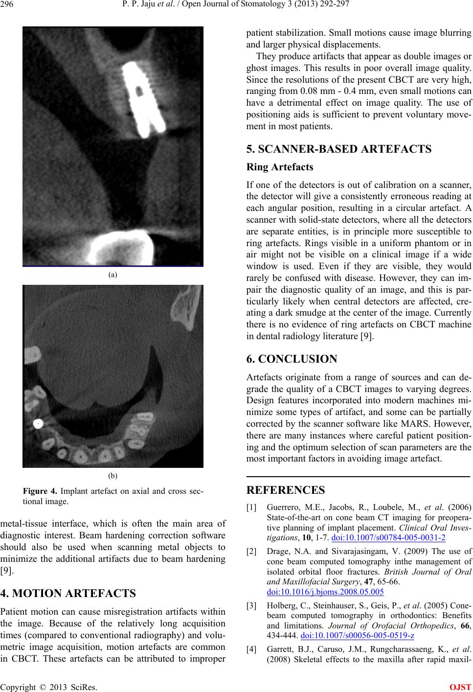
P. P. Jaju et al. / Open Journal of Stomatology 3 (2013) 292-297
Copyright © 2013 SciRes.
297
OJST
lary expansion assessed with cone-beam computed tomo-
graphy. American Journal of Orthodontics and Dento-
facial Orthopedics, 134, 8-9.
doi:10.1016/j.ajodo.2008.06.004
[5] Misch, K.A., Yi, E.S. and Sarment, D.P. (2006) Accuracy
of cone beam computed tomography for periodontal de-
fect measurements. Journal of Periodontology, 77, 1261-
1266. doi:10.1902/jop.2006.050367
[6] Honda, K., Arai, Y., Kashima, M., et al. (2004) Evalua-
tion of the usefulness of the limited cone-beamCT(3DX)
in the assessment of the thickness of the roof of the gle-
noid fossa of the temporomandibular joint. Dentomaxil-
lofacial Radiology, 33, 391-395.
doi:10.1259/dmfr/54316470
[7] Bianchi, S., Anglesio, S., Castellano, S., Rizzi, L. and
Ragona, R. (2001) Absorbed doses and risk in implant
planning: Comparison between spiral CT and cone beam
CT. Dentomaxillofacial Radiology, 30, S28.
[8] Tsiklakis, K., Donta, C., Gavala, S., Karayianni, K., Ka-
menopoulou, V. and Hourdakis, C.J. (2005) Dose reduc-
tion in maxillofacial imaging using low dose cone beam
CT. European Journal of Radiology, 56, 413-417.
doi:10.1016/j.ejrad.2005.05.011
[9] Barrett, J.F. and Keat, N. (2004) Artifacts in CT: Recog-
nition and avoidance. Radiographics, 24, 1679-1691.
doi:10.1148/rg.246045065
[10] Hunter, A.K. and McDavid, W.C. (2012) Characteriza-
tion and correction of cupping effect artefacts in cone
beam CT. Dentomaxillofacial Radiology, 41, 217-223.
doi:10.1259/dmfr/19015946
[11] Miracle, A.C. and Mukerji, S.K. (2009) Conebeam CT of
the head and neck, Part 1: Physical principles. American
Journal of Neuroradiology, 30, 1088-1095.
doi:10.3174/ajnr.A1653
[12] McDavid, W.D., Waggener, R.G., Payne, W.H. and Den-
nis, M.J. (1975) Spectral effects on three-dimensional re-
construction from x-rays. Medical Physics, 2, 321-324.
doi:10.1118/1.594200
[13] Brooks, R.A. and Di Chiro, G. (1976) Beam hardening in
X-ray reconstructive tomography. Physics in Medicine
and Biology, 21, 390-398.
doi:10.1088/0031-9155/21/3/004
[14] Lehr, J.L. (1983) Truncated-view artifacts: Clinical im-
portance on CT. American Journal of Roentgenology, 141,
183-191. doi:10.2214/ajr.141.1.183
[15] Bryant, J.A., Drage, N.A. and Richmond, S. (2008) Study
of the scan uniformity from an i-CAT cone beam CT
dental imaging system. Dentomaxillofacial Radiology, 37,
365-374. doi:10.1259/dmfr/13227258
[16] Katsumata, A., Hirukawa, A., Okumura, S., Naitoh, M.,
Fujishita, M., Ariji, E., et al. (2009) Relationship between
density variability and imaging volume size in cone-beam
computerized tomographic scanning of the maxillofacial
region: An in vitro study. Oral Surgery, Oral Medicine,
Oral Pathology, Oral Radiology, and Endodontics, 107,
420-425. doi:10.1016/j.tripleo.2008.05.049
[17] Jennings, R.J. (1988) A method for comparing beam-
hardening filter materials for diagnostic radiology. Me-
dical Physics, 15, 588-599. doi:10.1118/1.596210
[18] Meganck, J.A., Kozloff, K.M., Thornton, M.M., Broski,
S.M. and Goldstein, S.A. (2009) Beam hardening arti-
facts in micro-CT scanning can be reduced by X-ray
beam filtration and the resulting images can be used to
accurately measure BMD. Bone, 45, 1104-1116.
doi:10.1016/j.bone.2009.07.078
[19] Graham, S.A., Moseley, D.J., Siewerdsen, J.H., et al.
(2007) Compensators for dose and scatter management in
cone-beam computed tomography. Medical Physics, 34,
2691-2703. doi:10.1118/1.2740466
[20] Gupta, R., Grasruck, M., Suess, C., et al. (2006) Ultra-
high resolution flat-panel volume CT: Fundamental prin-
ciples, design architecture, and system characterization.
European Radiology, 16, 1191-1205.
doi:10.1007/s00330-006-0156-y
[21] Ning, R., Chen, B., Yu, R., et al. (2000) Flat panel detec-
tor-based cone-beam volume CT angiography imaging:
System evaluation. IEEE Transactions on Medical Imag-
ing, 19, 949-963. doi:10.1109/42.887842
[22] Neitzel, U. (1992) Grids or air gaps for scatter reduction
in digital radiography: A model calculation. Medical Phy-
sics, 19, 475-481. doi:10.1118/1.596836
[23] Siewerdsen, J.H., Mose ley, D. J., Bakhtiar, B., et al. (2004)
The influence of antiscatter grids on soft-tissue detec-
tability in cone-beam computed tomography with flat-
panel detectors. Medical Physics, 31, 3506-3520.
doi:10.1118/1.1819789
[24] Nickoloff, E.L., Lu, Z.F., Dutta, A., et al. (2007) Influ-
ence of flat-panel fluoroscopic equipment variables on
cardiac radiation doses. CardioVascular and Interven-
tional Radiology, 30, 169-176.
doi:10.1007/s00270-006-0096-6
[25] Orth, R.C., Wallace, M.J. and Kuo, M.D. (For the Tech-
nology Assessment Committee of the Society of Interven-
tional Radiology) (2008) C-arm cone-beam CT: General
principles and technical considerations for use in inter-
ventional radiology. Journal of Vascular and Interven-
tional Radiology, 19, 814-820.
doi:10.1016/j.jvir.2008.02.002
[26] Dorfler, A., Struffert, T., Engelhorn, T., et al. (2008) Ro-
tati onal flat-panel computed tomography in diagnostic
and interventional neuroradiology. Rofo, 180, 891-898.
[27] Jarry, G., Graham, S.A., Moseley, D.J., et al. (2006) Cha-
racterization of scattered radiation in kV CBCT images
using Monte Carlo simulations. Medical Physics, 33, 4320-
4329. doi:10.1118/1.2358324