Open Journal of Orthopedics, 2013, 3, 213-216
http://dx.doi.org/10.4236/ojo.2013.34039 Published Online August 2013 (http://www.scirp.org/journal/ojo)
Copyright © 2013 SciRes. OJO
213
Immediate Total Elbow Arthroplasty Following Olecranon
Osteotomy: A Case Report
Lee M. Reichel*, Cody D. Hillin, Charles A. Reitman
Department of Orthopedic Surgery, Baylor College of Medicine, Ben Taub General Hospital, Houston, USA.
Email: *leereichel@gmail.com
Received May 6th, 2013; revised June 8th, 2013; accepted June 22nd, 2013
Copyright © 2013 Lee M. Reichel et al. This is an open access article distributed under the Creative Commons Attribution License,
which permits unrestricted use, distribution, and reproduction in any medium, provided the original work is properly cited.
ABSTRACT
Consistent with an aging population, the overall number of distal humeral fractures in the elderly is increasing. Indica-
tions for application of acute total elbow arthroplasty (TEA) in the setting of distal humeral fractures are still being de-
fined. A variety of factors including chronologic age, physiologic age, bone quality, presence of pre-existing arthritis
and pre-existing medical conditions need to be considered. Optimally the decision to proceed with TEA verses open
reduction internal fixation (ORIF) is made preoperatively. The need to abandon ORIF may be not be apparent until after
fracture exposure, and the presence of an olecranon osteotomy makes performing TEA challenging. A case is presented
of acute conversion from ORIF to TEA following olecranon osteotomy, utilizing internal fixation bridging the ulnar
component and its cement mantle.
Keywords: Distal Humerus Fracture; Elderly; Total Elbow Arthroplasty; Olecranon Osteotomy
1. Historical Perspective
In younger patients, a generally accepted set of principles
have evolved for distal humeral fracture management,
but best treatment practices in the elderly have not been
clearly [1]. Recent debate has focused on whether elderly
patients benefit more from open reduction internal fix-
ation verses TEA for displaced intra-articular fractures.
Treatment strategies for elderly patients are important as
the absolute number of distal humeral fractures in pa-
tients greater than 60 years old is increasing secondary to
population dynamics [2,3]. Mckee et al. performed a ran-
domized, prospective trial comparing outcomes of ORIF
verses TEA in the elderly and found that at two years
patients who underwent TEA had more predictable and
better functional outcomes than the ORIF group and thus
supported TEA as a preferred alternative for fractures not
amenable to stable internal fixation [4].
It is generally accepted that olecranon osteotomy is
contra-indicated when performing TEA in order to main-
tain competency of the elbow extensor mechanism. Other
authors have alluded to conversion of attempted ORIF to
TEA, but have not given specifics on how this was done,
or if this was done following an olecranon osteotomy
[4,5]. Olecranon osteotomy allows maximal visualization
of the distal humerus, but is clearly a disadvantage if a
conversion to TEA is needed. Marra et al. reviewed a
series of 24 patients who presented at an average time of
39 months with established fractures or nonunions of the
olecranon from various causes including trauma, prior
olecranon osteotomy, and pathologic fractures who were
converted to TEA or had prior TEA [6]. These patients
were treated with excision and triceps advancement,
tension band with or without bone graft, or suture fix-
ation. One patient in their series had failure of their
tension band construct and underwent revision with a
non-bridging dorsal plate at the level of the ulnar com-
ponent and bone grafting. There have been no reports to
our knowledge on immediate conversion to TEA follow-
ing olecranon osteotomy in attempted ORIF of intra-
articular distal humerus fractures.
We present a case of a 60-year-old female acutely con-
verted to TEA following attempted ORIF of a com-
minuted displaced intra-articular distal humerus fracture
(Figure 1) utilizing an olecranon osteotomy. Periarticular
plate and screw fixation bridging the ulnar component of
the TEA and its cement mantle was used to repair the
olecranon osteotomy site, restoring the extensor me-
chanism.
The patient had medical history of acromegaly, trans-
phenoidal pituitary resection, and was compliant with
*Corresponding author.
Immediate Total Elbow Arthroplasty Following Olecranon Osteotomy: A Case Report
Copyright © 2013 SciRes. OJO
214
chronic steroid replacement. She presented six days after
falling off her porch for evaluation of left elbow and
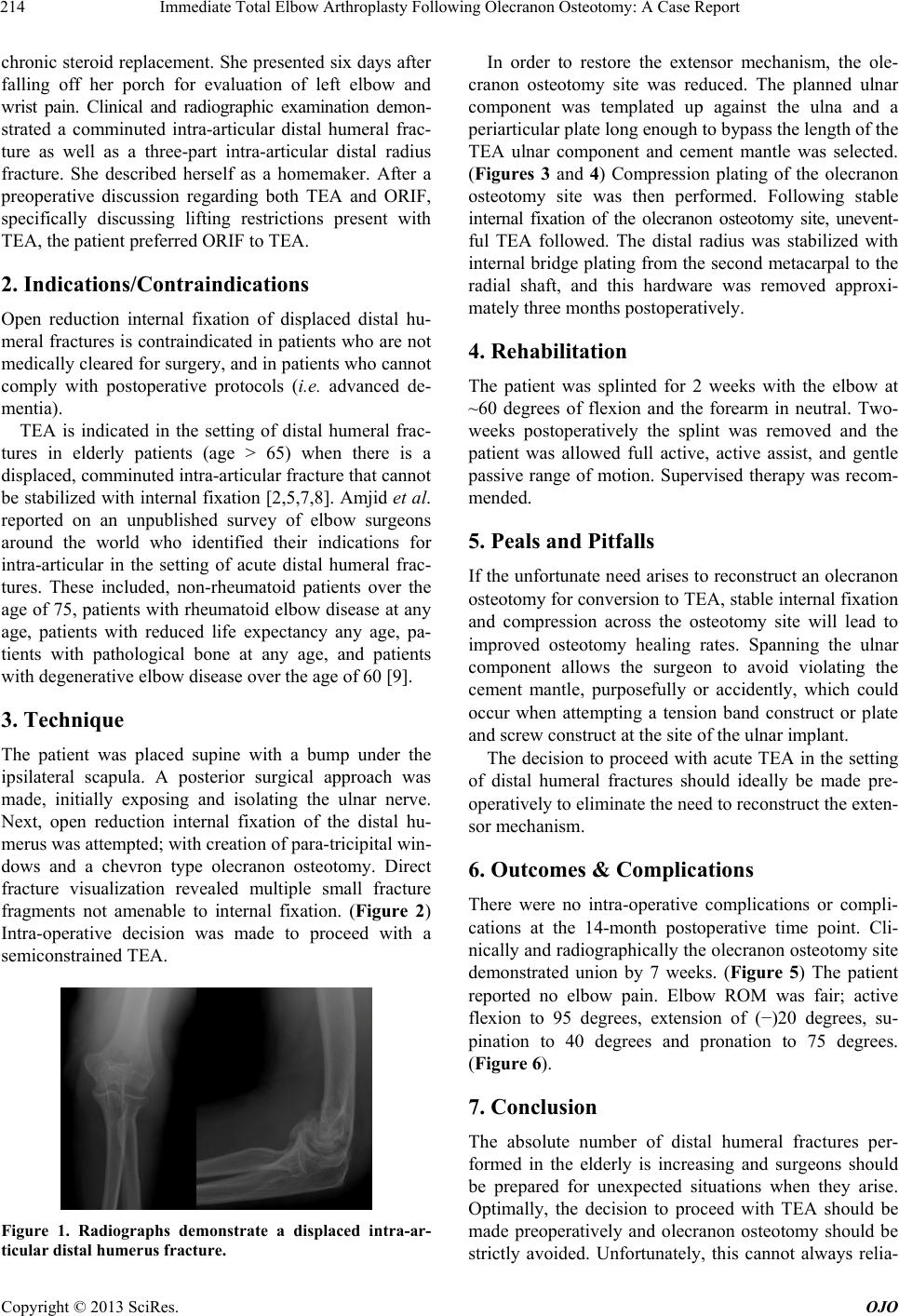
wrist pain. Clinical and radiographic examination demon-
strated a comminuted intra-articular distal humeral frac-
ture as well as a three-part intra-articular distal radius
fracture. She described herself as a homemaker. After a
preoperative discussion regarding both TEA and ORIF,
specifically discussing lifting restrictions present with
TEA, the patient preferred ORIF to TEA.
2. Indications/Contraindications
Open reduction internal fixation of displaced distal hu-
meral fractures is contraindicated in patients who are not
medically cleared for surgery, and in patients who cannot
comply with postoperative protocols (i.e. advanced de-
mentia).
TEA is indicated in the setting of distal humeral frac-
tures in elderly patients (age > 65) when there is a
displaced, comminuted intra-articular fracture that cannot
be stabilized with internal fixation [2,5,7,8]. Amjid et al.
reported on an unpublished survey of elbow surgeons
around the world who identified their indications for
intra-articular in the setting of acute distal humeral frac-
tures. These included, non-rheumatoid patients over the
age of 75, patients with rheumatoid elbow disease at any
age, patients with reduced life expectancy any age, pa-
tients with pathological bone at any age, and patients
with degenerative elbow disease over the age of 60 [9].
3. Technique
The patient was placed supine with a bump under the
ipsilateral scapula. A posterior surgical approach was
made, initially exposing and isolating the ulnar nerve.
Next, open reduction internal fixation of the distal hu-
merus was attempted; with creation of para-tricipital win-
dows and a chevron type olecranon osteotomy. Direct
fracture visualization revealed multiple small fracture
fragments not amenable to internal fixation. (Figure 2)
Intra-operative decision was made to proceed with a
semiconstrained TEA.
Figure 1. Radiographs demonstrate a displaced intra-ar-
ticular distal humerus fracture.
In order to restore the extensor mechanism, the ole-
cranon osteotomy site was reduced. The planned ulnar
component was templated up against the ulna and a
periarticular plate long enough to bypass the length of the
TEA ulnar component and cement mantle was selected.
(Figures 3 and 4) Compression plating of the olecranon
osteotomy site was then performed. Following stable
internal fixation of the olecranon osteotomy site, unevent-
ful TEA followed. The distal radius was stabilized with
internal bridge plating from the second metacarpal to the
radial shaft, and this hardware was removed approxi-
mately three months postoperatively.
4. Rehabilitation
The patient was splinted for 2 weeks with the elbow at
~60 degrees of flexion and the forearm in neutral. Two-
weeks postoperatively the splint was removed and the
patient was allowed full active, active assist, and gentle
passive range of motion. Supervised therapy was recom-
mended.
5. Peals and Pitfalls
If the unfortunate need arises to reconstruct an olecranon
osteotomy for conversion to TEA, stable internal fixation
and compression across the osteotomy site will lead to
improved osteotomy healing rates. Spanning the ulnar
component allows the surgeon to avoid violating the
cement mantle, purposefully or accidently, which could
occur when attempting a tension band construct or plate
and screw construct at the site of the ulnar implant.
The decision to proceed with acute TEA in the setting
of distal humeral fractures should ideally be made pre-
operatively to eliminate the need to reconstruct the exten-
sor mechanism.
6. Outcomes & Complications
There were no intra-operative complications or compli-
cations at the 14-month postoperative time point. Cli-
nically and radiographically the olecranon osteotomy site
demonstrated union by 7 weeks. (Figure 5) The patient
reported no elbow pain. Elbow ROM was fair; active
flexion to 95 degrees, extension of ()20 degrees, su-
pination to 40 degrees and pronation to 75 degrees.
(Figure 6).
7. Conclusion
The absolute number of distal humeral fractures per-
formed in the elderly is increasing and surgeons should
be prepared for unexpected situations when they arise.
Optimally, the decision to proceed with TEA should be
made preoperatively and olecranon osteotomy should be
strictly avoided. Unfortunately, this cannot always relia-
Immediate Total Elbow Arthroplasty Following Olecranon Osteotomy: A Case Report
Copyright © 2013 SciRes. OJO
215
Figure 2. Distal humerus fracture fragments visualized following olecranon osteotomy.
Figure 3. TEA and olecranon osteotomy repair using bridge
plate.
Figure 4. Intra-operative radiographs following bridging
internal fixation and total elbow arthro plasty.
Figure 5. 14-month follow-up radiographs demonstrating ole-
cranon osteotomy site union.
Figure 6. 14-month follow -up clinical photos demonstrating
final range of motion.
bly be predetermined, and cases may arise following an
olecranon osteotomy when the distal humeral fracture
may not be reconstructible. This case presents a method
to address the olecranon osteotomy (bypassing the ulnar
component and its cement mantle) and perform acute
TEA.
REFERENCES
[1] J. B. Jupiter, U. Neff, P. Holzach and M. Allgöwer, “In-
tercondylar Fractures of the Humerus. An Operative
Approach,” The Journal of Bone and Joint Surgery Ame-
rican Volume, Vol. 67, No. 2, 1985, pp. 226-239.
[2] A. Nauth, M. D. McKee, B. Ristevski, J. Hall and E. H.
Schemitsch, “Distal Humeral Fractures in Adults,” The
Journal of Bone and Joint Surgery American Volume, Vol.
93, No. 7, 2011, pp. 686-700. doi:10.2106/JBJS.J.00845
[3] M. Palvanen, P. Kannus, S. Niemi and J. Parkkari, “Secu-
lar Trends in Distal Humeral Fractures of Elderly Women:
Nationwide Statistics in Finland between 1970 and 2007,”
Bone, Vol. 46, No. 5, 2010, pp. 1355-1358.
doi:10.1016/j.bone.2009.11.025
[4] M. D. McKee, C. J. H. Veillette, J. A. Hall, E. H. Sche-
mitsch, L. M. Wild, R. McCormack, et al., “A Multi-
center, Prospective, Randomized, Controlled Trial of Open
Reduction—Internal Fixation versus Total Elbow Arthro-
plasty for Displaced Intra-Articular Distal Humeral Frac-
tures in Elderly Patients,” Journal of Shoulder and Elbow
Surgery/American Shoulder and Elbow Surgeons, Vol. 18
No. 1, 2009, pp. 3-12.
[5] M. A. Frankle, D. Herscovici, T. G. DiPasquale, M. B. Vasey
and R. W. Sanders, “A Comparison of Open Reduction
Immediate Total Elbow Arthroplasty Following Olecranon Osteotomy: A Case Report
Copyright © 2013 SciRes. OJO
216
and Internal Fixation and Primary Total Elbow Arthro-
plasty in the Treatment of Intraarticular Distal Humerus
Fractures in Women Older than Age 65,” Journal of Or-
thopaedic Trauma, Vol. 17, No. 7, 2003, pp. 473-480.
doi:10.1097/00005131-200308000-00001
[6] G. Marra, B. F. Morrey, S. H. Gallay, M. D. McKee and
S. O’Driscoll, “Fracture and Nonunion of the Olecranon
in Total Elbow Arthroplasty,” Journal of Shoulder and
Elbow Surgery/American Shoulder and Elbow Surgeons,
Vol. 15, No. 4, 2006, pp. 486-494.
[7] T. K. Cobb and B. F. Morrey, “Total Elbow Arthroplasty
as Primary Treatment for Distal Humeral Fractures in
Elderly Patients,” The Journal of Bone and Joint Surgery.
American Vo lume, Vol. 79, No. 6, 1997, pp. 826-832.
[8] R. Gambirasio, N. Riand, R. Stern and P. Hoffmeyer, “To-
tal Elbow Replacement for Complex Fractures of the
Distal Humerus. An Option for the Elderly Patient,” The
Journal of Bone and Joint Surgery British Volume, Vol.
83, No. 7, 2001, pp. 974-978.
doi:10.1302/0301-620X.83B7.11867
[9] A. Ali, S. Shahane and D. Stanley, “Total Elbow Arthro-
plasty for Distal Humeral Fractures: Indications, Surgical
Approach, Technical Tips, and Outcome,” Journal of
Shoulder and Elbow Surgery/American Shoulder and El-
bow Surgeons, Vol. 19, No. 2, 2010, pp. 53-58.