Paper Menu >>
Journal Menu >>
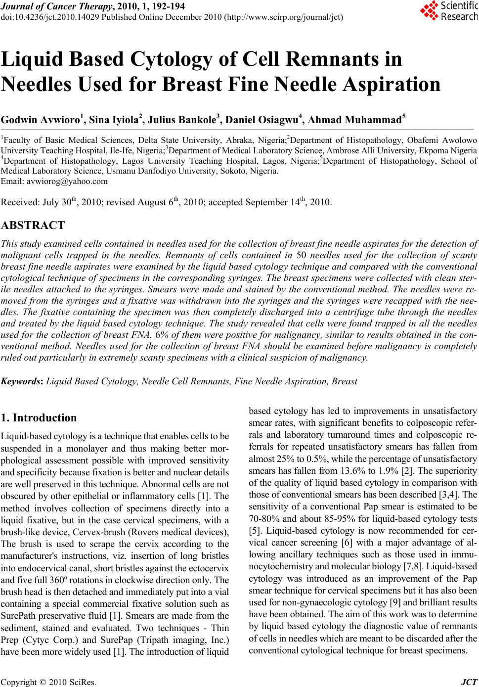 Journal of Cancer Therapy, 2010, 1, 192-194 doi:10.4236/jct.2010.14029 Published Online December 2010 (http://www.scirp.org/journal/jct) Copyright © 2010 SciRes. JCT Liquid Based Cytology of Cell Remnants in Needles Used for Breast Fine Needle Aspiration Godwin Avwioro1, Sina Iyiola2, Julius Bankole3, Da n i e l Os i a g w u 4, Ahmad Muhammad5 1Faculty of Basic Medical Sciences, Delta State University, Abraka, Nigeria;2Department of Histopathology, Obafemi Awolowo University Teaching Hospital, Ile-Ife, Nigeria;3Department of Medical Labor atory Science, Ambrose Alli University, Ekpoma Nig eria 4Department of Histopathology, Lagos University Teaching Hospital, Lagos, Nigeria;5Department of Histopathology, School of Medical Laboratory Science, Usmanu Danfodiyo University, Sokoto, Nigeria. Email: avwiorog@yahoo.com Received: July 30th, 2010; revised August 6th, 2010; accepted September 14th, 2010. ABSTRACT This study examined cells contain ed in needles used fo r the collection of breast fine needle asp irates for the d etection of malignant cells trapped in the needles. Remnants of cells contained in 50 needles used for the collection of scanty breast fine needle aspirates were examined by the liquid based cytology technique and compared with the conventional cytological technique of specimens in the corresponding syringes. The breast specimens were collected with clean ster- ile needles attached to the syringes. Smears were made and stained by the conventional method. The needles were re- moved from the syringes and a fixative was withdrawn into the syringes and the syringes were recapped with the nee- dles. The fixative containing the specimen was then completely discharged into a centrifuge tube through the needles and treated by the liquid based cytology technique. The study revealed that cells were found trapped in all the needles used for the collection of breast FNA. 6% of them were positive for malignancy, similar to results obtained in the con - ventional method. Needles used for the collection of breast FNA should be examined before malignancy is completely ruled out particularly in extremely scanty specimens with a clinica l suspicion of malignancy. Keywords: Liquid Based Cytology, Needle Cell Remnants, Fine Needle Aspiration, Breast 1. Introduction Liquid-based cytology is a technique that enables cells to be suspended in a monolayer and thus making better mor- phological assessment possible with improved sensitivity and specificity because fixation is better and nuclear details are well preserved in this technique. Abnormal cells are not obscured by other epithelial or inflammatory cells [1]. The method involves collection of specimens directly into a liquid fixative, but in the case cervical specimens, with a brush-like device, Cervex-brush (Rovers medical devices), The brush is used to scrape the cervix according to the manufacturer's instructions, viz. insertion of long bristles into endocervical canal, short bristl es against the ectocervix and five full 360º rotations in cl ockwise direction only. The brush head is then detached and im mediately put int o a vial containing a special commercial fixative solution such as SurePath preservative fluid [1]. Smears are made from the sediment, stained and evaluated. Two techniques - Thin Prep (Cytyc Corp.) and SurePap (Tripath imaging, Inc.) have been more widely used [1]. The int roduction of l iquid based cytology has led to improvements in unsatisfactory smear rates, with significant benefits to colposcopic refer- rals and laboratory turnaround times and colposcopic re- ferrals for repeated unsatisfactory smears has fallen from almost 25% to 0.5%, while the percent age of unsatisfactory smears has fallen from 13.6% to 1.9% [2]. The superiority of the quality of liquid based cytology in comparison with those of conventional smears has been described [3,4]. The sensitivity of a conventional Pap smear is estimated to be 70-80% and about 85-95% for liquid-based cytology tests [5]. Liquid-based cytology is now recommended for cer- vical cancer screening [6] with a major advantage of al- lowing ancillary techniques such as those used in immu- nocytochemistry and m olecular biology [7,8]. Liquid-based cytology was introduced as an improvement of the Pap smear technique for cervical specime ns but it has also been used for non-gynaecologic cytology [9] and brilliant results have been obtained. The aim of this work was to determine by liquid based cytology the diagnostic value of remnants of cells in needles which are meant to be discarded after the conventional cytological technique for breast specime ns.  Liquid Based Cytology of Cell Remnants in Needles Used for Breast Fine Needle Aspiration Copyright © 2010 SciRes. JCT 193 2. Materials and Methods Fifty breast aspirates received in needles and syringes were studied. The needles were removed from the sy- ringes after the smears were made by the conventional method. 10ml of liquid based fixative was then aspirated into the syringe and the needle was attached to the syringe. The fixative was then expelled through the needle forcing the cells to be released with the fixative. This was col- lected in a centrifuge tube and allowed to fix for 2 hours and was ce ntrif uged fo r about 10 min utes at 100 0 rpm a nd the supernatant removed. 5 ml of a cleaning solution was added to the sediment and spun for 10 minutes. The su- pernatant was removed and a cellular base added and mixed with vortex for 1 minute. Smears were made from the sediment and stained with haematoxylin and eosin (H&E) by applying Harris haematoxylin for 4 minutes. The smear was then rinsed in water, differentiated in 1% acid-alcohol for 3 seconds, rinsed in water, blued in Scott’s solution for 2 minutes and counterstained in 1% eosin for 30 seconds. Smears were then dehydrated through ascending grades of alcohol, cleared in xylene and coverslipped on DPX. The smears made by the con- ventional method were also stained with H&E and ex- amined under the X40 microscope objective. 3. Results The smears from four patients had a clean background with a uniform cell distribution with very few cases of overlapping of cells. The struct ure of the cells and nuclear appearance were well preserved with a good resolution which enabled a clearer and ac curate identification of cells under the microscope. Cells in sheets were also retained without disrupting their diag n ost i c val ue. 4. Discussion Breast carcinomas are the most frequent tumors in women. Table 1. Appearance and percentage of positive smears in remnants of cells in needles compared with the conventional method. Appearance of smear Posi- tive smears Nega- tive smears % of positive smears LBC of remnants of cells in nee- dles Clean background with good morphological assessment. Good fixa- tion and well preserved nuclear details. 3 47 6 Conven- tional method Similar to liquid based cytology of remnants of cells in needles except with pinkish backg round with some overlapping cells 3 47 6 Figure 1. Smears of cell remnants made from four needles treated by liquid based technique (H&E X40). In several countries, mammogr aphic screen ing allows the early diagnosis of tumors. However, when a tumor is suspected, morphological analysis alone can establish the diagnosi s of carci noma. I n this context, guided fine needle aspiration has become increasingly popular for obtaining tissue specimens for the diagnosis of malignant breast diseases [10]. Liquid-based cytology in breast FNA had a good correlation with conventional preparation of breast FNA with the advantages of easier and less time con- suming evaluation of cell morphology (clear background, no overlapping, smaller area to screen), reproducibility, a factor of great importance to quality control; and possi- bility of adjunctive investigations such as immunocytol- ogy and flow cytometry on the same material [11]. This has been corroborated with the addition that quantitative analysis of HER-2 mRNA correlated with the results of immunohistochemistry in cancer patients [12]. We have found similar morphological characteristics in our study with the ability to produce multiple clean smears from a single needle containing cells from breast specimen. This study has revealed that cells of diagnostic value are often trapped in the needles used for the collection of breast aspirates and should be examined before malignancy is completely ruled out, particularly in extremely scanty breast specimens where clinical diagnosis suggests ma- lignancy. A major disadvantage of extracting cells from used needles is the risk of accidental puncture of the fin- gers with such used needles which may contain virulent bacteria and viruses. For this reason, extreme care should be taken when handling used needles [13]. REFERENCES [1] A. N. Kavatkar, C. A. Nagwanshi and S. M. Dabak,  Liquid Based Cytology of Cell Remnants in Needles Used for Breast Fine Needle Aspiration Copyright © 2010 SciRes. JCT 194 “Study of a Manual Method of Liquid-Based Cervical Cytology,” Indian Journal of Pathology & Microbiology, Vol.51, No. 2, 2008, pp. 190-4. [2] ARW Williams, “Liquid-Based Cytology and Conven- tional Smears Compared over Two 12-Month Periods,” Cytopathology. Vol.17, No. 2, 2006, pp. 82-85. [3] B. Weynand, M. Berlière, E. Haumont, F. Massart, A. Pourvoyeur, P. Beri, et al. “A New Liquid-Based Cytology Technique: Thermo Shandon’s Papspin,” Acta Cytologica Vol. 47, No. 2, 2003, pp. 149-153. [4] C. Bergeron and F. Fagnani, “Performance of a New Liq- uid-based Cervical Screening Technique in the Clinical Setting of a Large F rench labora tory,” Acta Cytologica Vol. 47, No. 5, 2003, pp. 753-761. [5] D. Davey and R.J. Zarbo, “Introduction and Commentary, Strategic Science Symposium.” Archives of Path ology and Laboratory Medicine, Vol. 12No. 5, 2003, pp. 927-929. [6] C. Garbar, C. Mascaux and V. Fontaine, “Efficiency of an Inexpensive Liquid-Based Cytology Performed by Cyto- centrifugations: A Comparative Stud y Using the Histology as Reference Standard,” CytoJournal, Vol. 2, No. 15, 2005, pp. 15. [7] M. Bibbo, W. J. Klump, J. DeCecco and A. J. Kovatich, “Procedure for Immunocytochemical Detection of P16INK4A Antigen in Thin-layer, Liquid-Based Speci- mens,” Acta Cytologica , Vol. 46, No. 1, 2002, pp. 25-29. [8] L. Luzzatto, C. Van Haaften and M. E. Boon, “Prolifera- tion Patterns of Cervical Cells as Visualized in Leiden liquid Cytology Slides,” Diagnostic Cytopathology, Vol. 31, No. 1, 2004, pp. 5-9. [9] R. S. Hoda, “Non-gynecologic Cytology on Liquid Based Preparations: A Morphologic Review of Facts and Arti- facts,” Diagnostic Cytopathology, Vol. 35, No. 10, 2007, pp. 621-634. [10] H. Sartelet, E. Lagonette, M. Lorenzato, I. Duval, C. Lechki, C. Rigaud et al., “Comparison of Liquid Based Cytology and Histology for the Evaluation of HER-2 Status Using Immunostaining and CISH in Breast Carci- noma,” Journal of Clinical Pathology, Vol. 58, No. 8, 2005, pp. 864-871. [11] S. Veneti, D. Daskalopoulou, S. Zervoudis, E. Papasoti- riou and L. Ioannidou-Mouzaka, “Liquid-Based Cytology in Breast Fine Needle Aspiration. Comparison with the Conventional Smear,” Acta Cytologica, Vol. 47, No. 2, 2003, pp. 188-92. [12] K. Komatsu, Y. Nakanishi, T. Seki, A. Yoshino, F. Fuchi- noue, S. Amano, A. Komatsu, M. Sugitani and N. Nemoto, “Application of Liquid-Based Preparation to Fine Needle Aspiration Cytology in Breast Cancer,” Acta Cytologica, Vol. 52, No. 5, 2008, pp. 591-6. [13] Avwioro O. G., “Textbook of Histochemistry and Tissue Pathology,” 2nd Edition, Claverianun Press, Ibadan, Nige- ria. |