H. KHADRI ET AL. 245
of the particles of the materials is an efficient and reliable
tool for reinforcing their biocompatibility. In fact, nano-
technology helps in overcoming the limitations of size
and can change the outlook of the world regarding sci-
ence [20]. Studies reveal that the antimicrobial activity of
the silver particles is due to their positive charge that
qualifies them in reacting with the negatively charged
proteins on the cell membranes and thus contributing to
their antimicrobial activities [21-23]. Many reports have
suggested the efficacy of silver nanoparticles of their
antimicrobial activities and to mention some as follows.
Kim et al. [24] have obtained positive results against E.
coli and S. aureus where as a more profound effect was
seen against E. coli and comparably a milder effect against
S. aureus. Kim et al. [24] also obtained strong antifungal
activity for silver nanoparticles against Yeast cells. They
further observed a concentration dependant toxicity of
silver nanoparticles against bacteria with nanoparticles
ranging from 3.3 nM to 6.6 nM [7]. The antifungal ef-
fects of silver nanoparticles were estimated against eight-
een plant pathogenic fungi that included genera of Py-
thium, Fusarium, Alternar ia, Bo trytis, Cladosporium,
Corynespora, Cylindrocarpon, Stemphylium etc., by Kim
et al. [24]. The antifungal activity of the synthesized
nanoparticles has been tested against different fungal
plant pathogens namely Aspergillus niger (Collar rot),
Aspergillus falvus (Root rot), Rhizoctonia bataticola (Dry
rot), Sclerotium rolfsii (Stem rot) and Alternaria macro-
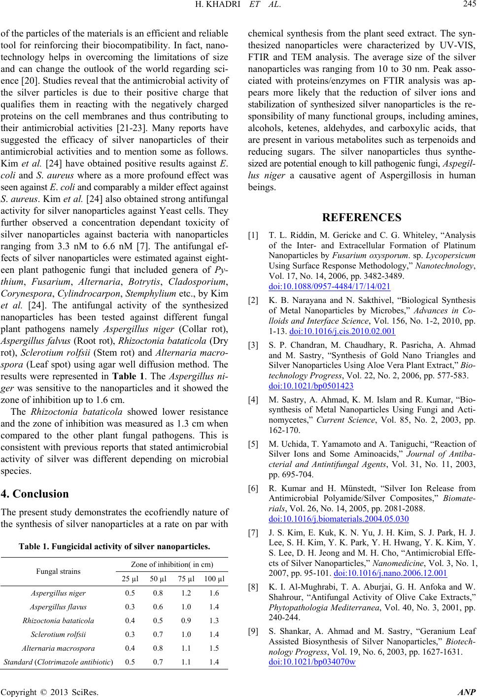
spora (Leaf spot) using agar well diffusion method. The
results were represented in Table 1. The Aspergillus ni-
ger was sensitive to the nanoparticles and it showed the
zone of inhibition up to 1.6 cm.
The Rhizoctonia bataticola showed lower resistance
and the zone of inhibition was measured as 1.3 cm when
compared to the other plant fungal pathogens. This is
consistent with previous reports that stated antimicrobial
activity of silver was different depending on microbial
species.
4. Conclusion
The present study demonstrates the ecofriendly nature of
the synthesis of silver nanoparticles at a rate on par with
Table 1. Fungicidal activity of silver nanoparticles.
Zone of inhibition( in cm)
Fungal strains
25 µl 50 µl 75 µl100 µl
Aspergillus niger 0.5 0.8 1.2 1.6
Aspergillus flavus 0.3 0.6 1.0 1.4
Rhizoctonia bataticola 0.4 0.5 0.9 1.3
Sclerotium rolfsii 0.3 0.7 1.0 1.4
Alternaria macrosp o r a 0.4 0.8 1.1 1.5
Standard (Clotrimazole antibiotic) 0.5 0.7 1.1 1.4
chemical synthesis from the plant seed extract. The syn-
thesized nanoparticles were characterized by UV-VIS,
FTIR and TEM analysis. The average size of the silver
nanoparticles was ranging from 10 to 30 nm. Peak asso-
ciated with proteins/enzymes on FTIR analysis was ap-
pears more likely that the reduction of silver ions and
stabilization of synthesized silver nanoparticles is the re-
sponsibility of many functional groups, including amines,
alcohols, ketenes, aldehydes, and carboxylic acids, that
are present in various metabolites such as terpenoids and
reducing sugars. The silver nanoparticles thus synthe-
sized are potential enough to kill pathogenic fungi, Aspegil-
lus niger a causative agent of Aspergillosis in human
beings.
REFERENCES
[1] T. L. Riddin, M. Gericke and C. G. Whiteley, “Analysis
of the Inter- and Extracellular Formation of Platinum
Nanoparticles by Fusarium oxysporum. sp. Lycopersicum
Using Surface Response Methodology,” Nanotechnology,
Vol. 17, No. 14, 2006, pp. 3482-3489.
doi:10.1088/0957-4484/17/14/021
[2] K. B. Narayana and N. Sakthivel, “Biological Synthesis
of Metal Nanoparticles by Microbes,” Advances in Co-
lloids and Interface Science, Vol. 156, No. 1-2, 2010, pp.
1-13. doi:10.1016/j.cis.2010.02.001
[3] S. P. Chandran, M. Chaudhary, R. Pasricha, A. Ahmad
and M. Sastry, “Synthesis of Gold Nano Triangles and
Silver Nanoparticles Using Aloe Vera Plant Extract,” Bio-
technology Progres s, Vol. 22, No. 2, 2006, pp. 577-583.
doi:10.1021/bp0501423
[4] M. Sastry, A. Ahmad, K. M. Islam and R. Kumar, “Bio-
synthesis of Metal Nanoparticles Using Fungi and Acti-
nomycetes,” Current Science , Vol. 85, No. 2, 2003, pp.
162-170.
[5] M. Uchida, T. Yamamoto and A. Taniguchi, “Reaction of
Silver Ions and Some Aminoacids,” Journal of Antiba-
cterial and Antintifungal Agents, Vol. 31, No. 11, 2003,
pp. 695-704.
[6] R. Kumar and H. Münstedt, “Silver Ion Release from
Antimicrobial Polyamide/Silver Composites,” Biomate-
rials, Vol. 26, No. 14, 2005, pp. 2081-2088.
doi:10.1016/j.biomaterials.2004.05.030
[7] J. S. Kim, E. Kuk, K. N. Yu, J. H. Kim, S. J. Park, H. J.
Lee, S. H. Kim, Y. K. Park, Y. H. Hwang, Y. K. Kim, Y.
S. Lee, D. H. Jeong and M. H. Cho, “Antimicrobial Effe-
cts of Silver Nanoparticles,” Nanomedicine, Vol. 3, No. 1,
2007, pp. 95-101. doi:10.1016/j.nano.2006.12.001
[8] K. I. Al-Mughrabi, T. A. Aburjai, G. H. Anfoka and W.
Shahrour, “Antifungal Activity of Olive Cake Extracts,”
Phytopathologia Mediterranea, Vol. 40, No. 3, 2001, pp.
240-244.
[9] S. Shankar, A. Ahmad and M. Sastry, “Geranium Leaf
Assisted Biosynthesis of Silver Nanoparticles,” Biotech-
nology Progress, Vol. 19, No. 6, 2003, pp. 1627-1631.
doi:10.1021/bp034070w
Copyright © 2013 SciRes. ANP