Open Journal of Ophthalmology, 2013, 3, 51-53
http://dx.doi.org/10.4236/ojoph.2013.33013 Published Online August 2013 (http://www.scirp.org/journal/ojoph) 51
MRI Findings in Post-Operative Bilateral Posterior
Ischemic Optic Neuropathy
Nirali P. Bhatt1*, Robert E. Morales2, Michaela K. Mathews1
1Department of Ophthalmology and Visual Sciences, University of Maryland School of Medicine, Baltimore, USA; 2Department of
Diagnostic Radiology and Nuclear Medicine, University of Maryland School of Medicine, Baltimore, USA.
Email: *niralipbhatt@gmail.com
Received April 19th, 2013; revised May 20th, 2013; accepted June 15th, 2013
Copyright © 2013 Nirali P. Bhatt et al. This is an open access article distributed under the Creative Commons Attribution License,
which permits unrestricted use, distribution, and reproduction in any medium, provided the original work is properly cited.
ABSTRACT
A 62-year-old female complained of vision loss following multiple abdominal surgeries for mesenteric ischemia. The
patient’s visual acuity was no light perceptio n (NLP) in the right eye and hand motion (HM) at 1’ in the left eye. Both
pupils were unreactive and no relative afferent pupillary defect was noted. Anterior segment and fundus examination
were unremarkable. T1 and T2 weighted MRI imaging of the brain was normal but diffusion weighted imaging (DWI)
revealed areas of bright signal within both intraorbital optic nerves, confirming the diagnosis of posterior ischemic optic
neuropathy.
Keywords: Optic Nerve; Post-Operative Posterior Ischemic Optic Neuropathy; MRI; DWI
1. Case Report
A 62-year-old Hispanic female with a previou s history of
diabetes, hypertension, and glaucoma underwent re-
peated laparotomies and bowel resections for mesenteric
ischemia. During the procedures, several hypotensive
episodes were recorded. After the last surgery, the patien t
complained of having difficulty seeing objects in front of
her. The patient’s visual acuity was found to be reduced
to no light perception (NLP) in the right eye and hand
motion (HM) at 1’ in the left eye. Both pupils were un-
reactive to light. No relative afferent pupillary defect was
present. The patient had normal ocular motility and
alignment. Confrontational visual fields could not be
obtained reliably. Anterior segment examination showed
moderate cataracts in both eyes. Both optic nerves
showed a cup-to-disc ratio of 0.4, without pallor or
edema. The retina, macula, periphery and retinal vessels
were unremarkable. Histopathology of the surgical
specimen as well as laboratory studies revealed no evi-
dence of vasculitis. The patient underwent MRI and
magnetic resonan ce angiography ( MRA) of the brain. No
acute infarct was identified within the brain parenchyma
with findings suggestive of chronic small vessel ischemic
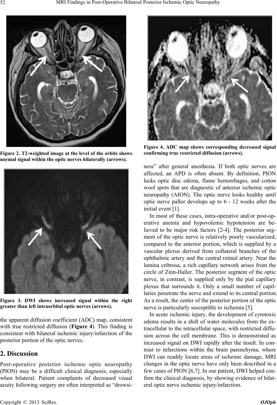
change in the white matter (Figure 1). No evidence of
advanced edema within the optic nerves was identified
(Figure 2). MRA of the brain showed no major branch
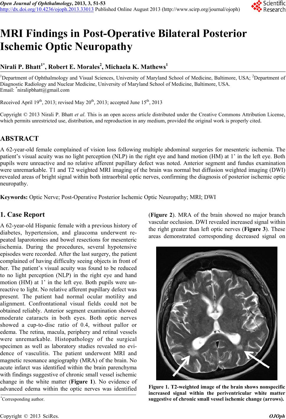
vascular occlusion. DWI revealed increased signal within
the right greater than left optic nerves (Figure 3). These
areas demonstrated corresponding decreased signal on
Figure 1. T2-weighted image of the brain shows nonspecific
increased signal within the periventricular white matter
suggestive of chronic small vessel ischemic change (arrows).
*Corresponding a uthor.
Copyright © 2013 SciRes. OJOph
MRI Findings in Post-Operative Bilateral Posterior Ischemic Optic Neuropathy
52
Figure 2. T2-weighted image at the level of the orbits shows
normal signal within the optic nerves bilaterally (arrows).
Figure 3. DWI shows increased signal within the right
greater than left intraorbital optic nerves (arrows).
the apparent diffusion coefficient (ADC) map, consistent
with true restricted diffusion (Figure 4). This finding is
consistent with bilateral ischemic injury/infarction of the
posterior po rti on of t he opti c ner ves.
2. Discussion
Post-operative posterior ischemic optic neuropathy
(PION) may be a difficult clinical diagnosis, especially
when bilateral. Patient complaints of decreased visual
acuity following surgery are often interp reted as “dro wsi-
Figure 4. ADC map shows corresponding decreased signal
confirming true restricte d diffusion (ar r ows).
ness” after general anesthesia. If both optic nerves are
affected, an APD is often absent. By definition, PION
lacks optic disc edema, flame hemorrhages, and cotton
wool spots that are diagnostic of anterior ischemic optic
neuropathy (AION). The optic nerve looks healthy until
optic nerve pallor develops up to 6 - 12 weeks after the
initial event [1].
In most of these cases, intra-operative and/or post-op-
erative anemia and hypovolemic hypotension are be-
lieved to be major risk factors [2-4]. The posterior seg-
ment of the optic nerve is relatively poorly vascularized,
compared to the anterior portion, which is supplied by a
vascular plexus derived from collateral branches of the
ophthalmic artery and the central retinal artery. Near the
lamina cribrosa, a rich capillary network arises from the
circle of Zinn-Haller. The posterior segment of the optic
nerve, in contrast, is supplied only by the pial capillary
plexus that surrounds it. Only a small number of capil-
laries penetrate the nerve and extend to its central portion.
As a result, the center of the posterior portion of the op tic
nerve is particularly susceptible to ischemia [5].
In acute ischemic injury, the development of cytotoxic
edema results in a shift of water molecules from the ex-
tracellular to the intracellular space, with restricted diffu-
sion across the cell membrane. This is demonstrated as
increased signal on DWI rapidly after the insult. In con-
trast to infarctions within the brain parenchyma, where
DWI can readily locate areas of ischemic damage, MRI
changes in the optic nerve have only been described in a
few cases of PION [6,7]. In our patient, DWI helped con-
firm the clinical diagnosis, by showing evidence of bilat-
eral optic nerve ischemic injury/infarction.
Copyright © 2013 SciRes. OJOph
MRI Findings in Post-Operative Bilateral Posterior Ischemic Optic Neuropathy
Copyright © 2013 SciRes. OJOph
53
REFERENCES
[1] S. Dunker, H. Y. Hsu, J. Sebag and A. A. Sadun, “Pe-
rioperative Risk Factors for Posterior Ischemic Optic
Neuropathy,” Journal of the American College of Surgeons,
Vol. 194, No. 6, 2002, pp. 705-710.
doi:10.1016/S1072-7515(02)01210-3
[2] S. S. Hayreh, “Posterior Ischaemic Optic Neuropathy:
Clinical Features, Pathogenesis, and Management,” Eye,
Vol. 18, 2004, pp. 1188-1206.
doi:10.1038/sj.eye.6701562
[3] L. A. Lee, N. J. Newman, T. A. Wagner, J. R. Dettori and
N. J. Dettori, “Postoperative Ischemic Optic Neuropathy,”
Spine, Vol. 35, No. 9, 2010, pp. S105-S116.
[4] N. J. Newman, “Perioperative Visual Loss after Nonocu-
lar Surgeries,” American Journal of Ophthalmology, Vol.
145, No. 4, 2008, pp. 604-610.
doi:10.1016/j.ajo.2007.09.016
[5] V. Purvin and B. Kuzma, “Intraorbital Optic Nerve Signal
Hyperintensity on Magnetic Resonance I maging Sequences
in Perioperative Hypotensive Ischemic Optic Neuropa-
thy,” Journal of Neuro-Ophthalmology, Vol. 25, No. 3,
2005, pp. 202-204.
doi:10.1097/01.wno.0000177295.52468.5b
[6] S. R. Sadda, M. Nee, N. R. Miller, V. Biousse, N. J.
Newman and A. Kouzis, “Clinical Spectrum of Posterior
Ischemic Optic Neuropathy,” American Journal of Oph-
thalmology, Vol. 132, No. 5, 2001, pp. 743-750.
doi:10.1016/S0002-9394(01)01199-0
[7] M. S. Vaphiades, “Optic Nerve Enhancement in Hypoten-
sive Ischemic Optic Neuropathy,” Journal of Neuro-Oph-
thalmology, Vol. 24, No. 3, 2004, pp. 235-236.
doi:10.1097/00041327-200409000-00011