Surgical Science, 2013, 4, 359-364
http://dx.doi.org/10.4236/ss.2013.48071 Published Online August 2013 (http://www.scirp.org/journal/ss)
A Qualitative Morphological Comparison of Two
Haemostatic Agents in a Porcine Liver Trauma Model
Kenneth E. Coenye1*, Claire Bourgain2, Claudia Keibl3, Sylvia Nürnberger3,4,
Martijn van Griensven3
1Departments for Surgery and Emergency Medicine, AZ Jan Portaels, Vilvoorde, Belgium
2Department for Pathological Anatomy, Free University Brussels (VUB), Brussels, Belgium
3Ludwig Boltzmann Institute for Experimental and Clinical Traumatology in the Research Centre of the AUVA, Vienna, Austria
4Department of Traumatology, Medical University, Vienna, Austria
Email: *kcoenye@azjanportaels.be
Received June 1, 2013; revised July 2, 2013; accepted July 10, 2013
Copyright © 2013 Kenneth E. Coenye et al. This is an open access article distributed under the Creative Commons Attribution Li-
cense, which permits unrestricted use, distribution, and reproduction in any medium, provided the original work is properly cited.
ABSTRACT
Background: Many surgical haemostatic agents are available which aim to reduce morbidity and mortality of bleeding,
as well as associated costs. We used qualitative techniques to compare the effectiveness of two topical gelatine-based
haemostatic agents in a porcine liver trauma model. Methods: We compared the activity of Floseal® (with human or
bovine thrombin), Surgiflo® and Surgiflo® plus 5000 IU bovine thrombin per 10 mL of product (n = 6 test sites/group).
Different clinical scenarios were evaluated in two anaesthetised piglets; normal physiological conditions (normotension
and normothermia), and a status of hypotension, hypothermia and haemodilution, requiring inotropic support. Laparo-
tomy was performed, and five identical stab wounds were made on each liver lobe. Four of these wounds were ran-
domly treated with one of the agents under investigation, while one wound was kept as an untreated control. Haemosta-
sis was observed during and immediately following surgery. After euthanisation, wounds were examined (blinded for
treatment) macroscopically, microscopically and using scanning electron microscopic evaluation. Results: Surgiflo®
produced some degree of clotting in 1/3 applications in both piglets. Co-treatment with thrombin resulted in a minor
improvement in performance in the animal with normal physiological conditions (some degree of clotting in all
wounds); no improvement was seen in the animal in a status of hypotension, hypothermia and haemodilution. Floseal®
induced clotting in all wounds for both piglets. Microscopic and scanning electron microscope examination of the stab
wounds demonstrated that Floseal® created a stable, dense agglomerate of gelatine and fibrin, which firmly adhered to
the adjacent liver tissue. In wounds treated with Surgiflo® or Surgiflo® plus thrombin, the gelatine contained more air
bubbles, resulting in less fibrin inclusion in the clot and weaker adhesion of the clot to the liver tissue. Conclusion:
Floseal® created a dense and stable blood clot, even in a piglet with hypotension, hypothermia and haemodilution.
Keywords: Haemostasis; Coagulation; Thrombin; Fibrin; Gelatine; Sealant; Clot; Surgery; Trauma; Wound Healing
1. Introduction
Techniques for controlling haemostasis during surgery
have evolved over recent decades, from application of
local pressure with moist gauze, to electrocautery, su-
tures, staples and haemostatic patches. Many topical agents
designed to help control haemostasis during surgery have
been licensed in the past decade. Surgical haemostatic
agents can be collagen-based, gelatine-based, cellulose-
based, polysaccharide-based or fibrin-based, and can also
be inorganic in nature [1].
When classic haemostasis fails and surgeons need to
transfuse critically ill patients, or administer platelets or
plasma products, morbidity and mortality rates increase,
as do the costs associated with the surgery [2,3]. The
latest commercially available agents claim to have an
immediate effect in areas where haemostasis control is
difficult or dangerous, thus reducing morbidity, mortality
and associated costs. Haemostasis has been achieved in
even the most severe trauma situations by using gela-
tine-based flowable agents [4].
In this experiment we set out to determine the effect-
tiveness of two of these gelatine-based agents, with or
without the addition of thrombin, by means of observa-
tional and morphological evaluation in a clinically rele-
*Corresponding author.
C
opyright © 2013 SciRes. SS
K. E. COENYE ET AL.
360
vant pig model. Both the agents functioned via two me-
chanisms: activation of the intrinsic coagulation path-
way as a result of contact between blood and the gelatine
surface; and tamponading of bleeding by the gelatine as
it swells following contact with blood. The added throm-
bin also directly induces fibrin formation [5,6].
2. Methods
We compared the activity of two variants of Floseal®
(Baxter Biosurgery Inc., Deerfield, Illinois, USA), a
cross-linked gelatine matrix with either human (2500 IU)
or bovine (5000 IU) thrombin, with Surgiflo® (Johnson
&Johnson Inc., New Brunswick, New Jersey, USA),
which contains only gelatine. To compare fibrin clot for-
mation induced by the two products, in addition to using
Surgiflo® according to the manufacturer’s instructions,
we also used it with 5000 IU bovine thrombin per 10 mL
of agent added, instead of the saline solution with which
it is intended to be used. In order to get a better under-
standing of the structure of the two gelatine agents, Sur-
giflo® and Floseal® particles were analysed morphologi-
cally using scanning electron microscopy (SEM) before
application to the wounds.
2.1. In Vivo Analysis of the Ability of Surgiflo®
and Floseal® to Control Haemostasis
The effect of Surgiflo® and Floseal® was evaluated in
two piglets. The first piglet provided a model of normal
physiological conditions (normotension and normother-
mia), while the second animal was in a status of hypo-
tension, hypothermia and haemodilution, requiring ino-
tropic support) (Table 1). In order to maintain mean ar-
terial pressure (MAP) in the second piglet, 9000 mL of
crystalloid and colloid fluids were infused over 180 min-
utes, compared with only 7000 mL of crystalloids in the
first piglet.
All experiments were approved by the animal protocol
review board of the City Government of Vienna. Both
piglets were premedicated with an intramuscular inject-
tion of a mixture of 30 mg xylazine, 25 mg ketamine,
10 mg tiletamine, 10 mg zolazepam and 5 mg butorpha-
nol, and were then intubated. Jugular, carotid and urinary
catheters were inserted and general anaesthesia was main-
tained with inhaled oxygen and isoflurane (1.5% - 2%)
and intravenous sufentanil (0.008 mg/kg/h) and alcu-
ronium (0.8 mg/kg/h). A laparotomy was performed in
each piglet, then five identical stab wounds approxima-
tely 3 cm apart from each other, were inflicted consecu-
tively in each of the three liver lobes by making a cross-
like incision of 12 mm wide and 10 mm deep with a self-
made stab device consisting of four perpendicular num-
ber 10 surgical blades in the shape of a cross.
Table 1. Physiological conditions of the animals during the
experiment.
Parameter
Piglet 1
(Physiological
conditions)
Piglet 2
(Hypotension,
hypothermia and
haemodilution)
Weight (kg) 30.5 30.6
Sex male male
Mean arterial pressure start
(mmHg) 90 75
Mean arterial pressure end
(mmHg) 75 65
Heart rate start (bpm) 110 95
Heart rate end (bpm) 80 146
Mean temperature (˚C) 36.5 32.0
Total volume infused (mL) 7000 9000
Colloids No Yes*
Inotropic drugs No Yes**
Urine output (mL) 1400 3000
Bpm: beats per minute, *3000 mL Voluven®, **diluted noradrenalin and pure
L-adrenalin to maintain blood pressure.
Each of the four agents was randomised to one stab
wound per lobe (Table 2). A fifth stab wound inflicted to
each lobe was treated with 2 × 2 minutes of gentle com-
pression without the application of a haemostatic agent,
to serve as a control. 5 mL of agent was applied to each
wound (corresponding to an entire syringe of Floseal®
and half a syringe of Surgiflo®). Photographs were taken
of each step of the procedure. After agents were applied
to the bleeding site, soft compression with moist gauze
was applied for exactly 2 minutes. The gauze was then re-
moved and an initial observation of haemostasis recorded.
If bleeding was still present, pressure was reapplied for a
further 2 minutes. Next, any excess product was removed
by gently rinsing with 10 mL of saline solution. Again,
observations of haemostasis were recorded.
After euthanising the piglets in deep anaesthesia, the
liver lobes were resected and tissue specimens of 1 × 1 ×
1 cm around each stab wound were excised. These
blocks were fixated in 4% buffered formaldehyde solu-
tion. In each animal, five tissue blocks from one liver
lobe were bisected to provide one sample for histology
and one sample for SEM from each block. The samples
for SEM were then dehydrated in a gradient series of
alcohol and chemically dried with hexamethyldisilazane.
Histological paraffin sections were stained with haema-
toxylin and eosin (H&E). Histology and SEM were used
to quantitatively evaluate the quality and distribution of
he gelatine, the amount of firin, the integration of the t
b
Copyright © 2013 SciRes. SS
K. E. COENYE ET AL.
Copyright © 2013 SciRes. SS
361
Table 2. Randomisation of agents.
Piglet 1
(Physiological conditions)
Piglet 2
(Hypotension, hypothermia and haemodilution)
Wound LL ML RL LL ML RL
1 Floseal® bT Floseal® bT Surgiflo® bT Surgiflo® bT Floseal® hT Floseal® hT
2 Surgiflo® bT Surgiflo® bT Floseal® hT Floseal® bT Floseal® bT Surgiflo®
3 Floseal® hT Surgiflo® Surgiflo® Surgiflo® Surgiflo® Surgiflo® bT
4 Surgiflo® Floseal® hT Floseal® bT Floseal® hT Surgiflo® bT Floseal® bT
5 Control Control Control Control Control Control
LL: left liver lobe, ML: medial liver lobe, RL: right liver lobe, hT: 2500 IU human thrombin, bT: 5000 IU bovine thrombin.
gelatine into the fibrin network and the quantity of blood
cells in the defect. The pathologists analysing the sam-
ples were blinded to both the agent used and the origin of
the specimen.
3. Results
3.1. Scanning Electron Microscopy Analysis of
Agents before Application to the Wound Figure 1. SEM of particles before application to the wound:
a) Floseal®; b) Surgiflo® (bar = 500 μm).
The mean particle size of both agents ranged from 100 to
500 µm (Figure 1). There were, however, important dif-
ferences when comparing their geometrical and topog-
raphic characteristics; Floseal® particles were compact,
flat and elongated with an irregular outline and Surgiflo®
particles were spongy, more spherical in shape and were
sometimes locally interconnected, creating a kind of net-
work.
Figure 2. Perioperative view (piglet 2): left: immediate
haemostasis with Floseal®; right: continued bleeding after
application of Surgiflo®.
3.2. In Vivo Analysis of the Ability of Surgiflo®
and Floseal® to Control Haemostasis
Wounds treated with Floseal® stopped bleeding after the
first two minutes of compression in both animals; a lump
of clotted gelatine was visible on top of the wound but
the actual blood clot was not visible on the surface of the
liver. In the piglet under normal physiological conditions,
Surgiflo® produced a physiological blood clot in one out
of three wounds; with the addition of thrombin, a degree
of clotting was observed in all 3 wounds. In contrast, in
the second piglet with hypotension, hypothermia and
haemodilution, both Surgiflo® and Surgiflo® with throm-
bin each produced some degree of clotting in just one out
of three applications. Rinsing away the surplus agent
resulted in bleeding recommencing in the Surgiflo® and
Surgiflo® with thrombin wounds that had clotted in the
second animal (Figure 2). All control stab wounds con-
tinued bleeding after 2 × 2 minutes of compression and
were either packed or sutured with Vicryl® (size 4/0) to
prevent important blood loss. Table 3 gives an overview
of the observations made after excess product was rinsed
away.
3.3. Microscopic Evaluation of the Wounds
The H&E stained microscopic samples showed clear
differences between the two agents in terms of the size
and structure of the gelatine, the presence of red blood
cells and fibrin in the wound and in the adherence of the
gelatine to the liver surface (Figure 3). The gelatine ma-
trix created by Surgiflo® was more porous and surrounded
by red blood cells. The particles were not strongly ad-
herent to the liver surface and there was limited fibrin
within the wound. In comparison, the dense gelatine par-
ticles in the Floseal® samples were closely adherent to
the hepatocytes and the amount of fibrin in the Floseal®-
treated wounds was higher. There was no difference be-
tween the Floseal® samples prepared with 2500 IU hu-
man thrombin or 5000 IU bovine thrombin. Adding
thrombin to Surgiflo® did not appear to change the effec-
tiveness of the agent in either piglet, as determined by
microscopic evaluation.
K. E. COENYE ET AL.
362
Table 3. Observations of haemostasis after excess product removed by rinsing.
Product Piglet 1
(Physiological conditions)
Piglet 2
(Hypotension, hypothermia and haemodilution)
LL ML RL LL ML RL
Floseal® hT + + + + + +
Floseal® bT + + + + + +
Surgiflo® /
Surgiflo® bT / /
Control
hT: 2500 IU human thrombin, bT: 5000 IU bovine thrombin, LL: left lobe, ML: medial lobe, RL: right lobe, : no haemostasis, : some haemostasis, blood
leaking, +: good haemostasis, dry.
Figure 3. H&E stain (piglet 2): left: Transition of the liver
to the Floseal® (bovine thrombin) filled defect: large gela-
tine particles (long arrow) are well attached to the defect
margin. The interspace between the particles is filled with a
few red blood cells (short arrow) and fibrin material (block
arrow); right: Defect margin of the wound filled with Sur-
giflo® (bovine thrombin) shows the dominance of the blood
cells (short arrow) over the cross section area of the gelatine
particles (long arrow), there is very little fibrin present
(HES, original magnification 100×).
The SEM findings were similar to those obtained with
the light microscope. There was a clear demonstration of
the adhesion of the gelatine in Floseal® to the liver sur-
face and the formation of a dense fibrin network around
the particles. The network became continuously looser
towards the middle of the defect, where the gelatine par-
ticles were lying almost free in the cavity of the wound.
The spongy particles in Surgiflo® did not prevent further
bleeding, resulting in a larger amount of red blood cells
and less production of fibrin within the wound. In
wounds treated with Floseal®, gelatine was present all the
way down to the base of the defect, but the gelatine in
Surgiflo® appeared to be pushed out towards the liver
surface by the blood emerging from the wound (Figure
4). The results with Floseal® were comparable between
piglets, whereas with Surgiflo® there was increased loss
of the agent from the wounds after washing in the second
piglet with hypotension, hypothermia and haemodilution
compared with the first piglet (physiological conditions).
4. Discussion
Whereas the qualitative observations of the haemostasis
Figure 4. Scanning electron microscope (piglet 1): a-b-c:
Floseal® bovine thrombin: a) The overview image shows the
complete filling of the wound down to the bottom of the
defect. b) Magnification of the area indicated in a) shows
the continuous transition of the liver tissue and the
defect-filling material. The dense particles adhere well to
the defect margin of the liver and are surrounded by a
dense fibrin network. c) Magnification of the area indicated
in b) shows the tight adhesion of the dense fibrin network to
the gelatine particles. d-e-f: Surgiflo® bovine thrombin: d)
Overview image of the Surgiflo®-filled defect with large
holes in the filling material. e) Magnification of the area
indicated in d) shows the spongy morphology of the par-
ticles and a distinct margin of the liver defect. f) A magni-
fication of the indicated area in image e) visualises the
surrounding of the irregular particles which mainly consist
of blood cells and short fibrin fibrils. (a&d: bar = 1 mm,
b&e: bar = 100 μm, c&f: bar = 20 μm).
obtained with Floseal® and Surgiflo® could be influen-
ced by user or observer bias as observers were not
blinded to the characteristic picture of the agent while
being applied, the histological and SEM findings, as in-
terpreted by a pathologist blinded to the agent applied,
showed a clear difference between the two products.
In Floseal®-filled incisions, a strongly adherent gela-
tine-fibrin agglomerate was found from the defect mar-
gins down to the bottom of the lesion. The centre of the
clot was almost blood-free and gelatine particles were
Copyright © 2013 SciRes. SS
K. E. COENYE ET AL. 363
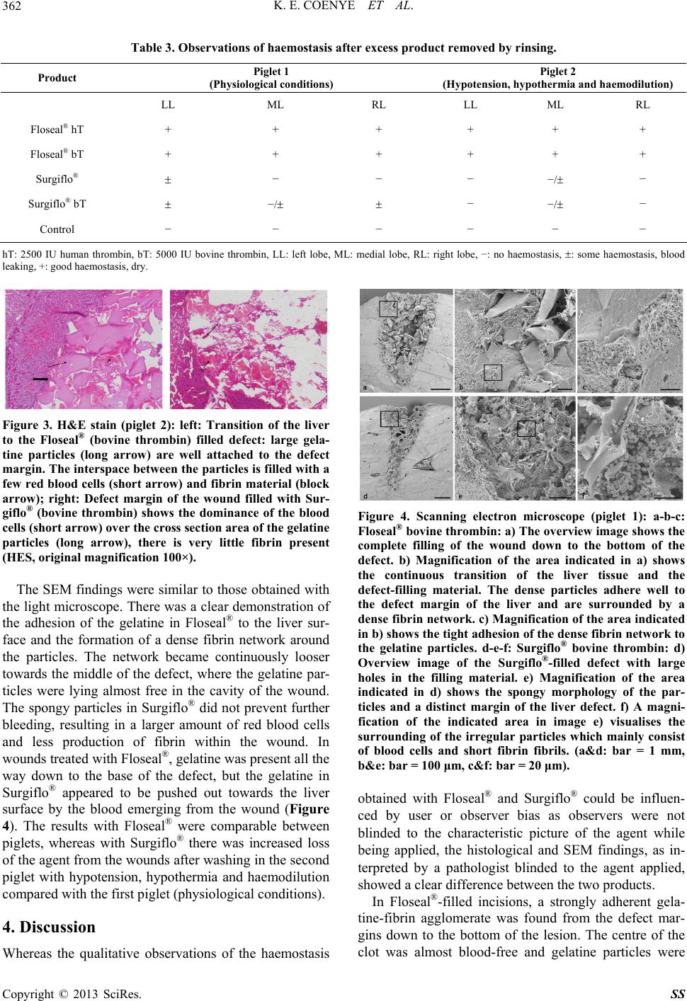
lying more or less freely. This suggests an immediate
initiation of blood coagulation occurring directly at the
site where the blood was leaking into the wound. SEM
imaging demonstrated that the large gelatine particles of
Floseal® were held against the wound surface by fibrin
strands, which were effectively created through fibrino-
gen activation by the thrombin present in the agent. The
activity of the agent did not appear to be influenced by
whether the added thrombin was of human or bovine
origin and whether the dose was 2500 IU or 5000 IU.
The flat surfaces of the Floseal® particles covered the
wound surface and were kept in place by the rapidly
formed fibrin. These mechanisms are likely to sustain the
clotting process, resulting in effective haemostasis.
With Surgiflo®, the particles were less well integrated
into the fibrin and were mainly found at the top of the
wound. Furthermore, the inner region of the wound was
mainly filled with erythrocytes with only a sparse fibrin
network present, suggesting less effective haemostasis.
The Surgiflo® particles did not seem to activate clotting
rapidly, hence allowing the wound to continue bleeding.
This blood appeared to push the gelatine particles up and
out of the wound, allowing them to wash away. The
spongy morphology of Surgiflo® may have caused air to
be trapped inside the particles, resulting in a less effec-
tive covering of the wound surface that failed to initiate
clotting, and causing the gelatine to float on the surface
of the upwelling blood. Similar observations were made
during sample processing, where Surgiflo® particles
floated on the top of the solution, whereas Floseal® par-
ticles sank immediately.
In the animal with hypotension, hypothermia and haemo-
dilution, requiring inotropic support), Floseal® was as ef-
fective under normal physiological conditions, provid-
ing effective haemostasis in all three wounds. In contrast,
Surgiflo® was completely washed out of the wound, and
only a minor degree of clotting was observed in one
wound after two rounds of compression.
These results have implications for clinical practice as
effective haemostasis is linked to improved surgical out-
comes. Absorbable topical haemostatic agents provide a
viable alternative where conventional methods are inef-
fective or impractical. However their benefits must be
carefully considered against potential adverse effects.
Although rare, these include anaemia, atrial fibrillation,
infection, rash, hypotension, respiratory distress, confu-
sion, arrhythmias, arterial thrombosis, and fever [7]. In
addition, expansion of a topical haemostatic agent within
the treated region can result in complications such as
pressing nerves in surrounding tissue against bone or
hard tissue [8]. Surgeons should consider the maximum
swell volume of the product used and its potential effect
on surrounding areas. Despite these considerations, topi-
cal agents remain a powerful tool to achieve haemostasis
during surgical procedures.
There are some limitations to this study. Firstly, this is
a qualitative study in 2 piglets consisting of 6 lesions per
group and 12 lesions per type of gelatine material, and no
quantitative techniques or statistical assessments were
undertaken. In addition, this study only considers one
measure of haemostasis—the presence or absence of
clotting—and does not incorporate any other aspects of
the clinical evaluation of haemostasis, such as the assess-
ment of disease symptoms and response to treatment.
The amount of blood lost from the wounds was not mea-
sured, and therefore the clinical significance of this blood
loss, both from treated and control wounds, is uncertain.
It is also unclear whether each stab wound on each liver
lobe caused similar degrees of blood loss and whether the
damage to these sites affected other wound sites, for
example by reducing blood flow. Finally, basic coagula-
tion tests were not undertaken in either animal, making it
difficult to speculate why Surgiflo® was not effective in
the animal with hypotension, hypothermia and haemoidi-
lution. Despite these limitations, differences between the
two scenarios could be clearly observed.
5. Conclusion
With regard to the results of this qualitative experiment,
Floseal® was able to create a dense and stable blood clot,
even in a piglet with hypotension, hypothermia and hae-
modilution. Floseal® may therefore offer some benefits
compared with Surgiflo®, which in this experiment re-
sulted in comparatively reduced clotting, particularly when
haemostasis was impaired. Further experiments in ani-
mals and humans would be needed before one product
could be recommended over the other for specific clini-
cal situations and applications.
6. Acknowledgements
The test animals, theatre time and products used, were
provided by the Ludwig Boltzmann Institute. The Uni-
versity Hospital of Brussels and the Medical University
of Vienna provided the logistics for the microscopical
studies. The Cell Imaging and Ultrastructure Research
Unit at the University of Vienna provided the equipment
for the electron microscopic investigations.
Medical writing assistance in editing the manuscript
was provided by Fishawack Communications Ltd and
funding for their support was provided by Baxter Bio-
surgery Inc.
6.1. Competing Interests
The authors of this article wish to state the following
conflict of interest with products discussed in this article:
Dr Coenye and Prof. van Griensven have received fees
from Baxter Bioscience Inc. for consultancy, not in rela-
tion to the experiment discussed in this article; Dr Bour-
Copyright © 2013 SciRes. SS
K. E. COENYE ET AL.
Copyright © 2013 SciRes. SS
364
gain, Dr Keibl and Mrs Nürnberger report no potential
conflicts of interest.
6.2. Authors’ Contributions
KEC initiated and conducted the experiment. CK and
MvG participated in the design of the study. CK was also
responsible for the animals before and during the obser-
vations. CB conducted the light microscopy examina-
tions of the samples. SN conducted the scanning electron
microscope study.
6.3. Authors’ Information
This study was purely investigator-driven and resulted
from the absence of any comparison between the agents
observed and clinically observed differences between the
products.
REFERENCES
[1] H. Seyednejad, M. Imani, T. Jamieson and A. M. Sei-
falian, “Topical Haemostatic Agents,” British Journal of
Surgery, Vol. 95, No. 10, 2008, pp. 1197-1225.
doi:10.1002/bjs.6357
[2] G. J. Murphy, B. C. Reeves, C. A. Rogers, S. I. Rizvi, L.
Culliford and G. D. Angelini, “Increased Mortality,
Postoperative Morbidity, and Cost After Red Blood Cell
Transfusion in Patients Having Cardiac Surgery,” Circu-
lation, Vol. 116, No. 22, 2007, pp. 2544-2552.
doi:10.1161/CIRCULATIONAHA.107.698977
[3] A. Shander, “Financial and Clinical Outcomes Associated
with Surgical Bleeding Complications,” Surgery, Vol.
142, Suppl. 4, 2007, pp. S20-S25.
doi:10.1016/j.surg.2007.06.025
[4] M. Leixnering, J. Reichetseder, A. Schultz, M. Figl, E.
Wassermann, H. Thurnher and H. Redl, “Gelatine Throm-
bin Granules For Hemostasis in a Severe Traumatic Liver
and Spleen Rupture Model in Swine,” Journal of Trau-
ma-Injury Infection & Critical Care, Vol. 64, No. 2, 2008,
pp. 456-461. doi:10.1097/TA.0b013e3180340de1
[5] M. C. Oz, D. M. Cosgrove, B. R. Badduke, J. D. Hill, M.
R. Flannery, R. Palumbo and N. Topic, “Controlled
Clinical Trial of a Novel Hemostatic Agent in Cardiac
Surgery. The Fusion Matrix Study Group,” The Annals of
Thoracic Surgery, Vol. 69, No. 5, 2000, pp. 1376-1382.
doi:10.1016/S0003-4975(00)01194-2
[6] R. A. De la Torre, S. L. Bachman, A. A. Wheeler, K. N.
Bartow and J. S. Scott, “Hemostasis and Hemostatic
Agents in Minimally Invasive Surgery,” Surgery, Vol.
142, No. 4, 2007, pp. S39-S45.
doi:10.1016/j.surg.2007.06.023
[7] Baxter Healthcare SA, “Floseal Instructions for Use,”
Zurich.
[8] S. Samadrula, “Topical Hemostatic Agents in Surgery: A
Surgeon’s Perspective,” AORN Journal, Vol. 88, No. 3,
2008, pp. S2-S11. doi:10.1016/S0001-2092(08)00586-3