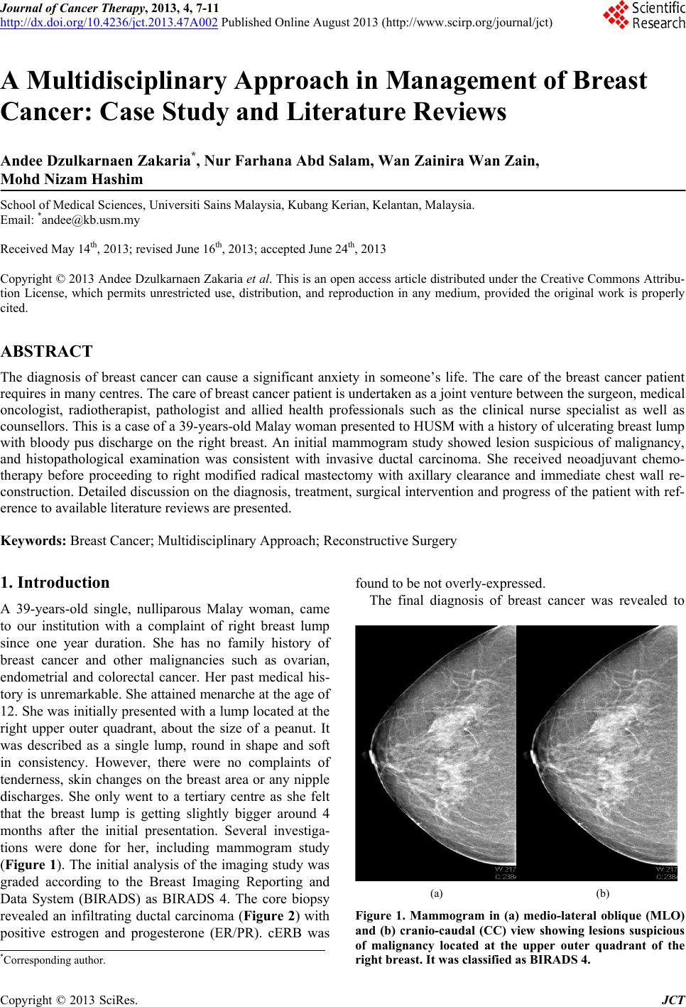
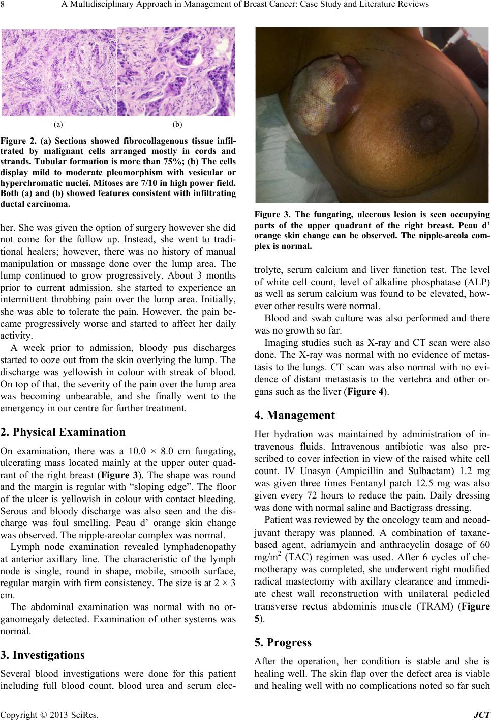
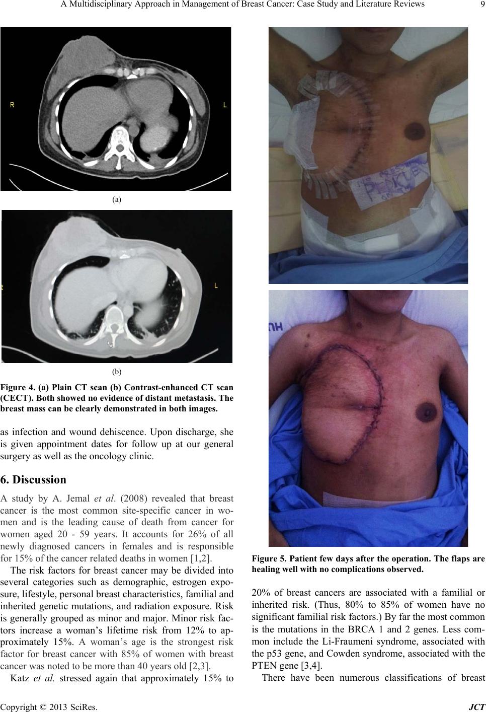
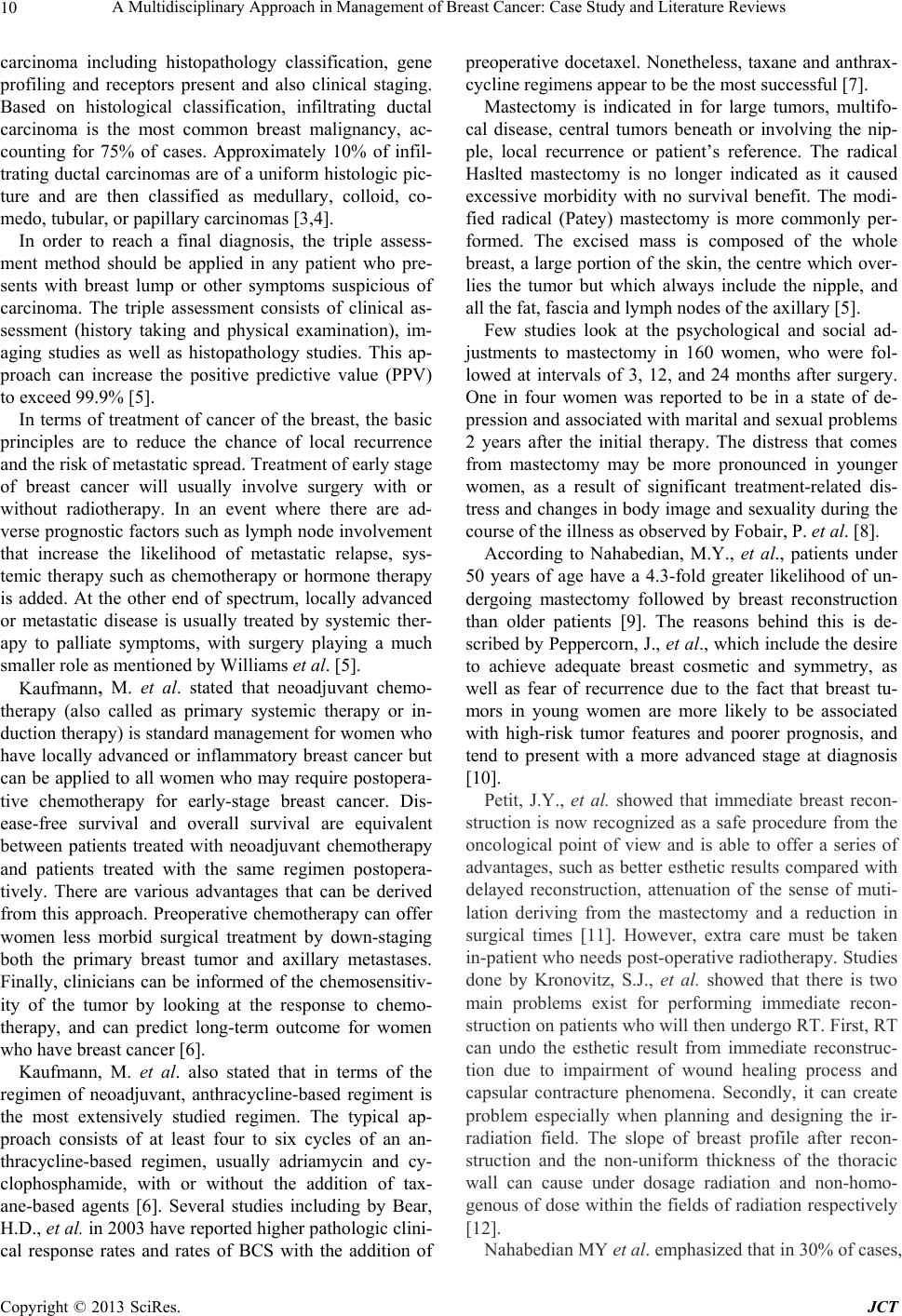
A Multidisciplinary Approach in Management of Bre a s t Cancer: Case Study and Literature Revi ews
Copyright © 2013 SciRes. JCT
11
particularly following RT, reconstruction with autologous
tissue is to be preferred [9]. The main myocutaneous
flaps used in breast reconstruction are the transverse rec-
tus abdominis myocutaneous (TRAM) flap introduced by
Hartrampf in 1982 and used both as a pedicle flap or free
(anastomosis between the inferior epigastric artery and
the thoracodorsal artery, subscapular or internal breast),
combined with or without prosthesis [9,13,14].
Pedicle TRAM flap uses abdominal muscle, fat and
skin tissue vascularized by the rectus muscle pedicle to
reconstruct the breast mound. Grossly, in the pedicle
TRAM flap, excess skin, subcutaneous fat and rectus
muscle from the infraumbilical area are transferred
through a subcutaneous tunnel to the ipsilateral or con-
tralateral mastectomy site. The flap is then rotated,
shaped into a breast mound, and sutured; the umbilicus
and the abdominal skin are sutured into its new position
and the abdomen skin is sutured as in an abdominoplasty.
Despite the loss of muscle function after a pedicle TRAM
flap harvest, it is still possible for patients to become
pregnant and carry a pregnancy to term, as well as to
achieve a normal vaginal delivery [15] .
Similar with our patient that in the free TRAM flap the
skin, subcutaneous fat, deep inferior epigastric artery, and
a small portion of the rectus muscle and fascia from the
infraumbilical area are transferred to the chest defect,
were epigastric vessels are reattached to either thora-
codorsal or internal thoracic vessels via microsurgery.
This technique allows the relocation of larger amounts of
tissue with a lesser risk of fat necrosis. Hence, it may be a
better procedure in patients with risk factors such as
smoking, diabetes mellitus, and obesity [15].
7. Conclusion
Multidisciplinary approach is paramount and effective in
breast cancer management. Despite the life changing di-
agnosis of breast cancer, reconstructive surgery opens an
avenue of possibility to encourage surgery when needed,
and improves the qua lity of life, physically and mentally.
8. Consent
Informed consent obtained from patient.
REFERENCES
[1] A. Jemal, R. Siegel, E. Ward, Y. Hao, J. Xu and T. Mur-
ray, “Cancer Statistics, 2008,” A Cancer Journal for Cli-
nicians, Vol. 58, No. 2, 2008, pp. 71-96.
doi:10.3322/CA.2007.0010
[2] N. N. Baxter, B. A. Virnig, S. B. Durham, et al., “Trends
in the Treatment of Ductal Carcinoma in Situ of the
Breast,” Journal of the National Cancer Institute, Vol. 96,
No. 6, 2004, pp. 443-448. doi:10.1093/jnci/djh069
[3] V. L. Katz and D. Detters, “Lentz: Comprehensive Gy-
necology Mosby an Imprint of Elsevier. Chapter 15. Di-
agnosis and Treatment of Benign and Malignant Breast
Diseases,” 6th Edition, Elsevier, Philadelphia, 2012.
[4] S. J. Katz, P. M. Lantz, N. K. Janz, et al., “Patient In-
Volvement in Surgery Treatment Decisions for Breast
Cancer,” Journal of Clinical Oncology, Vol. 23, No. 24,
2005, pp. 5526-5533. doi:10.1200/JCO.2005.06.217
[5] N. S. Williams, C. J. K. Bulstrode and P. R. O’Connell,
“Bailey and Love’s Short Practice of Surgery Textbook,”
25th Edition, Edward Arnold, London, 2008.
[6] M. Kaufmann, G. N. Hortobagyi, A. Goldhirsch, et al.,
“Recommendations from an International Expert Panel on
the Use of Neoadjuvant (Primary) Systemic Treatment of
Operable Breast Cancer: An Update,” Journal of Clinical
Oncology, Vol. 24, No. 12, 2006, pp. 1940-1949.
doi:10.1200/JCO.2005.02.6187
[7] H. D. Bear, S. Anderson, A. Brown, et al., “The Effect on
Tumor Response of Adding Sequential Preoperative Do-
cetaxel to Preoperative Doxorubicin and Cyclophos-
phamide: Preliminary Results from National Surgical
Adjuvant Breast and Bowel Project Protocol B-27,”
Journal of Clinical Oncology, Vol. 21, No. 22, 2003, pp.
555-558. doi:10.1200/JCO.2003.12.005
[8] P. Fobair, S. L. Stewart, S. Chang, C. D’Onofrio, P. J.
Banks and J. R. Bloom, “Body Image and Sexual Prob-
lems in Young Women with Breast Cancer,” Psychoon-
cology, Vol. 15, No. 7, 2006, pp. 579-594.
doi:10.1002/pon.991
[9] M. Y. Nahabedian, “Breast Reconstruction: A Review and
Rationale for Patient Selection,” Plastic and Recon-
structive Surgery, Vol. 124, No. 1, 2009, pp. 55-62.
doi:10.1097/PRS.0b013e31818b8c23
[10] J. Peppercorn, “Breast Cancer in Women under 40,” On-
cology, Vol. 23, No. 6, 2009, pp. 465-474.
[11] J. Y. Petit, M. Rietjens and C. Garusi, “Breast Recon-
structive Techniques in Cancer Patients: Which Ones,
When to Apply, Which Immediate and Long Term Risks?”
Journal of Hematology & Oncology, Vol. 38, No. 3, 2001,
pp. 231-239.
[12] S. J. Kronovitz and G. L. Robb, “Controversies Regard-
ing Immediate Reconstruction: Aesthetic Risks of Radia-
tion,” In: S. L. Spear, Ed., Surgery of the Breast, 2nd Edi-
tion, Lippincott Williams & Wilkins, Philadelphia, 2006,
pp. 679-699.
[13] S. Al Benna, “Female Plastic and Reconstructive Sur-
geons’ Personal Decision Making for Breast Cancer
Treatment and Reconstruction,” Archives of Gynecology
and Obstetrics, Vol. 284, No. 3, 2011, pp. 737-741.
doi:10.1007/s00404-010-1721-9
[14] A. K. Alderman, S. T. Hawley , J. Wa ljee, M. Mujahid, M.
Morrow and S. J. Katz, “Understanding the Impact of
Breast Reconstruction on the Surgical Decision-Making
Process for Breast Cancer,” Cancer, Vol. 112, No. 3,
2008, pp. 489-494. doi:10.1002/cncr.23214
[15] M. Marín-Gutzke and A. Sánchez-Olaso, “Reconstructive
Surgery in Young Women with Breast Cancer,” Breast
Cancer Research and Treatment, Vol. 123, Suppl. 1, 2010,
pp. 67-74.