Vol.2, No.12, 1369-1374 (2010)
doi:10.4236/ns.2010.212167
Copyright © 2010 SciRes. Openly accessible at http:// www.scirp.org/journal/NS/
Natural Science
Assessment of a short phylogenetic marker based on
comparisons of 3' end 16S rDNA and 5' end 16S-23S ITS
nucleotide sequences on the genus Xanthomonas
Sabarimatou Yakoubou1,2, Jean-Charles Côté1*
1Agriculture and Agri-Food Canada, Research Centre, Québec, Canada; *Corresponding Author: Jean-Charles.Cote@agr.gc.ca
2Département des Sciences Biologiques, Université du Québec à Montréal, Québec, Canada
Received 13 August 2010, revised 15 September 2010, accepted 20 September 2010.
ABSTRACT
A short phylogenetic marker previously used in
the reconstruction of the Class γ-proteobacteria
was assessed here at a lower taxa level, species
in the genus Xanthomonas. This maker is 224
nucleotides in length. It is a combination of a
157 nucleotide sequence at the 3' end of the 16S
rRNA gene and a 67 nucleotide sequence at the
5' end of the 16S-23S ITS sequence. A total of 23
Xanthomonas species were analyzed. Species
from the phylogenetically related genera Xylella
and Stenotrophomonas were included for com-
parison purposes. A bootstrapped neighbor-
joining phylogenetic tree was inferred from
comparative analyses of the 224 bp nucleotide
sequence of all 30 bacterial strains under study.
Four major Groups were revealed based on the
topology of the neighbor-joining tree, Group I to
IV. Group I and II contained the genera Steno-
trophomonas and Xylella, respectively. Group III
included five Xanthomonas species: X. theicola,
X. sacchari, X. albineans, X. transluscens and X.
hyacinthi. This group of Xanthomonas species
is often referred to as the hyacinthi group. Group
IV contained the other 18 Xanthomonas species.
The overall topology of the neighbor-joining
tree was in agreement with currently accepted
phylogenetic. The short phylogenetic marker
used here could resolve species from three dif-
ferent Xanthomonadacea genera: Stenotro-
phomonas, Xylella and Xanthomonas. At the
level of the Xanthomonas genus, distant spe-
cies could be distinguished, and whereas some
closely-related species could be distinguished,
others were undistinguishable. Pathovars could
not be distinguished. We have met the resolving
limit of this marker: pathovars and very closely
related species from same genus.
Keywords: 16S rRNA; 16S-23S ITS; Phylogeny;
Xanthomonas
1. INTRODUCTION
The genus Xanthomonas comprises 27 species. These
species are primarily characterized by the production of
xanthomonadins, a water-insoluble yellow pigment, and
the production of an exo-polysaccharide, the xanthan
gum, which is used as a thickening, stabilizing and gel-
ling agent in food, pharmaceutics, cosmetics and oil in-
dustries [1,2]. Most Xanthomonas species are plant
pathogens [3]. They cause diseases on several economi-
cally important plants including crucifers, Solanaceae,
citrus, cotton, cereals, ornamentals, fruit and nut trees
[3,4]. It is estimated that at least 124 monocotyledons
and 268 dicotyledons are infected by Xanthomonas spe-
cies [4-6].
Up to the mid-90’s, the classification of Xanthomonas
species and isolates was based on phenotypic data. The
main criteria for the creation of new species rested on
host specificity. The taxa “pathovar” was added to dis-
tinguish Xanthomonas species at the infrasubspecific
level. Some species, e.g. X. axonopodis, X. transluscens
or X. campestris, comprised more than ten, 40 and 125
pathovars, respectively [3]. Several methods were used
in an attempt to classify Xanthomonas species and
pathovars: restriction fragment-length polymorphism
(RFLP) [7,8], protein profiles [9] and fatty acid methyl
ester profiles [10-11]. Vauterin et al. reorganized the
classification of the genus Xanthomonas based on
DNA-DNA hybridization [12]. They revealed 20 DNA
homology groups which they considered genomic spe-
cies. Since then, other approaches based on different
nucleotide sequences have been used to study the phy-
logeny of Xanthomonas: the 16S rRNA gene [13], a
multilocus sequence typing (MLST), [14], the 16S-23S
intergenic spacer [15], the repetitive palindromic-based
S. Yakoubou et al. / Natural Scienc e 2 (2010) 1369-1374
Copyright © 2010 SciRes. Openly accessible at http:// www.scirp.org/journal/NS/
1370
polymerase chain reaction fingerprinting (Rep-PCR)
[16], the gyrB gene [17] and a multilocus sequence
analysis (MLSA) [18]. Seven additional Xanthomonas
species have now been described [19-25].
In a recent study [26], a short 232 bp nucleotide se-
quence “marker” was used to reconstruct the phylogeny
of the Class γ-proteobacteria. This 232 bp marker was a
combination of the last 157 bp at the 3’ end of the 16S
rRNA gene and the first 75 bp at the 5’ end of the
16S-23S rRNA internal transcribed spacer (ITS). We
showed that the 157 bp sequence was highly conserved
among closely related species. Owing to its higher rate
of nucleotide substitutions, the 75 bp added discriminat-
ing power among species from same genus and closely
related genera from same family. This marker could re-
construct the phylogeny of the species, genera, families
and Orders within the Class γ-proteobacteria in accor-
dance with the accepted classification.
In the current study, we further assess the resolving
power of this marker at a much lower taxa level: species
within the genus Xanthomonas.
2. MATERIALS AND METHODS
2.1. Bacterial Species and Strains
A total of 25 Xanthomonas strains from 23 species
were analyzed. Four Xylella fastidiosa strains and one
Stenotrophomonas maltophila stain were added for
comparison purposes. They were selected on the basis
that their complete genome sequences were freely
available in GenBank, at the National Center for Bio-
technology Information (NCBI) completed microbial ge-
nomes database (http://www.ncbi.nlm.nih.gov/genomes/
lproks.cgi, August 2009). All bacterial strains and their
GenBank accession numbers are listed in Table 1.
2.2. Sequences Analysis
First, the 16S rRNA and the 16S–23S ITS sequences
of the 30 bacterial strains under study were retrieved
from GenBank. Second, the 16S rRNA gene nucleotide
sequences were aligned using ClustalW [27] (data not
shown). The length of the nucleotide sequence most
conserved was determined at 157 bp. Third, the 16S-23S
ITS were aligned using ClustalW (data not shown). The
length of the nucleotide sequence most conserved was
determined at 67 bp. These two most conserved nucleo-
tide sequences, the 157 bp at the 3’ end of 16S, and the
67 bp at the 5’ end of 16S-23S ITS, were combined into
a single 224 bp sequence for each bacterial species and
strain under study. This 224 bp sequence will be used
here as a phylogenetic marker for the Xanthomonas spe-
cies and related genera under study.
2.3. Phylogenetic Analyses
A neighbor-joining tree was constructed [28] based on
the alignment of the 224 bp sequence of the 30 bacterial
strains under study. The tree was bootstrapped using
1,000 random samples of sites from the alignment, all
using CLUSTAL W software [27] at the DNA Data Bank
of Japan (DDBJ) (http://clustalw.ddbj.nig.ac.jp/tope.html),
with the Kimura’s parameter method [29]. The neighbor-
joining tree was drawn using TreeView (version 1.6.6)
[30,31].
3. RESULTS AND DISCUSSION
A bootstrapped neighbor-joining tree based was in-
ferred from the alignment of the 224 bp sequence of all
25 Xanthomonas species and pathovars, four Xylella
fastidiosa strains and Stenotrophomonas maltophila un-
der study (Figure 1). Four Groups, Group I to IV, were
revealed at the 95% nucleotide sequence identities.
Group I contains Stenotrophomonas maltophila. Group
II encompasses all four Xylella fastidiosa strains. They
share 99% nucleotide sequence identities. Group III in-
cludes five Xanthomonas species: X. theicola, X. sac-
chari, X. albineans, X. transluscens and X. hyacinthi.
They share at least 96% nucleotide sequence identities.
This group of Xanthomonas species is often referred to
as the hyacinthi group [15]. Our results are in agreement
with the first identification of the hyacinthi group based
on the homology of their 16S rRNA [13], 16S-23S ITS
[15] and gyrB nucleotide sequences [17] and MLSA [18].
Group IV contains 18 Xanthomonas species. These spe-
cies share at least 95% nucleotide sequence identities.
Six species can be distinguished: X. axonopodis, X.
codiaei, X. fragariae, X. campestris, X. cassavae and X.
melonis. Xanthomonas perforans, X. euvesicatoria and X.
alfalfae are grouped together and appear undistinguish-
able. These species share 100% nucleotide sequence
identities. The grouping of these six species is in agree-
ment with the work of Parkinson et al. [18] based on
comparison of gyrase B gene sequences. Furthermore,
the grouping of X. perforans, X. euvesicatoria and X.
alfalfae is in agreement with the work of Young et al.
[18] based on MLSA. Xanthomonas hortorum and X.
vasicola, and X. oryzae and X. bromi are grouped to-
gether, respectively, and appear undistinguishable. Both
pair of species share 99% and 100% nucleotide sequence
identities, respectively. Five other Xanthomonas species,
X. gardneri, X. vesicatoria, X. cucurbitae, X. arboricola
and X. pisi are grouped together and appear undistin-
guishable. These species share 100% nucleotide se-
quence identities. The three X. campestris strains appear
undistiguishable. They share 100% nucleotide sequence
identities. The three X. campestris strains appear un-
S. Yakoubou et al. / Natural Scienc e 2 (2010) 1369-1374
Copyright © 2010 SciRes. http://www.scirp.org/journal/NS/Openly accessible at
137
1371
distiguishable. They share 100% nucleotide sequence
identities.
Of the 23 Xanthomonas species under study, 15 spe-
cies or group of species could be distinguished by the
224 bp sequence used as marker. Very closely related
species, such as those in Group IV could not be dinstin-
guished. Pathovars could not be distinguished, as exem-
plified by the three X. camp estris pathovars. The overall
Table 1. Bacterial species used in this study.
Genera Species Pathovars/Strain GenBank accession no.
Stenotrophomonas maltophilia R551-3 NC_01107
Xanthomonas albilineans LMG 494T X95918
alfalfae F1 AF442741
arboricola pv. juglandis LMG 747 T Y10757
axonopodis LMG 538 T X95919
bromi LMG 947 T AF209754
campestris pv. campestris ATCC 33913 T NC_003902
campestris pv. campestris B100 NC_010688
campestris pv. campestris 8004 NC_007086
cassavae LMG 673 T AF209756
codiaei LMG 8678 T Y10765
cucurbitae LMG 690 T Y10760
euvesicatoria 85-10 NC_007508
fragariae LMG 708 T X95920
gardneri CNPH496 AY288083
hortorum LMG 733 T Y10759
hyacinthi LMG 739 T Y10754
melonis LMG 8670 T Y10756
oryzae pv. oryzae MAFF 311018 NC_007705
perforans CNPH411 AY288081
pisi LMG 847 T Y10758
Sacchari LMG 471 T Y10766
Theicola LMG 8684 T Y10763
transluscens pv. graminis AY247064
vasicola LMG 736 T Y10755
vesicatoria LMG 911 T Y10761
Xylella fastidiosa 9a5c NC_002488
M12 NC_010513
M23 NC_010577
Temecula NC_004556
S. Yakoubou et al. / Natural Scienc e 2 (2010) 1369-1374
Copyright © 2010 SciRes. Openly accessible at http:// www.scirp.org/journal/NS/
1372
0.01
Xanthomonas melonis
Stenotrophomonas maltophilia
Xylella fastidiosa 9a 5c
Xylella fastidiosa M1 2
Xylella fastidiosaM23
Xylella fastidiosaTemecula1
100
100
100
Xanthomonas theicola
Xanthomonas sacchari
Xanthomonas albilineans
70
Xanthomonas translucens
Xanthomonas hyacinthi
60
97
59
Xanthomonas axonopodis
Xanthomonas codiaei
Xanthomonas perforans
Xanthomonas euvesicatoria str 85-10
Xanthomonas alfalfae
63
60
65
Xanthomonas fragariae
Xanthomonas campestris campestris B100
Xanthomonas campestris campestris8004
Xanthomonas campestris campestris ATCC 33913
88
80
Xanthomonas hortorum
Xanthomonas vasicola
56
Xanthomonas oryzae
Xanthomonas bromi
Xanthomonas cassavae
Xanthomonas gardneri
Xanthomonas vesicatoria
Xanthomonas cucurbitae
Xanthomonas arboricola
Xanthomonas pisi
Grou ps
I
II
III
IV
0.010.01
Xanthomonas melonis
Stenotrophomonas maltophilia
Xylella fastidiosa 9a 5c
Xylella fastidiosa M1 2
Xylella fastidiosaM23
Xylella fastidiosaTemecula1
100
100
100
Xanthomonas theicola
Xanthomonas sacchari
Xanthomonas albilineans
70
Xanthomonas translucens
Xanthomonas hyacinthi
60
97
59
Xanthomonas axonopodis
Xanthomonas codiaei
Xanthomonas perforans
Xanthomonas euvesicatoria str 85-10
Xanthomonas alfalfae
63
60
65
Xanthomonas fragariae
Xanthomonas campestris campestris B100
Xanthomonas campestris campestris8004
Xanthomonas campestris campestris ATCC 33913
88
80
Xanthomonas hortorum
Xanthomonas vasicola
56
Xanthomonas oryzae
Xanthomonas bromi
Xanthomonas cassavae
Xanthomonas gardneri
Xanthomonas vesicatoria
Xanthomonas cucurbitae
Xanthomonas arboricola
Xanthomonas pisi
Grou ps
I
II
III
IV
Figure 1. Bootstrapped neighbor-joining tree of the genus Xanthomonas species inferred from the alignment of the 224 bp marker.
topology of the neighbor-joining tree was, however, in
agreement with phylogenetic trees based on the 16S
rRNA [13] and the 16S-23S ITS [15].
In previous studies, we showed that a DNA sequence
from 3’ end 16S rRNA gene and 5’ end 16S-23S ITS could
be used as a marker in the reconstruction of phylogenies
in the Gram-positive genus Bacillus and closely-related
genera [32], the Gram-positive Order Bacillales [33],
and the Gram-negative Class γ-proteobacteria [26]. This
maker ranged in size from 220 bp to 232 bp. It contained
150-157 bp from the 3’ end of the 16S rRNA gene and
67-75 bp from the 5’ end of the 16S-23S ITS. The
150-157 bp from the 3’ end of the 16S rRNA gene was
often sufficient to distinguish bacterial Orders, families,
and species from different genera. This sequence was,
however, highly conserved among closely related spe-
cies. Owing to its higher rate of nucleotide substitutions,
the 67-75 bp from the 5’ end of the 16S-23S ITS added
resolving power among closely related species from
same genus. This marker had proven useful in recon-
structing the phylogenies of the genus Bacillus and
closely-related genera [32], the Order Bacillales [33] and
the Class γ-proteobacteria [26] in accordance with ac-
cepted phylogenies inferred from much more compre-
hensive datasets. This marker presented several advan-
tages over the use of the entire 16S rRNA gene or the
16S-23S ITS or the generation of extensive phenotypic
and genotypic data in phylogenetic analyses. We showed
that the method was simple, rapid, suited to large
screening programmes and easily accessible to most
S. Yakoubou et al. / Natural Scienc e 2 (2010) 1369-1374
Copyright © 2010 SciRes. Openly accessible at http://www.scirp.org/journal/NS/
137
1373
laboratories. It also proved very valuable in revealing
bacterial species which appeared misassigned and for
which additional characterization appeared warranted.
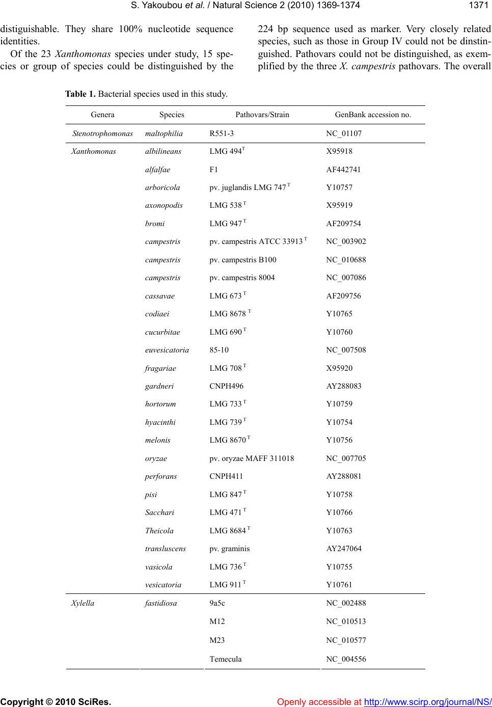
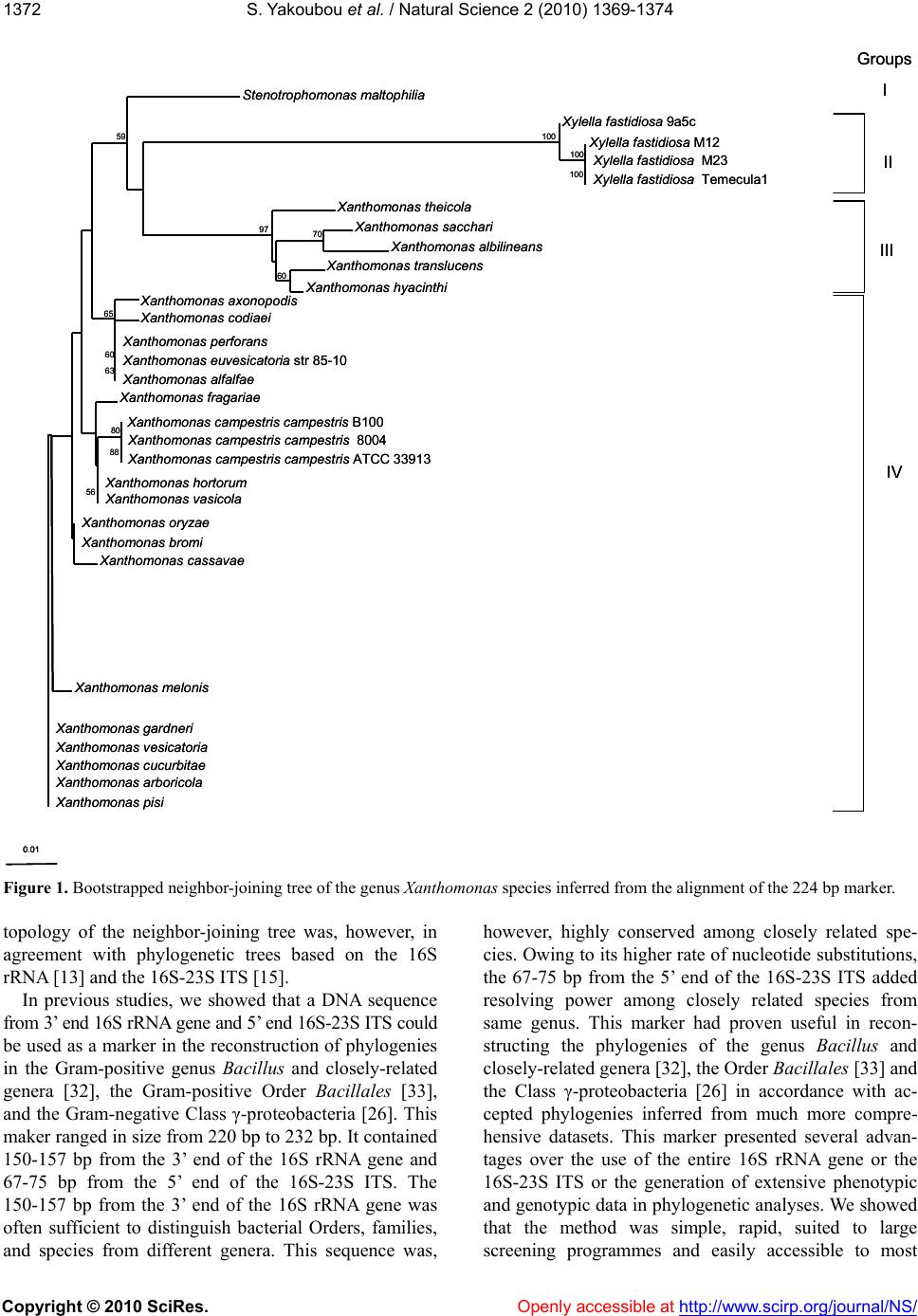
The resolving power of this marker has been further
analyzed here in a much deeper branch of the Class
γ-proteobacteria: the genus Xanthomonas. As expected,
we have shown here that this marker could resolve species
from three different Xanthomonadacea genera: St eno-
trophomonas, Xylella and Xanthomonas. At the level of
the Xanthomonas genus, distant species could be distin-
guished. However, although some closely-related species
could be distinguished, others were grouped together and
some were undistinguishable. Clearly, pathovars could
not be distinguished. We have met the resolving limit of
this marker: pathovars or very closely-related species.
4. CONCLUSION
A short DNA marker based on 3’ end 16S rDNA and
5’ end ITS, had been shown previously to be able to re-
construct the phylogeny of the Class γ-proteobacteria at
the Orders, families, genera and distantly-related species
levels This marker was analyzed here at a lower taxa
level. First, we have shown that this marker could cluster
species from same genera within the family Xanthomo-
nadacea. Next, at the genus Xanthomonas level, we have
shown that although the short DNA marker could dis-
tinguish several species, very closely-related species and
pathovars could not be distinguished. We have reached
the limit of the resolving power of the 224 bp sequence
as a phylogenetic marker: very closely-related species
and pathovars.
REFERENCES
[1] McNeely, W.H. and Kang, K.S. (1973) Xanthan and
some other biosynthetic gums. In: Whistler, R.L. and
BeMiller, J.N. Eds., 2nd Edition, Industrial Gums, Aca-
demic Press, New York, 473-497.
[2] Kennedy J.F. and Bradshaw, I.J. (1984) Production,
Properties, and Applications of Xanthan. In: Bushell M.
E., Ed., Progress in Industrial Microbiology, Elsevier,
Amsterdam, 319-371.
[3] Vauterin, L., Swings, J., Kersters, K., Gillis, M., Mew, T.
W., Schroth, M.N., Palleroni, N.J., Hildebrand, D.C.,
Stead, D.E. and other authors (1990) Towards an im-
proved taxonomy of Xanthomonas. International Journal
of Systematic Bacteriology, 40, 312-316.
[4] Hayward, A.C. (1993) The host of Xanthomonas. In
Swings J.G. and Civerolo E.L., Eds., Xanthomonas,
Chapman & Hall, London, 51-54.
[5] Leyns, F., De Cleene, M., Swing, J. and De Ley, J. (1984)
The host range of the genus Xanthomonas. Botanical re-
view, 50, 308-356.
[6] Vauterin, L., Hoste, B., Kersters, K. and Swings, J. (1995)
Reclassification of Xanthomonas. International Journal
of Systematic Bacteriology, 45, 472-489.
[7] Lazo, G.R., Roffey, R. and Gabriel, D.W. (1987)
Pathovars of Xanthomonas campestris are distinguishable
by restriction fragment-length polymorphism. Interna-
tional Journal of Systematic Bacteriology, 37, 144-221.
[8] Lazo, G.R., and Gabriel, D.W. (1987) Conservation of
plasmid DNA sequences and pathovar identification of
strains of Xanthomonas campestris. Phytopathology, 77,
448-453.
[9] Vauterin, L. Yang, P., Hoste, B., Vancanneyt, M.,
Civerolo, E.L., Swings, J. and Kersters, K. (1991) Dif-
ferentiation of Xanthomonas campestris pv. Citri strains
by sodium dodecyl; sulphate-polyacrylamide gel elec-
trophoresis of proteins, fatty acid analysis, and
DNA-DNA hybridization. International Journal of Sys-
tematic Bacteriology, 41, 535-542.
[10] Chase, A.R., Stall, R.E., Hodge, N.C. and Jones, J.B.
(1992) Characterization of Xanthomonas campestris
strains from aroids using physiological, pathological, and
fatty acid analyses. Phytopathology, 82, 754-759.
[11] Yang, P., Vauterin, L., Vancanneyt, M., Swings, J. and
Kersters, K. (1993) Application of fatty acid methyl es-
ters for the taxonomic analysis of the genus Xanthomo-
nas. Systematic and Applied Microbiology, 16, 47-71.
[12] Vauterin, L., Hoste, B., Kersters, K. and Swings, J. (1995)
Reclassification of Xanthomonas. International Journal
of Systematic Bacteriology, 45, 472-489.
[13] Hauben, L., Vauterin, L., Swings, J. and Moore, E. (1997)
Comparison of 16S ribosomal DNA sequences of all
Xanthomonas species. International Journal of System-
atic Bacteriology, 47, 328-335.
[14] Maiden, M.C.J., Bygraves, J.A., Feil, E.J., Morelli, G.,
Russell, J.E., Urwin, R., Zhang, Q., Zurth, K., Caugant,
D., Feavers, I.M., Achtman, M. and Spratt, B.G. (1998)
Multilocus sequence typing: A portable approach to the
identification of clones within populations of pathogenic
microorganisms. Proceedings of the National Academy of
Sciences, USA, 1998, 95, 3140-3145.
[15] Gonçalves, E.R. and Rosato, Y.B. (2002) Phylogenetic
analysis of Xanthomonas species based upon 16S–23S
rDNA intergenic spacer sequences. International Journal
of Systematic and Evolutionary Microbiology, 52, 355-
361.
[16] Rademaker, J.L.W., Louws, F.J., Schultz, M.H., Ross-
bach, U., Vauterin, L., Swings, J. and de Bruijn, F.J.
(2005) A comprehensive species to strain taxonomic
framework for Xanthomonas. Phytopathology, 95, 1098-
1111.
[17] Parkinson, N., Aritua, V., Heeney, J., Cowie, C., Bew, J.
and Stead, D. (2007) Phylogenetic analysis of Xantho-
monas species by comparison of partial gyrase B gene
sequences. International Journal of Systematic and Evo-
lutionary Microbiology, 57, 2881-2887.
[18] Young, J.M., Park, D.-C., Shearman, H.M. and Fargier, E.
(2008) A multilocus sequence analysis of the genus Xan-
thomonas. Systematic and Applied Microbiology, 31,
366-377.
[19] Trébaol, G., Gardan, C., Manceau, J., Tanguy, Y., Trilly, Y.
and Boury, S. (2000) Genomic and phenotypic charac-
terisation of Xanthomonas cynarae; a new species caus-
ing bacterial bract spot of artichoke (Cynara scolymus
L.). International Journal of Systematic and Evolution-
ary Microbiology, 50, 1471-1478.
S. Yakoubou et al. / Natural Scienc e 2 (2010) 1369-1374
Copyright © 2010 SciRes. http://www.scirp. org/journal/NS/Openly accessible at
1374
[20] Jones, J.B., Lacy, G.H., Bouzar, H., Stall, R.E. and
Schaad, N.W. (2004) Reclassification of the xanthomo-
nads associated with bacterial spot disease of tomato and
pepper. Systematic and Applied Microbiology, 27,
755-762.
[21] Euzéby, J. (2006) Validation of the publication of new
names and new combinations previously effectively, but
not validly published. Validation List no. 109. Interna-
tional Journal of Systematic and Evolutionary Microbi-
ology, 56, 925-927.
[22] Euzéby, J. (2007) List of New names and new combina-
tions previously effectively, but not validly, published.
Validation List no. 115. International Journal of System-
atic and Evolutionary Microbiology, 57, 893-897.
[23] Schaad, N.W., Postnikova, E., Lacy, G.H., Sechler, A.,
Agarkova, I., Stromberg, P.E., Stromberg, V.K. and Vi-
daver, A.K. (2005) Reclassification of Xanthomonas
campestris pv. citri (ex Hasse 1915) Dye 1978 forms A,
B/C/D, and E as X. smithii subsp. citri (ex Hasse) sp. nov.
nom. rev. comb. nov., X. fuscans subsp. aurantifolii (ex
Gabriel 1989) sp. nov. nom. rev. comb. nov., and X. al-
falfae subsp. citrumelo (ex Riker and Jones) Gabriel et al.,
1989 sp. nov. nom. rev. comb. nov.; X. campestris pv.
malvacearum (ex Smith 1901) Dye 1978 as X. smithii
subsp. smithii nov. comb. nov. nom. nov.; X. campestris
pv. alfalfae (ex Riker and Jones, 1935) Dye 1978 as X.
alfalfae subsp. alfalfae (ex Riker et al., 1935) sp. nov.
nom. rev.; and ‘‘var. fuscans’’ of X. campestris pv. phase-
oli (ex Smith, 1987) Dye 1978 as X. fuscans subsp. fus-
cans sp. nov. Systematic and Applied Microbiology, 28,
494-518.
[24] Schaad, N.W., Postnikova, E., Lacy, G., Sechler, A.,
Agarkova, I., Stromberg, P.E., Stromberg, V.K. and Vi-
daver, A.K. (2006) (Erratum) Emended classification of
Xanthomonad pathogens on citrus. Systematic and Ap-
plied Microbiology, 29, 690-695.
[25] Schaad, N.W., Postnikova, E., Lacy, G., Sechler, A.,
Agarkova, I., Stromberg, P. E., Stromberg, V.K. and Vi-
daver, A.K. (2007) Xanthomonas alfalfae sp. nov., Xan-
thomonas citri sp. nov. and Xanthomonas fuscans sp. nov.
In List of new names and new combinations previously
effectively, but not validly, published, Validation List no.
115. International Journal of Systematic and Evolution-
ary Microbiology, 57, 893-897.
[26] Yakoubou, S. and Côté, J.-C., (2010) Phylogeny of
γ-proteobacteria inferred from comparisons of 3' end 16S
rRNA gene and 5’ end 16S-23S ITS nucleotide sequences.
Natural Science, 2, 535-543.
[27] Thompson, J.D., Higgins, D.G. and Gibson, T.J. (1994)
Clustal W: improving the sensitivity of progressive mul-
tiple sequence alignment through sequence weighting,
position-specific gap penalties and weight matrix choice.
Nucleic Aci ds Research, 22, 4673-4680.
[28] Saitou, N. and Nei, M. (1987) The neighbour-joining
method: a new method of constructing phylogenetic trees.
Molecular Biology and Evolution, 4, 406-425.
[29] Kimura, M. (1983) The neutral theory of molecular evo-
lution. Cambridge University Press, UK.
[30] Page, R.D.M. (1996) TREEVIEW: An application to
display phylogenetic trees on personal computers. Com-
puter Application in the Biosciences, 12, 357-358.
[31] Page, R.D.M. (2000) TreeView—Tree drawing software
for Apple Macintosh and Windows. http://taxonomy.zoo-
logy.gla.ac.uk/rod/treeview.html.
[32] Xu, D. and Côté, J.-C. (2003) Phylogenetic relationships
between Bacillus species and related genera inferred
from comparison of 3’ end 16S rDNA and 5’ end
16S-23S ITS nucleotide sequences. International Journal
of Systematic and Evolutionary Microbiology, 53, 695-
704.
[33] Yakoubou, S., Xu, D. and Côté, J.-C. (2010) Phylogeny
of the Order Bacillales inferred from 3’ 16S rDNA and 5’
16S-23S ITS nucleotide sequences. Natural Science, 2
990-997.