 Food and Nutrition Sciences, 2013, 4, 124-130 http://dx.doi.org/10.4236/fns.2013.47A015 Published Online July 2013 (http://www.scirp.org/journal/fns) Effects of Carpio Decoction on the Structure of Kidney in Rats with Adriamycin-Induced Nephrosis Huaying Ning1,2, Hui Wu1,3, Xiaojian Tian1,4, Yunliang Guo1, Wei Shen1*, Xin Wang1, Zhou Zhen1 1Institute of Integrative Medicine, Qingdao University Medical College, Qingdao, China; 2Haihe Hospital of Tianjin, Tianjin, China; 3Affiliated Hospital of Suzhou University Medical College, Suzhou, China; 4Second Affiliated Hospital of Tianjin University of TCM, Tianjin, China. Email: *sw_qdu@163.com Received March 4th, 2013; revised April 4th, 2013; accepted April 17th, 2013 Copyright © 2013 Huaying Ning et al. This is an open access article distributed under the Creative Commons Attribution License, which permits unrestricted use, distribution, and reproduction in any medium, provided the original work is properly cited. ABSTRACT The aim of this study was to observe the effects of Cyprinus carpio decoction on the expression of aquaporins in rats with adriamycin-induced nephropathy and to explore the therapeutic mechanism on nephrotic edema. Total of 50 Wis- tar rats were randomly divided into normal group, model group, fosinopril group, Cyprinus carpio decoction treated with high dose group and low dose group consisting of 10 rats respectively. Nephropathy models were established by injecting adriamycin through tail vein and treated with Cyprinus carpio decoction. Urinary protein excretions in 12 h, serum albumin, total serum protein, serum sodium and potassium were measured by biochemical assay. The pathologi- cal changes and the expression of AQP1, AQP2, AQP3 in rat kidneys were respectively detected by HE stain and im- munohistochemiscal assay. The results indicated: 1) The urinary protein excretion in 12 h (proteinuria) increased sig- nificantly along the time longed modeling, while no significant increasing in Cyprinus carpio decoction treated group (F = 5.23 - 41.89, P < 0.05); 2) The serum albumin and total protein in model group were significantly lower than that in normal group, but that in Cyprinus carpio decoction treated group were higher than that in model group (F = 13.12 - 15.48, P < 0.05). The serum sodium and potassium in model group were higher than those in normal group, while that in Cyprinus carpio decoction treated group were higher than that in model group (F = 3.42 - 3.96, P < 0.05); 3) Renal glomerular capillaries congestive of group M rats were expansion. Glomerular mesangial cells and the basement mem- brane were diffuse hyperplasia, inflammatory cell infiltration. Glomerular mesangial cells and the basement membrane of Cyprinus carpio decoction with interventing groups reduced proliferation, less inflammatory cell infiltration; 4) In model group, the expressions of AQP1-3 in the renal tubule and collecting duct cells increased significantly than those in normal group, and those in Cyprinus carpio decoction treated group decreased than those in model group (F = 3.97 - 6.19, P < 0.05). It is suggested that Cyprinus carpio decoction could reduced the urinary protein excretion and alleviate pathological lesion and edema with adriamycin-induced nephropathy by decreasing the expressions of AQPs in kidneys. Keywords: Cyprinus carpio Decoction; Adriamycin Nephropathy; AQP1; AQP2; AQP3; Rats 1. Introduction Nephrotic syndrome (NS) is a clinical syndrome due to a series of pathophysiological changes of high glomerular filtration permeability and a large number of plasma pro- teins from the urine loss with edema, proteinuria, and low protein hyperlipidemia for the clinical features. Edema is related to water-sodium retention and no effective method to cure up to now. Water channel proteins (Aquaporin, AQP) are a family of transmembrane proteins through which the water molecules can pass specifically. Found so far, there are eight kinds of water channel proteins in kid- neys, which are important to renal re-absorption of water liquid and dilute urine concentration function. Abnormal expression of AQPs is one of the mechanisms water- sodium retention [1]. AQP1 was mainly expressed in the cell membrane surface of the renal proximal tubule and medullary descending thin section to reabsorb water from the original urine; AQP2 played an important role in water balance of the kidney, which was mainly distributed in the kidney distal tubule and the lumen side of the main col- lecting duct cells, controlled by arginine vasopressin (AVP) regulation [2]. AQP2 was an important protein to regulate *Corresponding author. Copyright © 2013 SciRes. FNS  Effects of Carpio Decoction on the Structure of Kidney in Rats with Adriamycin-Induced Nephrosis 125 the water permeability of renal collecting duct, and its ab- normal expression and regulation mechanism are closely related to physiological and pathophysiology in water re- gulation of the kidney. AQP3 may be the out-flow channel with renal re-absorption of water, mainly distributed in luminal side of collecting duct principal cells in the kid- ney. Cyprinus carpio was sweet in taste, mild in tropism, non-toxic, had a significant effect in treating various causes of edema documented in ancient and modern lit- erature. The people also often received good effect after the application of Cyprinus carpio decoction, but also had cases on treating edema with Cyprinus carpio decoction in clinical. This study was designed to explore the effect of Cyprinus carpio decoction on renal AQP expression nephropathy induced by adriamycin and investigate the mechanism treating renal edema of Cyprinus carpio de- coction in rats. 2. Materials and Methods 2.1. Experimental Animals Total of 50 healthy adult Wistar rats, male and female, 7W age, body weight in (180 ± 20) g, SPF grade, pur- chased from Shandong Lukang Pharmaceutical Co. Ltd (SLXK-Lu-2008-0002). The guidance suggestions for care of laboratory animals was followed according to the Guidelines for caring for experimental animals published by the Ministry of Science and Technology of the Peo- ple’s Republic of China. All the rats were fed adapted standard diet for 7 days in different cages with 12 h natu- ral light, cozy temperature, conformable humidity, freely drinking and eating and changed bedding every day. 2.2. The Nephropathy Model Induced by Adriamycin Adriamycin (doxorubicin hydrochloride) purchased from Zhejiang Hisun Pharmaceutical Co. Ltd. was diluted with saline solution for 2 mg/ml of injection. All rats feeding adaptively for 7 days and their 12 h-urine-protein were tested negatively, were divided randomly into normal group, model group and fosinopril group, Cyprinus car- pio decoction with high dose group and low dose group consisting of 10 rats respectively. The preparation me- thod of the nephropathy model induced by adriamycin in rats was referenced that of Bertani et al. [3,4]. The solu- tion of adriamycin was injected using micro-injection syringe in rat tail vein between needle and mouse tail vein for 30˚ after withdrawing a return of blood. The first injection of doxorubicin was at 4 mg/kg body weight. After 7 days the twice injection was at 3.5 mg/kg body weight in the same way. Then injection for 3 days it was a symbol of model of success that 12 h-urine-protein was tested for positive. Synchronize the control group was injected with normal saline. 2.3. The Preparation Method of Cyprinus carpio Decoction In this experiment, the fish was provided by Fisheries Research Institute, Henan Province, the Yellow River carp seed field (SC1043-2001). The fish was frozen im- mediately after harvest and thawed at room temperature before prepared Cyprinus carpio decoction, weighted, washed in cold water for three times, set in stainless steel pot, added distilled water to the total weight of fish and water for 5 times with the weight of the fish. After the decoction boiled 10 minutes, churned the fish in order to separated the bone and meat, fractured skull, then contin- ued to boiled with slow fire for churning once every 10 minutes. The pan was moved away from the heat source after the total weight of fish and water was 4 times the weight of fish (about 60 minutes). Using a 6-drug screen (100 meshes) filtered out the Cyprinus carpio decoction. The concentration of decoction was 25%. The condensed decoction in 25% concentration continued to be condens- ed with fast vacuum concentrator (Eppendorf Company, 5301), pre-selected temperature was 45˚C for 400% con- centration (including carp 4 g/ml). The decoction was packed in polyethylene plastic bags (100 ml/bag), disin- fected 60 minutes in 80˚C hot water, and cooled naturally, set −20˚C refrigerator to standby. 2.4. Intervention Experiments The intervention experiments started after 3 days in the 2nd doxorubicin injection and the 12 h-urine-protein test- ing positively (model success). Firstly, melting the Cy- prinus carpio decoction with thermostatic water-bath (36˚C) before using, then the rats were lavaged according to the following dose once a day for 21 days: 1) High-dose group: 22.50 g/kg body weight, Cypri- nus carpio decoction concentration for 4 g/ml. Equivalent adult (70 kg body weight) dose of 250 g, the dose the rats need were calculated in accordance with the conversion factor of 0.018 between rat and human body surface area; 2) Low-dose group: 11.25 g/kg weight, Cyprinus car- pio decoction concentration for 2 g/ml. Equivalent adult (70 kg body weight) dose of 125 g, the dose the rats need were calculated in accordance with the conversion factor of 0.018 between rat and human body surface area; 3) Fosinopril group: 0.9 mg/kg body weight. Fosino- pril sodium tablets (Mengnuo, Sino-American Shanghai Squibb Pharmaceuticals Ltd.) were fully crushed into fine powder to prepare the solution of 0.09 mg/ml con- centration with saline; 4) Normal group and model group: Synchronous given normal saline orally. 2.5. Observation Index 1) Histopathology: After collecting blood, the kidney Copyright © 2013 SciRes. FNS  Effects of Carpio Decoction on the Structure of Kidney in Rats with Adriamycin-Induced Nephrosis Copyright © 2013 SciRes. FNS 126 specimens was took out and washed the remnant blood with normal saline, removed the kidney capsule, sliced levelly the kidney for 2 parts along the center of the renal pedicle, then fixed in 4% paraformaldehyde, and pre- served in 4˚C. Conventional dehydration, transparent, dip wax, embedded, sliced thickness for 4 μm, patch. Sec- tions were conventional dewaxed, hydration, hematoxy- lin staining for 3 minutes, washing with water, eosin stain- ing for 1.5 minutes. Conventional dehydration, transpar- ent, mounted with neutral gum. The glomerular capillary endothelial cells, basement membrane, mesangial matrix, mesangial cells, tubular, interstitial and other changes of renal tissue were observe under light microscopy. 2) Immunohistochemistry: Took the kidney specimens to wash the remnant blood with normal saline, removed the kidney capsule, sliced levelly the kidney for 2 parts along the center of the renal pedicle, then placed in 4% paraformaldehyde, and preserved in 4˚C. Conventional dehydration, transparent, dip wax, embedded, sliced thick- ness for 4 μm, patch. Rabbit anti-rat AQP1-3 affinity- purified antibody, SABC kit, DAB chromogenic kit were purchased from Wuhan Boster Biological Engineering Co. Ltd. Take the slices at 60˚C oven to bake 12 h and conventionally dewax to water, operate according to kit instructions, DAB color 1 minutes, purple hematoxylin- stained 10 s, conventional dehydration, transparent, neu- tral resin were mounted. Brown-yellow granules were found in cell membrane and the cytoplasm with light microscope were considered as positive cells. Some sec- tions without primary antibody were alternative staining with 0.1 mol/LPBS to be a negative control. Each immu- nohistochemistry slice was randomly collected five high power field (400-fold) by two pathologists to quantitative analysis (Image-Pro Plus version 6.0) total area of posi- tive cells area and integrated optical density of positive staining area. Optical density value represented the aver- age expression, and the average optical density = IOD/ SUM area. The five images of each slice calculated the average was optical density measured values of the slice [5]. 2.6. Statistical Analysis Using SPSS 16.0 statistical software statistics the results and show in (χ ± S). The urinary protein excretion in 12 h in different groups at different times compared to use repeated measures analysis of variance (Repeated Meas- ures) and LSD between groups; the data of serum bio- chemical and the average optical density of immunohis- tochemical were analyzed by single-factor of variance (One-Way ANOVA) and LSD comparison between groups. Using Dunnett’ T3 and Dunnett’ C methods with hetero- scedasticity. P < 0.05 indicated a statistically significant difference. 3. Results 3.1. General Conditions Rats in normal group were in good spirits, shiny fur, move freely, responsive. The modeling rats had varying degrees of reduced feeding, weight loss, diarrhea, curled up and body hair was messy and dull after the first injec- tion of adriamycin. After the 2nd doxorubicin injection, the rats were evidently depressed, yellow fluffy body hair, loose stool, decreased appetite, weight loss, swollen limbs. In Cyprinus carpio decoction intervention groups, the rats increased food intake, gained weight, reduced edema gradually. Total of 11 rats were died in the experiment, 3 cases in the model group, 2 cases in fosinopril group, and 3 cases in Cyprinus carpio decoction treated with high dose group and low dose group respectively. Ascites were found in dead rats. 3.2. Urinary Protein Excretion in 12 h The urinary protein excretion in 12 h (12 h-urine-protein) had no significant differences among the groups before modeling (P > 0.05). The urinary protein excretion in 12 h of the model group rats heightened with prolong of time after modeling, but Cyprinus carpio decoction groups hadn’t obvious increasing. There was no significant dif- ference between the normal group and fosinopril group (P < 0.05). Analysis of variance suggested that the uri- nary protein excretion in 12 h at all measurement time points were significantly different (F = 41.89, P < 0.05), urinary protein changes were significantly different over time (F = 5.23, P < 0.05), and the difference of group effect was significant (F = 17.40, P < 0.05) (Table 1). Table 1. The urinary protein excretion in 12 h at different time point (± S x, mg/d). Time Groups n Before modeling After modeling 0 d After modeling 6 d After modeling 12 d After modeling 18 d After modeling 24 d Normal group 9 3.84 ± 2.54 4.34 ± 1.53 4.69 ± 2.89 5.54 ± 2.06 5.66 ± 2.60 4.98 ± 2.12 Model group 7 4.13 ± 2.79 10.19 ± 2.68a 12.37 ± 3.00a 16.61 ± 7.15a 19.20 ± 6.18a 20.09 ± 3.84a Fosinopril group 8 4.86 ± 3.12 10.00 ± 2.71a 12.64 ± 4.51a 9.69 ± 1.99a,b 10.49 ± 3.23a,b 10.57 ± 3.56a,b High doze group 7 2.50 ± 1.41 7.82 ± 1.30a,b 9.55 ± 1.99a 10.97 ± 2.71a,b 11.31 ± 2.57a,b 10.34 ± 1.44a,b Low doze group 7 2.62 ± 2.46 8.62 ± 2.23a 11.01 ± 2.87a 13.31 ± 3.39a,b 12.23 ± 3.05a,b 12.51 ± 2.76a,b Compared with normal group, aP < 0.05; Compared with model group, bP < 0.05. 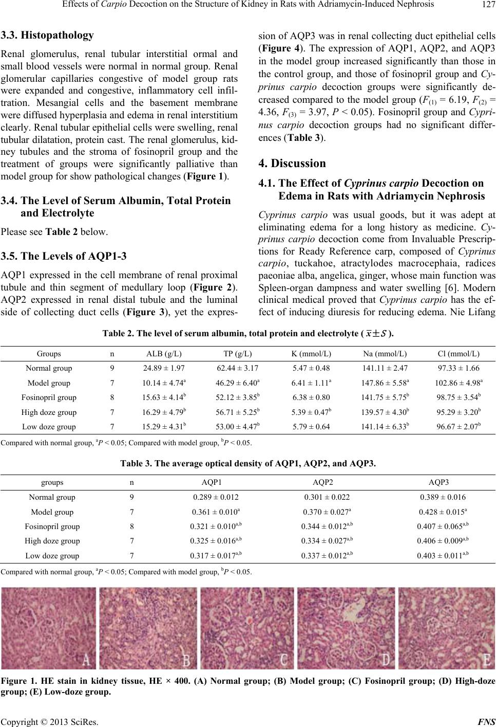 Effects of Carpio Decoction on the Structure of Kidney in Rats with Adriamycin-Induced Nephrosis 127 3.3. Histopathology Renal glomerulus, renal tubular interstitial ormal and small blood vessels were normal in normal group. Renal glomerular capillaries congestive of model group rats were expanded and congestive, inflammatory cell infil- tration. Mesangial cells and the basement membrane were diffused hyperplasia and edema in renal interstitium clearly. Renal tubular epithelial cells were swelling, renal tubular dilatation, protein cast. The renal glomerulus, kid- ney tubules and the stroma of fosinopril group and the treatment of groups were significantly palliative than model group for show pathological changes (Figure 1). 3.4. The Level of Serum Albumin, Total Protein and Electrolyte Please see Table 2 below. 3.5. The Levels of AQP1-3 AQP1 expressed in the cell membrane of renal proximal tubule and thin segment of medullary loop (Figure 2). AQP2 expressed in renal distal tubule and the luminal side of collecting duct cells (Figure 3), yet the expres- sion of AQP3 was in renal collecting duct epithelial cells (Figure 4). The expression of AQP1, AQP2, and AQP3 in the model group increased significantly than those in the control group, and those of fosinopril group and Cy- prinus carpio decoction groups were significantly de- creased compared to the model group (F(1) = 6.19, F(2) = 4.36, F(3) = 3.97, P < 0.05). Fosinopril group and Cypri- nus carpio decoction groups had no significant differ- ences (Table 3). 4. Discussion 4.1. The Effect of Cyprinus carpio Decoction on Edema in Rats with Adriamycin Nephrosis Cyprinus carpio was usual goods, but it was adept at eliminating edema for a long history as medicine. Cy- prinus carpio decoction come from Invaluable Prescrip- tions for Ready Reference carp, composed of Cyprinus carpio, tuckahoe, atractylodes macrocephaia, radices paeoniae alba, angelica, ginger, whose main function was Spleen-organ dampness and water swelling [6]. Modern clinical medical proved that Cyprinus carpio has the ef- fect of inducing diuresis for reducing edema. Nie Lifang Table 2. The level of serum albumin, total protein and electrolyte (± S x). Groups n ALB (g/L) TP (g/L) K (mmol/L) Na (mmol/L) Cl (mmol/L) Normal group 9 24.89 ± 1.97 62.44 ± 3.17 5.47 ± 0.48 141.11 ± 2.47 97.33 ± 1.66 Model group 7 10.14 ± 4.74a 46.29 ± 6.40a 6.41 ± 1.11a 147.86 ± 5.58a 102.86 ± 4.98a Fosinopril group 8 15.63 ± 4.14b 52.12 ± 3.85b 6.38 ± 0.80 141.75 ± 5.75b 98.75 ± 3.54b High doze group 7 16.29 ± 4.79b 56.71 ± 5.25b 5.39 ± 0.47b 139.57 ± 4.30b 95.29 ± 3.20b Low doze group 7 15.29 ± 4.31b 53.00 ± 4.47b 5.79 ± 0.64 141.14 ± 6.33b 96.67 ± 2.07b Compared with normal group, aP < 0.05; Compared with model group, bP < 0.05. Table 3. The average optical density of AQP1, AQP2, and AQP3. groups n AQP1 AQP2 AQP3 Normal group 9 0.289 ± 0.012 0.301 ± 0.022 0.389 ± 0.016 Model group 7 0.361 ± 0.010a 0.370 ± 0.027a 0.428 ± 0.015a Fosinopril group 8 0.321 ± 0.010a,b 0.344 ± 0.012a,b 0.407 ± 0.065a,b High doze group 7 0.325 ± 0.016a,b 0.334 ± 0.027a,b 0.406 ± 0.009a,b Low doze group 7 0.317 ± 0.017a,b 0.337 ± 0.012a,b 0.403 ± 0.011a,b Compared with normal group, aP < 0.05; Compared with model group, bP < 0.05. Figure 1. HE stain in kidney tissue, HE × 400. (A) Normal group; (B) Model group; (C) Fosinopril group; (D) High-doze group; (E) Low-doze group. Copyright © 2013 SciRes. FNS 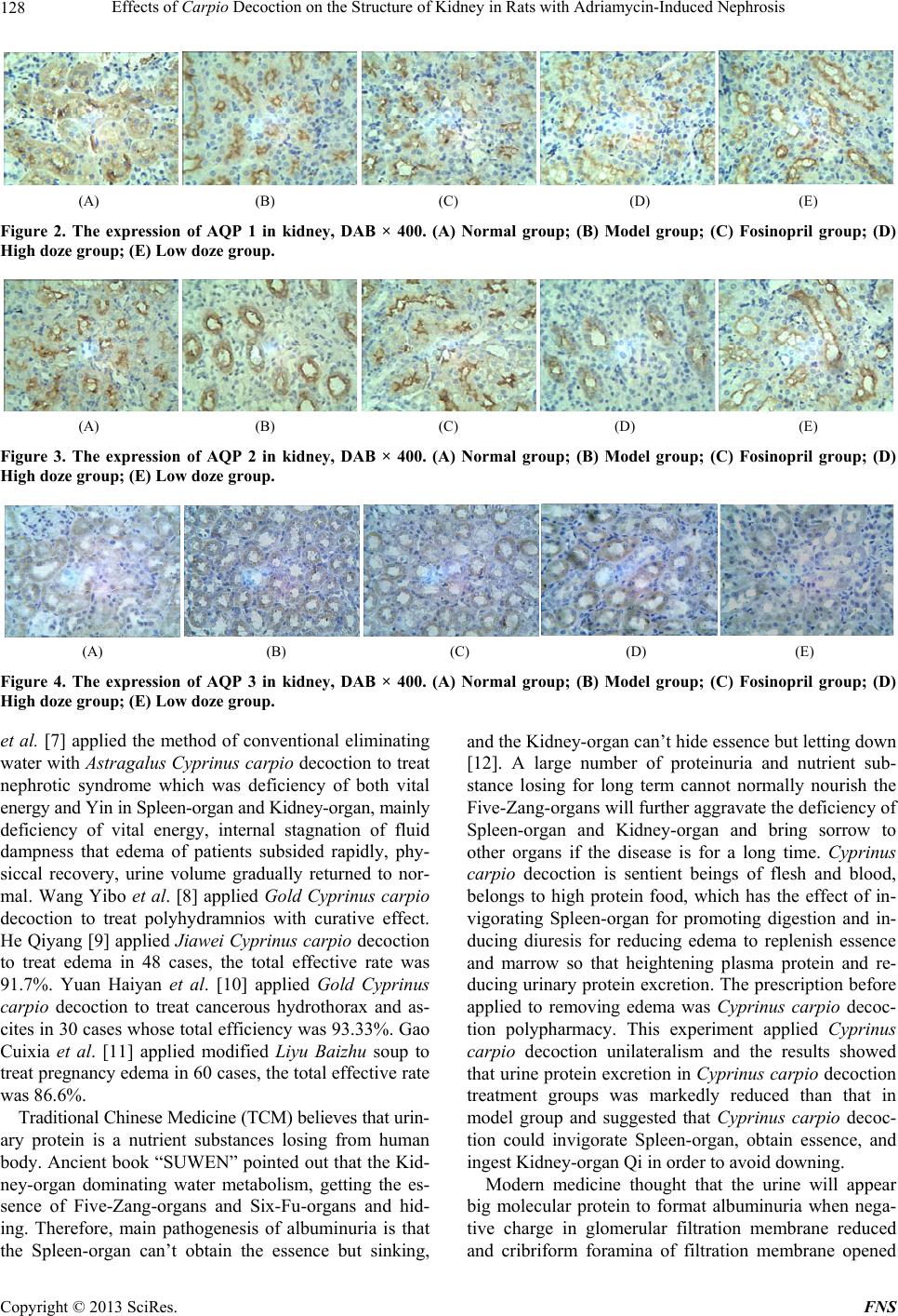 Effects of Carpio Decoction on the Structure of Kidney in Rats with Adriamycin-Induced Nephrosis 128 (A) (B) (C) (D) (E) Figure 2. The expression of AQP 1 in kidney, DAB × 400. (A) Normal group; (B) Model group; (C) Fosinopril group; (D) High doze group; (E) Low doze group. (A) (B) (C) (D) (E) Figure 3. The expression of AQP 2 in kidney, DAB × 400. (A) Normal group; (B) Model group; (C) Fosinopril group; (D) High doze group; (E) Low doze group. (A) (B) (C) (D) (E) Figure 4. The expression of AQP 3 in kidney, DAB × 400. (A) Normal group; (B) Model group; (C) Fosinopril group; (D) High doze group; (E) Low doze group. et al. [7] applied the method of conventional eliminating water with Astragalus Cyprinus carpio decoction to treat nephrotic syndrome which was deficiency of both vital energy and Yin in Spleen-organ and Kidney-organ, mainly deficiency of vital energy, internal stagnation of fluid dampness that edema of patients subsided rapidly, phy- siccal recovery, urine volume gradually returned to nor- mal. Wang Yibo et al. [8] applied Gold Cyprinus carpio decoction to treat polyhydramnios with curative effect. He Qiyang [9] applied Jiawei Cyprinus carpio decoction to treat edema in 48 cases, the total effective rate was 91.7%. Yuan Haiyan et al. [10] applied Gold Cyprinus carpio decoction to treat cancerous hydrothorax and as- cites in 30 cases whose total efficiency was 93.33%. Gao Cuixia et al. [11] applied modified Liyu Baizhu soup to treat pregnancy edema in 60 cases, the total effective rate was 86.6%. Traditional Chinese Medicine (TCM) believes that urin- ary protein is a nutrient substances losing from human body. Ancient book “SUWEN” pointed out that the Kid- ney-organ dominating water metabolism, getting the es- sence of Five-Zang-organs and Six-Fu-organs and hid- ing. Therefore, main pathogenesis of albuminuria is that the Spleen-organ can’t obtain the essence but sinking, and the Kidney-organ can’t hide essence but letting down [12]. A large number of proteinuria and nutrient sub- stance losing for long term cannot normally nourish the Five-Zang-organs will further aggravate the deficiency of Spleen-organ and Kidney-organ and bring sorrow to other organs if the disease is for a long time. Cyprinus carpio decoction is sentient beings of flesh and blood, belongs to high protein food, which has the effect of in- vigorating Spleen-organ for promoting digestion and in- ducing diuresis for reducing edema to replenish essence and marrow so that heightening plasma protein and re- ducing urinary protein excretion. The prescription before applied to removing edema was Cyprinus carpio decoc- tion polypharmacy. This experiment applied Cyprinus carpio decoction unilateralism and the results showed that urine protein excretion in Cyprinus carpio decoction treatment groups was markedly reduced than that in model group and suggested that Cyprinus carpio decoc- tion could invigorate Spleen-organ, obtain essence, and ingest Kidney-organ Qi in order to avoid downing. Modern medicine thought that the urine will appear big molecular protein to format albuminuria when nega- tive charge in glomerular filtration membrane reduced and cribriform foramina of filtration membrane opened Copyright © 2013 SciRes. FNS 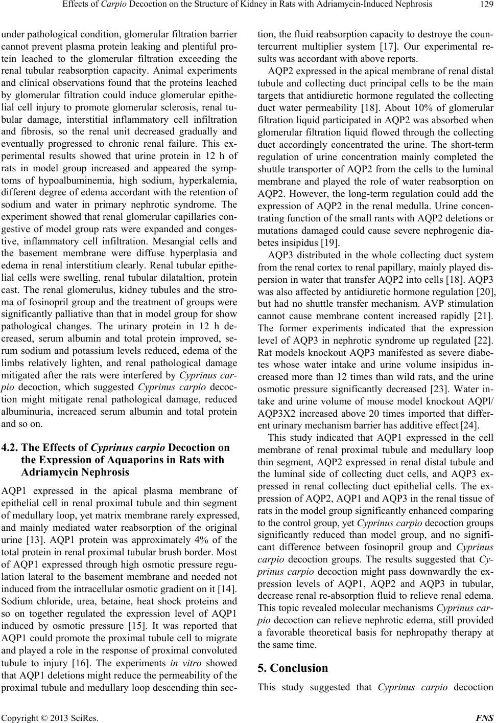 Effects of Carpio Decoction on the Structure of Kidney in Rats with Adriamycin-Induced Nephrosis 129 under pathological condition, glomerular filtration barrier cannot prevent plasma protein leaking and plentiful pro- tein leached to the glomerular filtration exceeding the renal tubular reabsorption capacity. Animal experiments and clinical observations found that the proteins leached by glomerular filtration could induce glomerular epithe- lial cell injury to promote glomerular sclerosis, renal tu- bular damage, interstitial inflammatory cell infiltration and fibrosis, so the renal unit decreased gradually and eventually progressed to chronic renal failure. This ex- perimental results showed that urine protein in 12 h of rats in model group increased and appeared the symp- toms of hypoalbuminemia, high sodium, hyperkalemia, different degree of edema accordant with the retention of sodium and water in primary nephrotic syndrome. The experiment showed that renal glomerular capillaries con- gestive of model group rats were expanded and conges- tive, inflammatory cell infiltration. Mesangial cells and the basement membrane were diffuse hyperplasia and edema in renal interstitium clearly. Renal tubular epithe- lial cells were swelling, renal tubular dilataltion, protein cast. The renal glomerulus, kidney tubules and the stro- ma of fosinopril group and the treatment of groups were significantly palliative than that in model group for show pathological changes. The urinary protein in 12 h de- creased, serum albumin and total protein improved, se- rum sodium and potassium levels reduced, edema of the limbs relatively lighten, and renal pathological damage mitigated after the rats were interfered by Cyprinus car- pio decoction, which suggested Cyprinus carpio decoc- tion might mitigate renal pathological damage, reduced albuminuria, increaced serum albumin and total protein and so on. 4.2. The Effects of Cyprinus carpio Decoction on the Expression of Aquaporins in Rats with Adriamycin Nephrosis AQP1 expressed in the apical plasma membrane of epithelial cell in renal proximal tubule and thin segment of medullary loop, yet matrix membrane rarely expressed, and mainly mediated water reabsorption of the original urine [13]. AQP1 protein was approximately 4% of the total protein in renal proximal tubular brush border. Most of AQP1 expressed through high osmotic pressure regu- lation lateral to the basement membrane and needed not induced from the intracellular osmotic gradient on it [14]. Sodium chloride, urea, betaine, heat shock proteins and so on together regulated the expression level of AQP1 induced by osmotic pressure [15]. It was reported that AQP1 could promote the proximal tubule cell to migrate and played a role in the response of proximal convoluted tubule to injury [16]. The experiments in vitro showed that AQP1 deletions might reduce the permeability of the proximal tubule and medullary loop descending thin sec- tion, the fluid reabsorption capacity to destroye the coun- tercurrent multiplier system [17]. Our experimental re- sults was accordant with above reports. AQP2 expressed in the apical membrane of renal distal tubule and collecting duct principal cells to be the main targets that antidiuretic hormone regulated the collecting duct water permeability [18]. About 10% of glomerular filtration liquid participated in AQP2 was absorbed when glomerular filtration liquid flowed through the collecting duct accordingly concentrated the urine. The short-term regulation of urine concentration mainly completed the shuttle transporter of AQP2 from the cells to the luminal membrane and played the role of water reabsorption on AQP2. However, the long-term regulation could add the expression of AQP2 in the renal medulla. Urine concen- trating function of the small rants with AQP2 deletions or mutations damaged could cause severe nephrogenic dia- betes insipidus [19]. AQP3 distributed in the whole collecting duct system from the renal cortex to renal papillary, mainly played dis- persion in water that transfer AQP2 into cells [18]. AQP3 was also affected by antidiuretic hormone regulation [20], but had no shuttle transfer mechanism. AVP stimulation cannot cause membrane content increased rapidly [21]. The former experiments indicated that the expression level of AQP3 in nephrotic syndrome up regulated [22]. Rat models knockout AQP3 manifested as severe diabe- tes whose water intake and urine volume insipidus in- creased more than 12 times than wild rats, and the urine osmotic pressure significantly decreased [23]. Water in- take and urine volume of mouse model knockout AQPl/ AQP3X2 increased above 20 times imported that differ- ent urinary mechanism barrier has additive effect [24]. This study indicated that AQP1 expressed in the cell membrane of renal proximal tubule and medullary loop thin segment, AQP2 expressed in renal distal tubule and the luminal side of collecting duct cells, and AQP3 ex- pressed in renal collecting duct epithelial cells. The ex- pression of AQP2, AQP1 and AQP3 in the renal tissue of rats in the model group significantly enhanced comparing to the control group, yet Cyprinus carpio decoction groups significantly reduced than model group, and no signifi- cant difference between fosinopril group and Cyprinus carpio decoction groups. The results suggested that Cy- prinus carpio decoction might pass downwardly the ex- pression levels of AQP1, AQP2 and AQP3 in tubular, decrease renal re-absorption fluid to relieve renal edema. This topic revealed molecular mechanisms Cyprinus car- pio decoction can relieve nephrotic edema, still provided a favorable theoretical basis for nephropathy therapy at the same time. 5. Conclusion This study suggested that Cyprinus carpio decoction Copyright © 2013 SciRes. FNS  Effects of Carpio Decoction on the Structure of Kidney in Rats with Adriamycin-Induced Nephrosis Copyright © 2013 SciRes. FNS 130 could reduce the urinary protein excretion and alleviate pathological lesion and edema with adriamycin-induced nephropathy by decreasing the expressions of AQPs in kidneys. 6. Acknowledgements This study was supported by grant-in-aids for The Na- tional Natural Science Foundation of China (grant No. 81072754). REFERENCES [1] S. J. Liu, Y. Gu, Y. R. Jiang, et al., “The Effect of Astra- galus on Renal Aquporins of Adriamycin Induced Neph- rotic Syndrome Rats,” Chinese Journal of Integrated Tra- ditional and Western Medicine, Vol. 5, No. 11, 2004, pp. 627-630. [2] E. Beitz and J. E. Schultz, “The Mammalian Aquaporin Water Channel Family: A Promising New Drug Target,” Current Medicinal Chemistry, Vol. 6, No. 6, 1999, pp. 457-467. [3] T. Bertani, A. Poggi, H. Pozzoni, et al., “Adriamycin- Induced Nephrotic Syndrome in Rats: Sequence of Patho- logic Events,” Laboratory Investigation, Vol. 46, No. 1, 1982, pp. 16-23. [4] W. N. Yang, L. H. Yu, S. W. Guo, et al., “Establishment of a Modified Model of Adriamycin Nephrosis in Rats,” Journal of Xi’an Jiaotong University (Medical Sciences), Vol. 30, No. 4, 2009, pp. 45-448,452. [5] P. Yu, H. Bu, H. Wang, et al., “Comparative Study on Image Analysis and Manual Counting of Immunohisto- chemistry,” Journal of Biomedical Engineering, Vol. 20, No. 2, 2003, pp. 288-290. [6] Y. K. Luo, “Gynecology of Traditional Chinese Medicine,” Shanghai Scientific & Technical Publishers, Shanghai, 1990, p. 115. [7] L. F. Nie, D. B. Shao and X. B. Lin, “The Experience of Astragalus Carp Soup with Nephritic Syndrome,” Liao- ning Journal of Traditional Chinese Medicine, Vol. 5, No. 1, 1985, pp. 22-23. [8] Y. B. Wang, “Gold Carp Soup Treats Polyhydramnios for 46 Cases,” Jiangsu Journal of Traditional Chinese Medi- cine, Vol. 24, No. 6, 2003, pp. 38-40. [9] Q. Y. He, “Carp Soup Treats Edema for 48 Cases,” Chi- nese Journal of Information on Traditional Chinese Me- dicine, Vol. 9, No. 4, 2002, pp. 62-64. [10] H. Y. Yuan, Y. Zhang and D. Q. Hong, “Gold Carp Soup Treats Malignant Pleural Effusion and Ascites for 30 Cases,” Practical Journal of Medicine, Vol. 18, No. 4, 2004, pp. 345-347. [11] C. X. Gao and C. Y. Zhang, “Observation of Efficacy on 60 Cases of Gestational Edema Treated with Modified Liyu Baizhu Soup,” World Journal of Integrated Tradi- tional and Western Medicine, Vol. 4, No. 2, 2009, pp. 123-124. [12] W. B. Hu, “The Effect of Buyang Huanwu Decoction in the Treatment of Chronic Nephropathy Proteinuria: Clini- cal Observation of 20 Cases,” Forum on traditional Chi- nese Medicine, Vol. 14, No. 6, 1999, pp. 38-40. [13] D. M. Zhang, Z. H. Xu, L. Xin, et al., “The Expression of AQP1, AQP2, AQP3 in Renal Tissues of Glomerular Dis- ease and Its Significance,” Chinese Journal of Immunol- ogy, Vol. 25, No. 2, 2009, pp. 84-86. [14] F. Umenishi, T. Narikiyo and R. W. Schrier, “Hypertonic Induction of Aquaporin-1 Water Channel Independent of Transcellular Osmotic Gradient,” Biochemical and Bio- physical Research Communications, Vol. 325, No. 2, 2004, pp. 595-599. doi:10.1016/j.bbrc.2004.10.076 [15] F. Umenishi, S. Yoshihara, T. Narikiyo, et al., “Modula- tion of Hypeitonicity-Induced Aquaporin-1 by Sodium Chloride, Urea, Betaine, and Heat Shock in Murine Renal Medullary Cells,” Journal of the American Society of Ne- phrology, Vol. 16, No. 3, 2005, pp. 600-607. doi:10.1681/ASN.2004030241 [16] M. Hara-Chikuma and A. S. Verkman, “Aquaporin-1 Fa- cilitates Epithelial Cell Migration in Kidney Proximal Tu- bule,” Journal of the American Society of Nephrology, Vol. 17, No. 1, 2006, pp. 39-45. doi:10.1681/ASN.2005080846 [17] C. L. Chou, M. A. Knepper, A. N. Van Hoek, et al., “Re- duced Water Permeability and Altered Ultrastructure in Thin Dascending Limb of Henle in Aquaprinl Null Mice,” Journal of Clinical Investigation, Vol. 103, No. 4, 1999, pp. 491-496. [18] M. Kuwahara, T. Asait, Y. Terada, et al., “The C-Termi- nal Tail of Aquaporin-2 Determines Apical Trafficking,” Kidney International, Vol. 68, No. 5, 2005, pp. 1999- 2009. doi:10.1111/j.1523-1755.2005.00654.x [19] S. Nielsin, T. H. Kwon, J. Frekiaar, et al., “Key Roles of Renal Aquaporins in Water Chalance and Water-Chalan- ce Disorders,” News in Physiological Science, Vol. 15, No. 2, 2000, pp. 136-143. [20] H. Liu and E. M. Wintour, “Aquaporins in Develop- ment—A Review,” Reproductive Biology and Endocri- nology, Vol. 3, No. 1, 2005, pp. 18-28. doi:10.1186/1477-7827-3-18 [21] B. M. Christensen, W. Wang and J. Frokiaer, “Axial Het- erogeneity in Basolateral AQP2 Localization in Rat Kid- ney: Effect of Vasopressin,” American Journal of Physi- ology—Renal Physiology, Vol. 284, No. 4, 2003, pp. F701-F717. [22] C. A. Ecelbarger, J. Terris, C. T. Frimdt, et al., “Aq- uaporin-3 Water Channel Location and Regulationin Rat Kidney,” American Journal of Physiology, Vol. 269, No. 5, 1995, pp. 663-672. [23] T. Ma, Y. Song, B. Yang, et al., “Nephrogenic Diabetes Insipidus in Mice Lacking Aquaporin-3 Water Channels,” Proceedings of the National Academy of Sciences of the United States of America, Vol. 97, No. 8, 2000, pp. 4386- 4391. doi:10.1073/pnas.080499597 [24] S. L. Chen, “Aquaporin Protein and Kidney Disease,” Chinese Journal of Pediatrics, Vol. 43, No. 9, 2005, pp. 711-713.
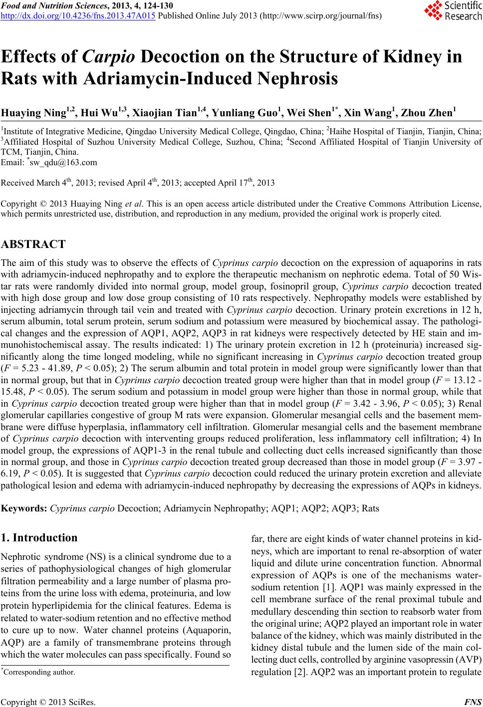
|