Open Journal of Respiratory Diseases, 2013, 3, 39-43
http://dx.doi.org/10.4236/ojrd.2013.32007 Published Online May 2013 (http://www.scirp.org/journal/ojrd)
Churg-Strauss Syndrome Revealed by Acute
Coronary Artery Disease: A Case Report*
Maurice Estivals1#, Marc Périé2, Daniel Colombier3, Bruno Farah4
1Department of Pulmonology, Hôpital du Pays d’Autan, Castres, France
2Department of Cardiology, CHT G. Bourret, Nouméa, New Caledonia
3Department of Radiology, Clinique Pasteur, Toulouse, France
4Department of Interventional Cardiology, Clinique Pasteur, Toulouse, France
Email: #m-estivals@chic-cm.fr
Received April 2, 2013; revised May 2, 2013; accepted May 9, 2013
Copyright © 2013 Maurice Estivals et al. This is an open access article distributed under the Creative Commons Attribution License,
which permits unrestricted use, distribution, and reproduction in any medium, provided the original work is properly cited.
ABSTRACT
Churg-Strauss syndrome (CSS) is a very rare small-vessel vasculitis. Clinical features include asthma, rhinitis and/or
sinusitis, and peripheral eosinophilia. Although cardiac findings are observed in 50% of cases, coronary artery disease is
rarely reported and even more rarely suggestive. The value of cardiac MRI for these patients is discussed here. A
52-year-old non-smoker male without family antecedents of cardiovascular disease presented with worsening of atypi-
cal asthma that developed 10 months earlier. A month before, he had been admitted to the ICU because of respiratory
distress and cardiogenic shock with chest pain. The angiogram revealed stenosis of the three main coronary arteries
requiring the placement of several stents. Follow-up cardiac assessments showed good results. General impairment,
unstable asthma associated with rhinitis, and eosinophilia suggested a systemic disease. The diagnosis of CSS was es-
tablished considering that five criteria of the American College of Rheumatology were found. Prednisolone was pre-
scribed at 1 mg/kg/day, which co mpletely suppressed all symptoms and eosinophilia. Cardiac MRI was performed two
months later and revealed a good control of myocardial lesions characterized by fibrosis beneath the anterior endocar-
dium and the median septum. Immunosuppressive treatment was then administered together with corticosteroid therapy.
These results suggest that acute coronary artery disease can reveal CSS in some cases. Here, the patient’s cardiac as-
sessment was normal apart from the acute episode, and cardiac MRI helped detect signs of myocarditis and establish a
prognosis of CSS.
Keywords: Vasculitis; Churg-Strauss Syndrome; Acute Coronary Artery Disease; Eosinophilia; Cardiac MRI
1. Introduction
According to the Chapel Hill classification, Churg-Strauss
syndrome (CSS) is a necrotizing vasculitis of small-to-
medium-sized vessels. It is a rare disease, with 0.5 - 6.8
million cases per year. Physiologically, it is characterized
by eosinophilia, an inflammatory disease, and by the
presence of neutrophilic p olynuclear anticytop lasmic an-
tibodies in 35% - 50% of patients. Symptoms include
asthma, rhinitis, sinus polyposis and mononeuritis multi-
plex. Cardiac findings are observed in about 50% - 60%
of cases in the form of pericarditis, myocarditis or
ischemic cardiopathy. These play an important role in
prognosis, as they are responsible for 48% of deaths.
Ischemic events are rare, and are rarely suggestive.
2. Case Report
A 52-year-old non-smoker male was admitted to the
Emergency Department in February 2009 with cardio-
genic shock occurring in the context of acute coronary
syndrome (ACS). An electrocardiogram revealed a lower
branch ST elevation with extensive lesion beneath the
anterior endocardium. Prior to this, the patient had suf-
fered from moderate persistent asthma for 10 months and
had been treated with a combination of fluticasone 250
mg/salmeterol (two puffs/day).
Emergency coronary angiography revealed occlusion
of the right coronary artery and the medial left anterior
descending (LAD) artery. Nitrates could not be used lo-
cally due to the initial cardiogenic shock. Stents were
placed on the LAD a rtery. However, iterative obstructions
led to multiple stenting of the LAD and the circumflex
arteries with several resuscitations due to cardiac arrest.
*Conflicts of interests: none to report.
#Corresponding author.
C
opyright © 2013 SciRes. OJRD
M. ESTIVALS ET AL.
40
The patient was th en transferred to intensive care with
mechanical ventilation for 48 hours and placement of an
intra-aortic bal loon pum p for three days. He was then sent
to Sydney for a coronary angiography and intracoronary
ultrasound analysis. The right coronary artery was found
to have been initially occluded but was completely cleared
by the local injection of nitrates. No atheroma was found
in the left network, which led to a diagnosis of coronary
artery spasms in a healthy arterial network in a man
without family history of cardiovascular disease.
Peripheral eosinophilia of 2000 polynuclear eosino-
phils was found. Suspected, but unproven, parasitosis was
treated with albendazole.
Three weeks after returning from Australia, the patient
was admitt ed to the Departm ent of Pulm onology due to a n
episode exacerbated asthma. He showed signs of chronic
rhinitis with occasionally purulent rhinorrhea linked to
heartburn-associated hyposmia. He reported a weight l oss
of 7 kg in the past year, although his weight had remained
stable since the coronary event. On pulmonary examina-
tion, he had diffuse wheezing and a prolonged expiratory
phase without stridor. The cardiovascular examination
was normal. ENT examination confirmed a mild rhinor-
rhea associated with posterior discharge. Late-onset
asthma associated with ENT findings, possible gastroe-
sophageal reflux (GER) and peripheral eosinophilia sug-
gested a systemic disease. Laboratory analyses revealed
pronounced peripheral eosinophilia with 16,900 polynu-
clear eosinophils, i.e., 68.6% of a total sample of 24.6 ×
10³ leucocytes.
Chest CT (Figure 1) revealed the presence of lingular
infiltrates. No mediastinal adenopathy, pleural effusion or
pericardial r eaction was found. Sliding hiatus h ernia was
also found.
Facial CT (Figure 2) confirmed sinusitis, mainly af-
fecting the frontal and maxillary areas associated with
chronic sinusitis.
Macroscopic bronchoscopic findings were normal
without vocal chord anomaly but with a marked inflam-
mation of the larynx. Bronchoalveolar lavage (BAL) re-
vealed eosinophilic alveolitis (560 white elements per
mm3 including 49% of eosinophils).
At the bacteriological, virological and mycological
levels, the BAL fluid was sterile.
Histologic examination of bronchial biopsies did not
find abnormal cells but confirmed a marked extravascular
eosinophilia of the sup erficial chorion.
Immunologi cal t ests (C AN, ANC A, rhe umatoi d factors,
quantitation of immunoglobulins, and IgE dosage), para-
sitic serology, aspergillus-specific IgE and allergy tests
were all negative.
Parasitological stool tests yielded negative results three
times.
PHmetry and esogastric fibroscopy confirmed the pre-
Figure 1. Chest CT. Appearance of isolated infiltrate of the
left upper lobe, bilateral bronchial syndrome.
Figure 2. Sinus CT. Appearance of frontal and bilateral
chronic maxillary sinusitis.
sence of GER. The electrocardiogram showed a sinus
rhythm without changes in repolarization.
The echocardiogram performed six weeks after the
initial episode revealed normal left ventricular function
and cardiac rhythm.
CSS diagnosis was established due to the fact that
Lanham’s three criteria were met. In addition, Churg-
Strauss vasculitis could explain the significant coronary
artery disease, in the absence any other cardi ovascular ri sk
factors.
Oral corticosteroid therapy was administered, starting
with a dosage of 1 mg/kg/day of prednisolone, together
with GER treatment and disease modifying inhaled ther-
apy (formoterol 48 µg/day and budesonide 1600 mg/day)
for asthma.
Rapid and co mplete suppression of the cough and ENT
symptoms (rhinorrhea and dysphonia) was observed.
After four weeks, a return to the previous weight was
noted, the asthma remained und er control and the lev el of
Copyright © 2013 SciRes. OJRD
M. ESTIVALS ET AL. 41
eosinophils returned to normal.
In April 2009, two months after the initial ACS, a
thorough cardiac checkup wa s performed. The patient was
found to be free of all symptoms of coronary disease, and
the electrocardiogram revealed normal results. The cor-
onal CT showed satisfactory stent permeability without
any sign of restenosis, in addition to a normal coronary
network. No parietal irregularity or calcification was
observed.
On cardiac magnetic resonance imaging (MRI), the left
ventricle (LV) was not dilated (LVEDV: 130 ml, indexed
75 ml/m2) and no thinning of the myocardial wall was
found on T1 sequences. There was no vascular dysfunc-
tion or pericardial discharge or thickening. A clear
subendocardial hypersignal was observed in the medial
septum and the anterior median areas on T2 STIR se-
quences, suggesting an edema.
Finally, diffuse abnormalities of delayed subendocar-
dial enhancement that did not correspond to coronary
systematization of the median anterior, median inferior
and lateral walls were found. These showed at some
points a nodular character, particularly on the inferior-
median wall.
The global and segmental function of the LV was
maintained with an LV ejection fraction (LVEF) of 63%
(minor axis), 55% (four cavities) and 56% (two cavities).
The perfus ion was het erogene ous with a sli ght defect i n
the median septal wall, suggesting microcirculatory issues
(Figure 3). It was concluded that the T2 sequence
anomalies and delayed enhancement were compatible
with myocardial injury within the scope of vasculitis and
probably associated with sub-endocardial fibrosis (Fig-
ures 4 and 5). LV function was maintained with LVEF at
around 60%.
Considering the myocardial injury observed on the
MRI, a decision was made to start immunosuppressive
treatment in association with corticosteroid therapy (aza-
thioprine 2 mg/kg/d ay).
3. Discussion
CSS is characterized by necrotizing vasculitis, which
affects both small and medium-sized blood vessels at the
pulmonary and systemic levels, and is associated with
intra- and extravascular granuloma, eosinophilia, and
infiltration of eosinophils at the tissue level in asthmatic
patients who often suffer from rhinitis or chronic sinusi-
tis [1]. It is a rare condition (0.5 to 6.8/106 per year). The
diagnostic criteria were established by Lanham [2] at the
Hammersmith Hospital in London.
Here, the diagnosis of CSS was established because
three of the criteria of this classification with a sensitivity
and specificity of 95% were found [2]. It should be noted
that although ENT and asthmatic symptoms were present,
the allergy results were negative and IgE levels were
Figure 3. Cardiac MRI. Short axis perfusion. First-pass per-
fusion sequence after bolus intravenous injection of Gado-
linium. Hyposignal in the anterior-septal wall suggesting
microcirculatory issues.
Figure 4. Cardiac MRI. DES 2 cav. 2. Delayed enhancement
sequence 10 min after intravenous injection of Gadolinium in
slices, two cavities long axis. Subendocardial contrast en-
hancement in two distinct areas: anterior-median and infe-
rior-median. A pseudo-nodular enhancement can be ob-
served in the latter.
normal. Asthma was moderated with normal respiratory
function under disease modifying treatment; it was never
corticodependent and was not treated with leukotriene
inhibitors. Other possible diagnoses were eliminated (Car-
rington’s disease, Loeffler’s syndrome, allergic broncho-
pulmonary aspergillosis, Wegener’s granu lomatosis, Ho dg -
kin’s lymphoma and drug-induced eosinophilic pneumo-
nia).
We did not consider here the po ssibility that treatment
with inhaled substances could have caused CSS [3], as
peripheral eosinophilia was not available at treatment
onset.
Copyright © 2013 SciRes. OJRD
M. ESTIVALS ET AL.
42
Figure 5 . Card iac MRI. T2 ST IR sho rt axis . Weighted black
blood T2 sequence with fat saturation. Subendocardial hy-
persigna l in t he an terior-s epta l and infer ior a re as s ugges ting
myocardial edema.
Our patient had an ANCA-negative phenotype, and
showed evidence of pulmonary infiltrates on CT images,
but no pleural effusion; bronchial biopsies revealed
marked extravascular eosinophilia on the superficial
chorion. According to the French Vascu litis Study Group
criteria [4], he was at risk of suffering a cardiac event.
Cardiac findings are present in about 50% of cases of
CSS [1,5] and are responsible for almost half of CSS
deaths [6]; they are sometimes difficult to identify. The
most frequent cardiac symptoms are pericarditis and myo-
carditis. Vascular problems and heart failure due to LV
dysfunction have also been described [7-10]. Several
studies have confirmed the benefits of cardiac MRI in the
early diagnosis of cardiac disease [5,11,12], while other
diagnostic techniques do not yield useful results.
Cardiac localizations are more frequent in the ANCA-
negative phenotype [4,5,12] as was the case in our ob-
servation. In the series published by Neumann et al. [5],
22 out of 49 patients showed clinical signs of cardiac
disease, ANCA were often negative and elevated eosi-
nophilia was often found. A total of 50% of patients had
LV dysfunction, 73% had vascular leakages and 41% had
pericarditis. Endomyocardial lesions were detected by
MRI in 59% of patients who then had a less favorable
diagnosis.
LV function was initially normal apart from the acute
episode, and there was no vascular injury or pericardial
effusion. The coronary angiography revealed a coronary
network free from atheroma and restenosis, and perme-
able stents. There were no unfavorable symptom and
initial treatment solely based on corticosteroid therapy
seemed justified, as in the case reported by Wagner et al.
[7].
The detection of cardiac injury by MRI had an impact
on treatment, as cardiomyopathy plays a role in four of
five factors that determine poor prognosis for CSS,
namely: age over 65 years, severe gastrointestinal injury,
renal failure with elevated serum creatinine levels (>150
µm/L), and heart disease [5,12-14].
However, due to the low number of cases describe d, it
seems premature to intensify CSS treatment based solely
on MRI results from asymptomatic patients [12].
Here, the patient had a favorable prognosis in terms of
ENT involvement, but a poor prognosis in terms of heart
disease [14]. Therefore, immunosuppressive treatment
was started.
Acute cor ona ry event s have been poo rly repo rted in t he
literature [7,9,10,15]. They rarely suggest CSS and no
case of multiple stenting cou ld be found in the literature.
The mechanisms mentioned are coronary spasms, which
are possi bly favo red more by the to xicity of eosinophi ls or
by myocardial granulomas than by the localization of
vasculitis at the level of the coronary arteries [15,16]. In
the case reported by Wagner et al., the patient’s history
suggested undiagnosed CSS. His phenotype was ANCA
negative, with very h igh initial eosinophilia Tanaka et al.
[12] reported a case of ACS due to a vasospasm with
significant eosinophilia (>5000/mm3) and ANCA-nega-
tive phenotype [15]. The authors suggested a link between
coronary vasospasm due to CSS and eosinophilia. It is
interesting to note that in the case reported here, eosino-
philia was not found during the ACS. Significant eosi-
nophilia was only found a month after the event (Figure
6).
What, then, is the mechanism of vasospasm?
The overall prognosis for patients with CSS remains
favorable (79% alive at 10 years) [13]. Heart disease is
Figure 6. Level of eosinophils in time.
Copyright © 2013 SciRes. OJRD
M. ESTIVALS ET AL.
Copyright © 2013 SciRes. OJRD
43
responsible for almost 50% of deaths [6], so intensive
initial treatment is recommended.
Two years after initial diagnosis, the patient remains
treated with azathioprine and small doses of corticoster-
oids (5 mg/day of prednisone). There has been no relapse
at the cardiovascular level, but respiratory function re-
mains below normal (LVEF = 3.4 liters at 100% of theo-
retical values, Tiffeneau = 73%, and mean peak expira-
tory flow 25/75 at 2.73 liters so 73% of theoretical values).
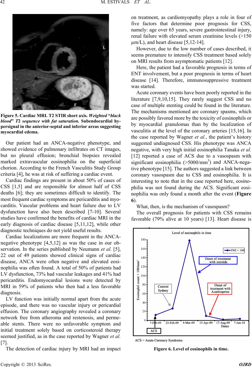
The level of eosinophils was at 6 80/mm3.
In conclusion, CSS can be accompanied and revealed
by ACS. The mechanism of ACS in the healthy coronary
network is a vasospasm that seems independent of the
coronary localization of the vasculitis, although its origins
remain uncertain. In such cases, cardiac MRI should be
systematically performed to identify myocardial injuries
that can be useful for the prognosis. This will ensure early
start of immunosuppressive therapy.
REFERENCES
[1] J. Churg and L. Strauss, “Allergic Granulomatosis, Aller-
gic Angiitis and Periarteritis Nodosa,” American Journal
of Pathology, Vol. 27, No. 2, 1951, pp. 277-301.
[2] J. C. Lanham, K. B. Elkon, C. D. Pusey and G. R. Hughes,
“Systemic Vasculitis with Asthma and Eosinophili a: A Cli-
nical Approach to the Churg-Strauss Syndrome,” Medi-
cine (Baltimore), Vol. 63, 1984, pp. 65-81.
doi:10.1097/00005792-198403000-00001
[3] C. Le Gall, S. Pham, S. Vignes, G. Garcia, H. Nunes, D.
Fichet, G. Simonneau, P. Duroux and M. Humbert, “In-
haled Corticosteroids and Churg-Strauss Syndrome: A
Report of Five Cases,” European Respiratory Journal,
Vol. 15, No. 5, 2000, pp. 978-981.
doi:10.1034/j.1399-3003.2000.15e29.x
[4] R. Sablé-Fourtassou, P. Cohen, A. Mahr, C. Pagnoux, L.
Mouthon, D. Jayne, D. Blockmans, J. F. Cordier, P. De-
laval, X. Puechal, D. Lauque, J. F. Viallard, A. Zoulim
and L. Guillevin, “Antineutrophil Cytoplasmic Antibod-
ies and the Churg-Strauss Syndrome,” Annals of Internal
Medicine, Vol. 143, No. 9. 2005, pp. 632-638.
doi:10.7326/0003-4819-143-9-200511010-00006
[5] T. Neumann, B. Manger, M. Schmid, C. Kroegel, A.
Hansch, W. A. Kaiser, D. Reinhardt, G. Wolf, G. Hein, G.
Mall, G. Schett and J. Zwerina, “Cardiac Involvement in
Churg-Strauss Syndrome: Impact of Endomyocarditis,”
Medicine (Baltimore), Vol. 88, No. 4, 2009, pp. 236-243.
doi:10.1097/MD.0b013e3181af35a5
[6] S. Hellemans, J. Dens and D. Knockaert, “Coronary In-
volvement in the Churg-Strauss Syndrome,” Heart, Vol.
77, 1997, pp. 576-578.
[7] A. D. Wagner, G. P. Meyer, M. Rihl, A. Rathmann, U.
Wittkop, H. Zeidler, H. Haller and J. Lotz, “Acute Coro-
nary Syndrome Associated with Churg-Strauss Syndrome,”
Vascular Health and Risk Management, Vol. 3, No. 5,
2007, pp. 775-779.
[8] C. R. McGavin, A. J. Marshall and C. T. Lewis, “Churg-
Strauss Syndrome with Critical Endomyocardial Fibrosis:
10 Years Survival after Combined Surgical and Medical
Management,” Heart, Vol. 87, No. 5, 2002, p. e5.
doi:10.1136/heart.87.5.e5
[9] P. Petrakopoulou, W. M. Franz, P. Boekstegers and M.
Weis, “Vasospastic Angina Pectoris Associate d with Ch u r g -
Strauss Syndrome,” Nature Clinical Practice Cardiovas-
cular Medicine, Vol. 2, 2005, pp. 484-489.
doi:10.1038/ncpcardio0299
[10] N. Kakouros, R. Bastiaenen, A. Kourliouros and L. An-
derson, “Churg-Strauss Pre senting as Acute Coronary Sy n-
drome: Sometimes It’s Zebras,” BMJ Case Reports, 2011.
[11] S. E. Petersen, A. Kardos and S. Neubauer, “Subendocar-
dial and Papillary Muscle Involvement in a Patient with
Churg-Strauss Syndrome, Detected by Contrast Enhanced
Cardiovascular Magnetic Resonance,” Heart, Vol. 91, No.
1, 2005, p. e9. doi:10.1136/hrt.2004.050070
[12] J. Marmursztejn, O. Vignaux, P. Cohen, P. Guilpain, C.
Pagnoux, H. Gouya, L. Mouthon, P. Legmann, D. Duboc
and L. Guillevin, “Impact of Cardiac Magnetic Reso-
nance Imaging for Assessment of Churg-Strauss Syn-
drome: A Cross-Sectional Study in 20 Patients,” Clinical
and Experimental Rheumatology, Vol. 27, No. 1, Suppl.
52, 2009, pp. S70-S76.
[13] L. Guillevin, F. Lhote, M. Gayraud, P. Cohen, B. Jar-
rousse, O. Lortholary , N. Thibult and P. Casassus, “Prog-
nostic Factors in Polyarteritis Nodosa and Churg-Strauss
Syndrome. A Prospective Study in 342 Patients,” Medi-
cine (Baltimore), Vol. 75, No. 1, 1996, pp. 17-28.
doi:10.1097/00005792-199601000-00003
[14] L. Guillevin, C. Pagnoux, R. Seror, A. Mahr, L. Mouthon
and P. Le Toumelin, “The Five Factors Score Revisited:
Assessment of Prognosis of Systemic Necrotizing Vascu-
litidies Based on the French Vasculitis Study Group
(FVSG),” Medicine (Baltimore), Vol. 90, No. 1, 2011, pp.
19-27. doi:10.1097/MD.0b013e318205a4c6
[15] M. Tanaka, N. Mise, N. Kurita, T. Suzuki, K. Hara, A.
Fujii, N. Uesugi and T. Sugimoto, “A Case of Churg-
Strauss Syndrome with Necrotizing Crescentric Glome-
rulonephritis Accompanied by Acute Coronary Syndrome
due to Vasospasm,” American Journal of Kidney Dis-
eases, Vol. 56, No. 2, 2010, pp. e5-e9.
doi:10.1053/j.ajkd.2010.03.011
[16] B. Dunogué, C. Pagnoux and L. Guillevin, “Churg-Stauss
Syndrome (CSS): Clinical Symptoms, Complementary In-
vestigations, Prognosis and Outcome, and Treatment,”
Seminars in Respiratory and Critical Care Medicine, Vol.
32, No. 3, 2011, pp. 298-309.
doi:10.1055/s-0031-1279826