Vol.2, No.11, 1308-1311 (2010)
doi:10.4236/health.2010.211194
Copyright © 2010 SciRes. http://www.scirp.org/journal/HEALTH/
Health
Openly accessible at
Experiences in total knee arthtroplasty after distal
femoral varus osteotomy
Bianca Röschke1, Wolfgang Hönle2, Alexander Schuh1*
1Research Unit Orthopaedics and General Surgery, Neumarkt Clinic, Neumarkt, Germany; *Corresponding Author:
Alexander.Schuh@klinikum.neumarkt.de;
2Department of Orthopaedic Surgery, Neumarkt Clinic, Neumarkt, Germany.
Received 21 July 2010; revised 20 August 2010; accepted 25 August 2010.
ABSTRACT
Introduction: There is only little information avai-
lable about total knee arthroplasty (TKA) follow-
ing distal femoral varus osteotomy (DFVO). The
aim of our study was to show our experiences
and mid-term results of TKA after a previous
DFVO. Material and method: In a retrospective
study we identified 36 consecutive patients who
had undergone TKA after a previous distal fe-
moral varus osteotomy. The average duration of
follow-up after the TKA was 8.2 years (min: 5.0,
max: 9.2). X-rays were taken in 2 planes before
TKA, 1 week after TKA and at latest follow-up.
Tibiofemoral alignment was measured on weight-
bearing long-leg anteroposterior radiographs. Ra-
diolucent lines at latest follow-up were docu-
mented. Functional evaluations were performed
preoperatively and postoperatively (at the time
of latest follow-up).
Results: The mean Knee Society knee score in-
creased from 42 points before the arthroplasty
to 91.3 points after the arthroplasty. The mean
Knee Society function score increased from
27.4 points preoperatively to 93.2 points post-
operatively. The mean overall Knee Society score
increased from 91.3 points preoperatively to
163.4 points postoperatively. The mean radio-
graphic alignment was 4.5˚ of valgus (10˚ of
varus to 19˚ of valgus) before TKA and 3.1˚ of
valgus (range, 3˚ of varus to 6˚ of valgus) at the
time of latest follow-up. Postoperative compli-
cations included one deep vein thrombosis with
non-lethal pulmonary embolism, one wound in-
fection requiring revision and one septic loos-
ening. Discussion: It is possible to perform TKA
following DFVO with good mid-term results. In
comparison to the literature there is no higher
risk of complications in TKA following DFVO in
comparison to primary TKA.
Keywords: Total Knee Arthroplasty; Distal Femoral
Varus Osteotomy; Result; Complication; Knee
Society Score
1. INTRODUCTION
Osteotomies around the knee for the purpose of un-
loading the affected compartment and correction of the
underlying deformity are a well recognised treatment al-
ternative for symptomatic osteoarthritis of the knee in
active patients 1. Varus deformity with medial com-
partment arthritis is more common than valgus deform-
ity with lateral compartment involvement 1,2. Several
studies reported good outcomes lasting more than a
decade for patients with varus deformity treated by high
valgus tibial osteotomy in appropriately selected patients.
For a valgus deformity of more than 12˚ or in situations
where the plane of the joint is deviated from the hori-
zontal by more than 10˚, a distal femoral varus osteot-
omy (DFVO) should be preferred 1. A recent study
indicated good mid-term success rates of DFVO even in
the presence of moderate patellofemoral arthritis at a
mean follow-up of 99 months 3. Edgerton et al. 4
reported on 23 patients (24 knees managed with DFVO)
and found 71% with good or excellent results at a mean
follow-up of 8.3 years. In a recent study by Kosashvili et
al. 1 33 consecutive DFVOs (31 patients) with a mini-
mum follow-up of ten years (mean 15.1, range 10-25)
were reviewed. Fifteen DFVOs were converted to total
knee arthroplasty (TKA) and one DFVO was awaiting
TKA. The authors came to the conclusion that DFVO is
reliably in delaying the need for TKA by more than a
decade as conversion to TKA in less than ten years oc-
curred in 6.1%. Success of primary TKA with knee os-
teoarthritis is well established, and about 85% of patients
are satisfied with the surgical outcome 5. In the past,
there have been reports of technical difficulties after
B. Röschke et al. / HEALTH 2 (2010) 1308-1311
Copyright © 2010 SciRes. http://www.scirp.org/journal/HEALTH/Openly accessible at
1309
failed high tibial head osteotomy that influenced out-
comes of knee replacement. There is only little informa-
tion available in TKA following DFVO 6-8. The aim
of our study was to show our experiences and mid-term
results of TKA after a previous DFVO.
2. MATERIAL AND METHODS
In a retrospective study we identified 36 consecutive
patients who had undergone TKA after a previous distal
femoral varus osteotomy. The average duration of fol-
low-up after the TKA was 8.2 years (min: 5.0 max: 9.2).
The study group included 14 men and 22 women who
had a mean age of 58.4 years (min: 27.8, max: 81.5) at
the time of the arthroplasty. 21 right knees and 14 left
knees were involved. TKA was performed at an average
of 9.2 years (min: 2.2, max: 35.8) after the osteotomies.
The average height of the patients was 156.9 cm (min:
152, max: 178), the average weight was 78.2 kg (min: 51,
max: 108). All osteotomies had been internally fixed wi-
th a lateral blade-plate. Removal of the plates was pre-
formed after 1-2 years after the osteotomy, at the time of
TKA no hardware removal was necessary.
15 knees had osteoarthritis grade 3, 21 knees grade 4
according to Kellgren and Lawrence 9 preoperatively.
In all cases an intramedullary femoral alignment guide
was used during TKA procedures. The operative notes
did not indicate any modification of the starting hole in
knees in which an intramedullary guide was used for
alignment.
In all cases an intraoperative joint aspiration was per-
formed, in one case an infection with Staph aureus could
be detected, without any clinical signs for infection pre-
operatively.
Prostheses included 9 FS Knees (Protek, Switzerland,
now Zimmer, Warsaw, Indiana) 2 Emotion Knees (Aes-
culap, Tuttlingen, Germany) and 25 PFC prostheses
(DePuy, Johnson and Johnson, Warsaw, Indiana).
All procedures were performed in an ultra-clean-air
theater (with antibiotic prophylaxis). During their stay at
the hospital, all patients were treated with low molecular
weight Heparin and compression stockings as a prophy-
laxis against deep vein thrombosis. For the duration of 6
weeks, partial weight bearing of 20 kg with the support
of lower arm crutches was required.
X rays were taken in 2 planes before TKA, 1 week af-
ter TKA and at latest follow-up. Tibiofemoral alignment
was measured on weightbearing long-leg anteroposterior
radiographs. Radiolucent lines at latest follow-up were
documented according to the Knee Society total knee
arthroplasty roentgenographic evaluation system 10.
Functional evaluations were performed preoperatively
and postoperatively (at the time of follow-up) with use
of the 200-point system of the Knee Society 11. The
two components of this scale, the 100-point knee score
and the 100-point function score, were extracted before
and after the arthroplasty by means of a chart review
performed after an average of 8.2 years of follow-up.
3. RESULTS
Mean operating time was 98.4 minutes (min: 55, max.
120).
Mean range of motion was Extension/Flexion 0/6/88˚
preoperatively and improved to Extension/Flexion 3/0/97˚
postoperatively.
The mean Knee Society knee score increased from 42
points (min: 15, max: 59) before the arthroplasty to 91.3
points (min: 85; max: 98) after the arthroplasty. The
mean Knee Society function score increased from 27.4
points (min: 15, max: 60) preoperatively to 93.2 points
(min: 50, max: 100 points) postoperatively. The mean
overall Knee Society score increased from 91.3 points
(min: 53, max: 127) preoperatively to 163.4 points (min:
110, max: 178) postoperatively.
3/36 cases had femoral notching intraoperatively. In
these cases partial weight bearing was requested for 10
weeks, further follow-up was uneventfull in all 3 cases.
Postoperative complications included one deep vein
thrombosis with non-lethal pulmonary embolism, one
wound infection requiring revision and one septic loos-
ening requiring 2 stage revision arthroplasty.
The mean radiographic alignment was 4.5˚ of valgus
(10˚ of varus to 19˚ of valgus) before TKA and 3.1˚ of
valgus (range, 3˚ of varus to 6˚ of valgus) at the time of
latest follow-up. At latest follow-up mean femoral angle
α was 97.1˚ (min: 88, max: 104). Mean tibial angle β
was 98.1˚ (min: 85, max: 95). Mean femoral flexion
angle γ was 3.9˚ (min: 1.2, max: 8). Mean tibial angle δ
was 89˚ (min: –3, max: 9).
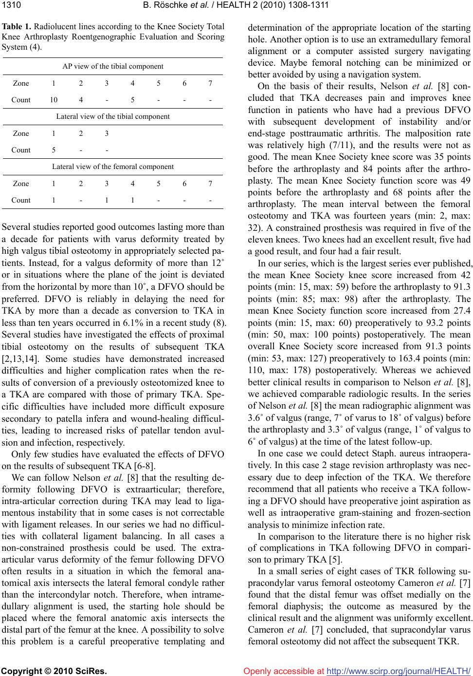
Radiolucent lines according to the Knee Society total
knee arthroplasty roentgenographic evaluation system
are shown in Table 1.
4. DISCUSSION
Distal femoral varus osteotomy (DFVO) is indicated
for patients with isolated lateral compartment os-
teoarthritis of the knee with associated valgus deformity
of the knee. Aim of this procedure is delaying TKA. But
DFVO can only be recommended if the results of TKA
following DFVO are comparable to primary TKA and if
there are not more complications. The ideal patient has
isolated lateral compartment arthritis with a moderate
valgus deformity, is physiologically young, has an oc-
cupation or activity level that makes arthroplasty less
appropriate, and has a normal body-mass index and sat-
isfactory range of motion and stability of the knee 8,12].
B. Röschke et al. / HEALTH 2 (2010) 1308-1311
Copyright © 2010 SciRes. http://www.scirp.org/journal/HEALTH/Openly accessible at
1310
Table 1. Radiolucent lines according to the Knee Society Total
Knee Arthroplasty Roentgenographic Evaluation and Scoring
System (4).
AP view of the tibial component
Zone 1 2 3 4 5 6 7
Count 10 4 - 5 - - -
Lateral view of the tibial component
Zone 1 2 3
Count 5 - -
Lateral view of the femoral component
Zone 1 2 3 4 5 6 7
Count 1 - 1 1 - - -
Several studies reported good outcomes lasting more than
a decade for patients with varus deformity treated by
high valgus tibial osteotomy in appropriately selected pa-
tients. Instead, for a valgus deformity of more than 12˚
or in situations where the plane of the joint is deviated
from the horizontal by more than 10˚, a DFVO should be
preferred. DFVO is reliably in delaying the need for
TKA by more than a decade as conversion to TKA in
less than ten years occurred in 6.1% in a recent study (8).
Several studies have investigated the effects of proximal
tibial osteotomy on the results of subsequent TKA
2,13,14. Some studies have demonstrated increased
difficulties and higher complication rates when the re-
sults of conversion of a previously osteotomized knee to
a TKA are compared with those of primary TKA. Spe-
cific difficulties have included more difficult exposure
secondary to patella infera and wound-healing difficul-
ties, leading to increased risks of patellar tendon avul-
sion and infection, respectively.
Only few studies have evaluated the effects of DFVO
on the results of subsequent TKA 6-8.
We can follow Nelson et al. 8 that the resulting de-
formity following DFVO is extraarticular; therefore,
intra-articular correction during TKA may lead to liga-
mentous instability that in some cases is not correctable
with ligament releases. In our series we had no difficul-
ties with collateral ligament balancing. In all cases a
non-constrained prosthesis could be used. The extra-
articular varus deformity of the femur following DFVO
often results in a situation in which the femoral ana-
tomical axis intersects the lateral femoral condyle rather
than the intercondylar notch. Therefore, when intrame-
dullary alignment is used, the starting hole should be
placed where the femoral anatomic axis intersects the
distal part of the femur at the knee. A possibility to solve
this problem is a careful preoperative templating and
determination of the appropriate location of the starting
hole. Another option is to use an extramedullary femoral
alignment or a computer assisted surgery navigating
device. Maybe femoral notching can be minimized or
better avoided by using a navigation system.
On the basis of their results, Nelson et al. 8 con-
cluded that TKA decreases pain and improves knee
function in patients who have had a previous DFVO
with subsequent development of instability and/or
end-stage posttraumatic arthritis. The malposition rate
was relatively high (7/11), and the results were not as
good. The mean Knee Society knee score was 35 points
before the arthroplasty and 84 points after the arthro-
plasty. The mean Knee Society function score was 49
points before the arthroplasty and 68 points after the
arthroplasty. The mean interval between the femoral
osteotomy and TKA was fourteen years (min: 2, max:
32). A constrained prosthesis was required in five of the
eleven knees. Two knees had an excellent result, five had
a good result, and four had a fair result.
In our series, which is the largest series ever published,
the mean Knee Society knee score increased from 42
points (min: 15, max: 59) before the arthroplasty to 91.3
points (min: 85; max: 98) after the arthroplasty. The
mean Knee Society function score increased from 27.4
points (min: 15, max: 60) preoperatively to 93.2 points
(min: 50, max: 100 points) postoperatively. The mean
overall Knee Society score increased from 91.3 points
(min: 53, max: 127) preoperatively to 163.4 points (min:
110, max: 178) postoperatively. Whereas we achieved
better clinical results in comparison to Nelson et al. 8,
we achieved comparable radiologic results. In the series
of Nelson et al. 8 the mean radiographic alignment was
3.6˚ of valgus (range, 7˚ of varus to 18˚ of valgus) before
the arthroplasty and 3.3˚ of valgus (range, 1˚ of valgus to
6˚ of valgus) at the time of the latest follow-up.
In one case we could detect Staph. aureus intraopera-
tively. In this case 2 stage revision arthroplasty was nec-
essary due to deep infection of the TKA. We therefore
recommend that all patients who receive a TKA follow-
ing a DFVO should have preoperative joint aspiration as
well as intraoperative gram-staining and frozen-section
analysis to minimize infection rate.
In comparison to the literature there is no higher risk
of complications in TKA following DFVO in compari-
son to primary TKA 5.
In a small series of eight cases of TKR following su-
pracondylar varus femoral osteotomy Cameron et al. 7
found that the distal femur was offset medially on the
femoral diaphysis; the outcome as measured by the
clinical result and the alignment was uniformly excellent.
Cameron et al. 7 concluded, that supracondylar varus
femoral osteotomy did not affect the subsequent TKR.
B. Röschke et al. / HEALTH 2 (2010) 1308-1311
Copyright © 2010 SciRes. http://www.scirp.org/journal/HEALTH/
1311
In our study TKA was performed at an average of 9.2
years (min: 2.2, max: 35.8) after DFVO. The average du-
ration of follow-up after TKA was 8.2 years. According
to our results we postulate that it is possible to perform
TKA following DFVO with good mid-term results. We
could not find any technical difficulties in TKA follow-
ing DFVO. Therefore we can recommend DFVO as a
reliable procedure in delaying the need for TKA in pa-
tients with osteoarthritis of the lateral compartment with
a valgus deformity.
Openly accessible at
REFERENCES
[1] Kosashvili, Y., Safir, O., Gross, A., Morag, G., Lakstein,
D. and Backstein, D. (2010) Distal femoral varus os-
teotomy for lateral osteoarthritis of the knee: A minimum
ten-year follow-up. International Orthopaedics, 34, 249-
254.
[2] van Raaij, T.M., Reijman, M., Furlan, A.D. and Verhaar,
J.A. (2009) Total knee arthroplasty after high tibial os-
teotomy. A systematic review. BMC Musculoskelet Dis-
ord, 20(10), 88.
[3] Wang, J.W. and Hsu, C.C. (2006) Distal femoral varus
osteotomy for osteoarthritis of the knee. Surgical tech-
nique”. The Journal of Bone and Joint Surgery, 88(Supp l),
100-108.
[4] Edgerton, B.C., Mariani, E.M. and Morrey, B.F. (1993)
Distal femoral varus osteotomy for painful genu valgum.
A five to 11 year follow up study. Clinical Orthopaedics
and Related Research, 288, 263-269.
[5] National Institutes of Health (2003) NIH consensus state-
ment on total knee replacement. NIH Consensus and
State-of-the-Science Statements, 20, 1-34.
[6] Beyer, C.A., Lewallen, D.G. and Hanssen, A.D. (1994)
Total knee arthroplasty following prior osteotomy of the
distal femur. American Journal of Knee Surgery, 7, 25-
30.
[7] Cameron, H.U. and Park, Y.S. (1997) Total knee replace-
ment after supracondylar femoral osteotomy. American
Journal of Knee Surgery, 10(2), 70-71.
[8] Nelson, C.L., Saleh, K.J., Kassim, R.A., Windsor, R.,
Haas, S., Laskin, R. and Sculco, T. (2003) Total knee ar-
throplasty after varus osteotomy of the distal part of the
femur. The Journal of Bone and Joint Surgery, 85(6),
1062-1065.
[9] Kellgren, J.H. and Lawrence, J.S. (1957) Radiological
assessment of osteoarthrosis. Annals of the Rheumatic
Diseases, 16(4), 494-502.
[10] Ewald, F.C. (1989) The Knee Society total knee arthro-
plasty roentgenographic evaluation and scoring system.
Clinical Orthopaedics and Related Research, 248, 9-12.
[11] Insall, J.N., Dorr, L.D., Scott, R.D. and Scott, W.N. (1989)
Rationale of the knee dociety clinical rating system. Cli-
nical Orthopaedics and Related Research, 248, 13-14.
[12] Pach, M., Uvízl, M., Holibka, R. and Zapletalová, J.
(2005) Varus supracondylar osteotomy of the femur-long-
term results. Acta Chirurgiae Orthopaedicae et Trauma-
tologiae Cechoslovaca, 72, 363-370.
[13] Katz, M.M., Hungerford, D.S., Krackow, K.A. and Len-
nox, D.W. (1987) Results of total knee arthroplasty after
failed proximal tibial osteotomy for osteoarthritis. The
Journal of Bone and Joint Surgery, 69(2), 225-233.
[14] Windsor, R.E., Insall, J.N. and Vince, K.G. (1988) Tech-
nical considerations of total knee arthroplasty after pro-
ximal tibial osteotomy. Journal of Bone and Joint Sur-
gery, 70(4), 547-555.