Vol.2, No.11, 1294-1298 (2010)
doi:10.4236/health.2010.211192
Copyright © 2010 SciRes. http://www.scirp.org/journal/HEALTH/
Health
Openly accessible at
The effect of sodium phosphate enema in patients
ingested foreign bodies: a prospective, randomized trial
——Sodium phosphate enema in patients ingested foreign bodies
Zeynep Ozkan1, Metin Kement1*, Mustafa Oncel1,2, Levent Kaptanoglu1, Cem Gezen1
1Department of General Surgery, Kartal Education and Research Hospital, Istanbul, Turkey; drzeynepozkan@yahoo.com;
*Corresponding Author: mkement@yahoo.com; oncel@hotmail.com; leventkaptanoglu@yahoo.com; cemgezen@hotmail.com;
2Gumushane Univercity College of Health Sciences, Gumushane, Turkey.
Received 14 August 2010; revised 20 August 2010; accepted 23 August 2010.
ABSTRACT
Background: Although foreign body ingestion is
a common problem, poor knowledge for the
treatment still remains a major obstacle, espe-
cially in preventing complications. Phosphate
enemas were applied for chronic constipation
and bowel cleansing for a long period of time.
This study aims to evaluate the effect of pho-
sphate enema on bowel movements in patients
who suffer from foreign body ingestion. Me-
thods: Forty consecutive patients admitted to
our emergency department for foreign body in-
gestion were randomized into two groups. Ca-
ses in Group SP received daily one dose of so-
dium phosphate enemas, while Group C did not.
Patients in both groups were followed up with
daily imaging and physical examinations. Demo-
graphics, number of X-rays, radiation dose, and
period of the foreign body passage were record-
ed and compared between two groups. Also, da-
ta of patients who ingested pins (Group P) were
compared with of those who ingested other ma-
terials (Group O). Results: There was no statis-
tical difference between groups (Group SP vs.
Group C) comparing demographics, number of
X-rays, total amount of radiation doses (p > 0.05
for all comparisons). The period of foreign body
passage was shorter in Group SP (3.4 ± 1.6 days
vs. 6.2 ± 3.8 days; p = 0.005). Also, patients who
ingested pins (Group P) were compared to pa-
tients who ingested other materials (Group O).
Pins were ingested by 23 (65%) patients, these
were older (16.6 ± 9.4 years vs. 4.8 ± 3.5 years; p
= 0.001) and generally females (78.3% vs. 29.4%;
p = 0.006). Contrary to the other materials, pins
were less frequently confirmed in stool of pa-
tients (p = 0.02). Conclusion: Sodium phosphate
enema use may hasten the passage of the for-
eign bodies. Ingestion of pins is the more com-
mon especially in turbaned young females. The
evacuation of pins is less recognized with direct
examination of the stool, so daily imaging may
be necessary for this group of patients.
Keywords: Sodium Phosphate Enema; Foreign
Body Ingestion
1. INTRODUCTION
The ingestion of foreign bodies is a common problem
among patients admitted to emergency units. Although
ingestion of a foreign body is generally associated with
little or no morbidity, and thus often underestimated;
serious complications, such as perforation, bleeding or
obstruction are also reported [1-3]. Most cases are pre-
school age children; however the aspiration or ingestion
of turban pins has been encountered among adolescent
girls in Islamic countries, where turban is worn as a kind
of head cover for religious intentions [3-6].
The heterogeneity in the type of ingested foreign body
has limited the development of an algorithm for mana-
gement. If the foreign body passes the pylorus, follow-
up is indicated to rule out complications until the mate-
rial is evacuated. However, the details of protocols, such
as whether or not daily radiographies are necessitated,
are still debated [7]. Since the majority of materials are
radio-opaque, a frontal radiograph may be helpful for the
diagnosis. Endoscopic removal is generally possible if
the material is located in the esophagus. Some have
suggested the extraction of the material with a Foley
catheter, since it is a cost-effective approach, or the use
of glucagon and water as an effective treatment for the
esophageal foreign bodies [8,9]. However, the best of
our knowledge, there have been no prospective studies
Z. Ozkan et al. / HEALTH 2 (2010) 1294-1298
Copyright © 2010 SciRes. http://www.scirp.org/journal/HEALTH/Openly accessible at
1295
for the evaluation of any treatment modality when the
ingested foreign material is located beyond the pylorus,
where endoscopic removal is almost impossible.
Sodium phosphate enema is useful as a laxative in the
relief of occasional constipation and as part of bowel
cleansing regimen in preparing the colon for surgery,
x-ray or endoscopic examination. It has been long used
for the treatment of chronic constipation [10]. Although
the effect of sodium phosphate enema on bowel move-
ments has never been evaluated, it generally induces
complete emptying of the left colon. Its use is safe even
in children, and side-effects have been reported in only a
small number of cases. This study aims to evaluate the
effect of sodium phosphate enema in patients who in-
gested foreign material.
2. MATERIALS AND METHODS
This study was approved by the Hospital Education
Planning Committee and conducted in Kartal Education
and Research Hospital between March 2003 and August
2005. Written informed consent was obtained from all
patients or from their parents if the patient was under 18
year-old.
The consecutive patients who admitted to our emer-
gency department with foreign body ingestion between
March 2003 and August 2005 were included in this study.
Patients younger than 6 month-old or pregnant were ex-
cluded because sodium phosphate enema or radiological
examinations might be unsafe. Patients were also excluded
if an endoscopic procedure or surgery was necessitated
due to esophageal material, extralumination of the foreign
body or the presence of acute abdomen at admission.
All enrolled patients were randomized with a comput-
erized algorithm into two groups. Patients in Group SP
received a single dose of sodium phosphate enema (135
ml for adults and 3 ml/kg for children; B.T. Enema Sol,
Yenisehir Drugs, Turkey) immediately after the admis-
sion to emergency unit and they were instructed to use a
single daily dose of sodium phosphate enema. Group C
was control group; the patients in this group did not re-
ceive any treatment and were only observed.
In this study, another analysis was made by comparing
the patients who ingested pins (Group P) with of those
who ingested other materials (Group O).
All patients were followed up with daily X-ray and
physical examination until the evacuation was confirmed
by the absence of the material in the X-rays or by direct
visualization of the foreign body in the stool.
These were obtained prospectively and compared with-
in the groups: Demographics, period between the inges-
tion and admission, characteristics of the materials, pe-
riod of the foreign body passage, number of X-ray ex-
aminations and exposed radiation dose. The data were
compared within Group SP and Group C. Also, data of
the patients who ingested pins (Group P) were compared
with of those who ingested other materials (Group O).
Statistics: All statistical analyses were done with SPSS
for Windows 10.0. The results were presented as percent-
tages, means, standard deviations and ranges. Student t-
test and Pearson’s Chi-square test were used for evalua-
tion of numeric and continuing values and a p value less
than 0.05 was considered significant.
3. RESULTS
Forty patients admitted to our emergency department
due to foreign body ingestion were randomized into two
groups. There were 23 (57.5%) females, and the median
age was 11 years old (1-39 years). Most patients were
under 10 years old (n = 21; 52.5%) or teenager (age be-
tween 10 and 20 years old) (n = 15; 37.5%), and only 6
(15.0%) patients were older than 20 years old. The mean
period between the ingestion and hospital admission was
113.5 ± 176 (20-900) minutes. The most commonly in-
gested material was pin (n = 23; 57.5%). The demo-
graphic data and the characteristics of the foreign mate-
rials were similar within the groups (Table 1). In study
period, none of our patients developed any complica-
tions such as bowel obstruction or perforation.
The passage period of the material was more than 5
days in 1 (5.0%) and 8 (40.0%) in Groups SP and C,
respectively, and significantly shorter in Group SP than
in Group C (p = 0.005) (Table 1). However, the number
of X-ray examinations and the total amount of radiation
given to the patients were similar in Groups SP and C
(Table 1).
In a retrospective analysis, the patients who ingested
pins (Group P; n = 23) were compared to those ingested
other materials (Group O; n = 17). Females were major-
ity population in Group P (p = 0.006) and they were
older than patients in Group O (p = 0.001) (Table 2).
The number of patients received sodium phosphate en-
ema treatment, the passage period of the material, num-
ber of X-ray examinations and doses of radiation were
similar within the groups. Foreign body could not be
visualized in the stool in more patients in Group P than
in those in Group O (p = 0.02).
4. DISCUSSION
Foreign body ingestion is quite common problem.
Most of the foreign bodies that reach the gastrointestinal
tract will pass spontaneously. However, 10% to 20% of
them will require nonoperative intervention such as en-
doscopy, and approximately 1% of them will require
surgery [11-13]. In most instances, the ingestion goes
Z. Ozkan et al. / HEALTH 2 (2010) 1294-1298
Copyright © 2010 SciRes. http://www.scirp.org/journal/HEALTH/Openly accessible at
1296
Table 1. Comparison of Group SP and Group C by demographics and clinical characteristics.
Group SP (n = 20) Group C (n = 20) p
Demographics
Age [Mean ± SD (range) in years] 11.1 ± 9.4 (1-33) 12.0 ± 8.5 (1-39) 0.85
Gender [Females (%)] 11 (55) 12 (60) 0.75
Admission Period [Median (range) in minutes] 120.5 ± 188.9 (20-900) 106.5 ± 166.8 (20-800) 0.80
Type of The Material
Pin (%) 10 (50) 13 (65)
Coin (%) 4 (20) 4 (20)
Screw (%) 2 (10) 0 (0)
Safety pin (%) 1 (5) 1 (5)
Earring (%) 1 (5) 1 (5)
Coil (%) 1 (5) 0 (0)
Peg (%) 1 (5) 0 (0)
Ring (%) 0 (0) 1 (5)
0.74
Passage Period of The Material [Mean ± SD (range) in days] 2.7 ± 1.3 (1-6) 5 ± 3.9 (1-18) 0.016*
Number of X-ray Examinations [Mean ± SD (range)] 2.5 ± 0.7 (1-4) 3.2 ± 1.8 (1-10) 0.12
Radiation Dose [Mean ± SD (range) in miliREM] 775 ± 217 (310-1210) 992 ± 558 (310-2100) 0.12
(*: Statistically significant. The data were given as means and ranges or percentages. The Student t-test and Pearson’s Chi-square test were used for evaluation
of numeric and continuing values and a p value less than 0.05 was considered significant).
Table 2. Comparison of Group P and Group O by demographics and clinical characteristics.
Group P (n = 23) Group O (n = 17) p
Demographics
Age [Mean ± SD (range) in years 16.6 ± 9.4 (1-39) 4.8 ± 3.5 (1-14) 0.001*
Gender [Females (%)] 18 (78.3) 5 (29.4) 0.006*
End of Follow-up
by the Direct Vision of the Material in the Stool (%) 7 (34.4) 12 (70.8)
by the Absence of the Material in the X-rays (%) 16 (69.6) 5 (29.4)
0.02*
Passage Period of The Material [Mean ± SD (range) in days] 3.7 ± 3.8 (1-18) 4 ± 2.4 (1-10) 0.73
Number of X-ray Examinations 3.1 ± 1.9 (1-10) 2.6 ± 0.9 (1-7) 0.32
Radiation Dose [Mean (range) in miliREM] 962 ± 589 (310-3100) 806 ± 279 (310-2170) 0.32
Number of patients received sodium phosphate enema treatment (%)13 (56.5) 7(41.2) 0.34
(*: Statistically significant. The data were given as means and ranges or percentages. The Student t-test and Pearson’s Chi-square test were used for evaluation
of numeric and continuing values and a p value less).
unrecognized or unreported until the onset of symptoms.
Children, the mentally impaired, or the psychiatric pa-
tients may present with refusal to eat, vomiting, choking,
drooling, wheezing, bloodstained saliva, or respiratory
distress. Erythema, tenderness, or crepitus in the neck
may be present with oropharyngeal or esophageal perfo-
ration. The abdomen should be examined for evidence of
small bowel perforation or peritonitis. These conditions
will require surgical intervention. Ventilation, airway com-
romise and the risk of aspiration should also be assessed. p
Z. Ozkan et al. / HEALTH 2 (2010) 1294-1298
Copyright © 2010 SciRes. http://www.scirp.org/journal/HEALTH/
1297
Openly accessible at
Radiographs can mostly identify foreign bodies and
free mediastinal or peritoneal air. The lateral projection
confirms location in the esophagus and may reveal the
presence of more than one coin. However, non-metallic
objects are not readily seen. A contrast examination should
not be performed routinely because of the risk of aspira-
tion and coating of the foreign body and esophageal mu-
cosa compromises subsequent endoscopy. If symptoms
are not clear or specific, a cautious contrast study may
be appropriate to clarify the presence of a foreign body
or its location. Computerized tomography may be useful
in some cases. Metal detectors can detect swallowed me-
tallic objects and may be of use as a screening tool espe-
cially in pediatric patients [14]. Persistent symptoms re-
lated to the esophagus in cases of suspected foreign body
ingestion should be pursued with endoscopy even after
an apparently unrevealing radiographic evaluation.
Management of foreign body ingestion is influenced
by many factors such as patient’s age and clinical condi-
tion; size, shape, and classification of the ingested mate-
rial; the anatomic location in which the object is lodged
[15]. The physician should decide whether endoscopic or
surgical intervention is necessary or not, what degree of
urgency is called for, and by what means. In medication,
smooth-muscle relaxation agents such as benzodiazepi-
nes, and nifedipine and those which improve peristaltic
activity such as glucagon and E-Z Gas may be used. But,
there is not convincing evidence in the literature that the
use of such agents changes clinical outcomes [16].
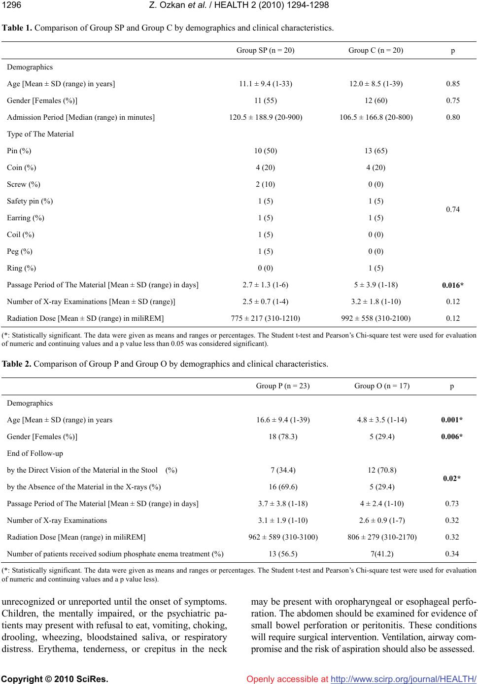
This study is the first to evaluate the effect of sodium
phosphate enema in patients who swallowed foreign ma-
terials. The results of the study indicate that the use of
enema shortens the period between the ingestion and eva-
cuation of the material, however, do not change the num-
ber of necessitated x-rays, or the radiation dose adminis-
trated to the patients. In our opinion, this shortening ef-
fect of enema was due to emptying of the rectum. Rectal
distension with feces produces an inhibitory effect on
jejunal and ileal peristalsis. This effect was termed as
recto-enteric reflex [17]. Rectal emptying by enema de-
creases rectal distension and in this way may inhibit
recto-enteric reflex.
Contrary to the literature, pins were the most com-
monly ingested material in our study, because most of
our patients were young Muslim girls who inadvertently
swallowed turban pins. Also, the proportion of children
was less than the western studies in our study because of
high number of young girls [1-3]. Thus we, retrospec-
tively, compared the characteristics between patients who
ingested pins versus other materials. We have found that
the pin ingestion mostly occurs in turbaned young girls.
Thus, in our opinion, pin ingestion is a completely dif-
ferent entity according to demographic evaluation. This
analysis revealed that direct visualization of the pins in
the stool is difficult, so the defecation of material was
inferred by the absence of pins on abdominal radiographs
in almost two-thirds of the patients in this group. Al-
though radiograph of stool would be an option to avoid
radiation exposure to young girls, daily radiography is
frequently necessary in patients who ingest pin.
5. CONCLUSIONS
Sodium phosphate enemas may be beneficial in pa-
tients who suffer from foreign body ingestion, since it is
completely safe, easy-to-use, well-accepted by the patients,
and it may hasten the pass over period of the materials.
As a subgroup of swallowed foreign materials, pin in-
gestion is more common among turbaned young girls in
our country.
REFERENCES
[1] Paul, R.I., Christoffel, K.K., Binns, H.J., Jaffe, D.M. and
the Pediatric Practice Research Group. (1993) Foreign
body ingestion in children: Risk of complication varies
with site of initial health care contact. Pediatrics, 91(1),
121-127.
[2] Hashmonai, M., Kaufman, T. and Schramek, A. (1978)
Silent perforations of the stomach and duodenum by
needles. Archives of Surgery, 113(12), 1406-1409.
[3] Cheng, W. and Tam, P.K.H. (1999) Foreign-body inges-
tion in children: Experience with 1,265 cases. Journal of
Pediatric Surgery, 34(10), 1472-1476.
[4] Kaptanoglu, M., Dogan, K., Onen, A. and Kunt, N. (1999)
Turban pin aspiration: A potential risk for young Islamic
girls. International Journal of Pediatric Otorhinolaryn-
gology, 48(2), 131-135.
[5] Shabb, B., Taha, A.M., Hamada, F. and Kanj, N. (1996)
Straight pin aspiration in young women. Journal of Trauma,
40(5), 827-828.
[6] Ucan, E.S., Tahaoglu, K., Mogolkoc, N., Dereli, S., Ba-
soz-demir, N., Basok, O., Turktas, H., Akkocoglu, A. and
Ates, M. (1996) Turban pin aspiration syndrome: A new
form of foreign body aspiration. Respiratory Medicine,
90(7), 427-428.
[7] Hodge III, D., Tecklenburg, F. and Fleisher, G. (1985)
Coin ingestion: Does every child need a radiograph? An-
nals of Emergency Medicine, 14(5), 443-446.
[8] Kelley, J.E., Leech, M.H. and Carr, M.G. (1993) A safe
and cost-effective protocol for the management of eso-
phageal coins in children. Journal of Pediatric Surgery,
28(7), 898-900.
[9] Robbins, M.I. and Shortsleeve, M.J. (1994) Treatment of
acute esophageal food impaction with glucagons, an ef-
fervescent agent, and water. American Journal of Roent-
genology, 162, 325-328.
[10] Davies, C. (2004) The use of phosphate enemas in the
treatment of constipation. Nursing Times, 100(18), 32-35.
[11] Webb, W.A. (1995) Management of foreign bodies of the
upper gastrointestinal tract: Update. Gastrointest Endosc,
Z. Ozkan et al. / HEALTH 2 (2010) 1294-1298
Copyright © 2010 SciRes. http://www.scirp.org/journal/HEALTH/Openly accessible at
1298
41(1), 39-51.
[12] Nandi, P. and Ong, G.B. (1978) Foreign body in the eso-
phagus: Review of 2394 cases. British Journal of Surgery,
65, 5-9.
[13] Vizcarrondo, F.J., Brady, P.G. and Nord, H.J. (1983) Fo-
reign bodies of the upper gastrointestinal tract. Gastro-
intest Endosc, 29(3), 208-210.
[14] Doraiswamy, N.V., Baig H. and Hallam, L. (1999) Metal
detector and swallowed metal foreign bodies in children.
Journal of Accident & Emergency, 16(2), 123-125.
[15] Ginsberg, G.G. (1995) Management of ingested foreign
objects and food bolus impactions. Gastrointest Endosc,
41(3), 33-38.
[16] Al-Haddad, M., Ward, E.M., Scolapio, J.S., Ferguson,
D.D. and Raimondo, M. (2006) Glucagon for the relief of
esophageal food impaction does it really work. Digestive
Diseases Sciences, 51(11), 1930-1933.
[17] Shafik, A. (2000) Effect of rectal distension on the small
intestine with evidence of a recto-enteric reflex. Hepato-
gastroenterology, 47(34), 1030-1033.