Forensic Medicine and Anatomy Research
Vol.1 No.3(2013), Article ID:34355,3 pages DOI:10.4236/fmar.2013.13009
Hypoplastic plexiform right anterior cerebral artery and absence of anterior communicating artery—A case report
![]()
Melaka Manipal Medical College (Manipal Campus), Manipal University, Manipal, India; *Corresponding Author: nayaksathish@gmail.com
Copyright © 2013 Satheesha Nayak Badagabettu et al. This is an open access article distributed under the Creative Commons Attribution License, which permits unrestricted use, distribution, and reproduction in any medium, provided the original work is properly cited.
Received 22 February 2013; revised 10 April 2013; accepted 17 April 2013
Keywords: Anterior Cerebral Artery; Circle of Willis; Brain; Variation; Anterior Communicating Artery
ABSTRACT
Anterior cerebral artery is the smaller terminal branch of the internal carotid artery. It is one of the arteries involved in the formation of the arterial circle of Willis at the base of the brain. Its hypoplasia or absence can cause serious problems during neurosurgery or in the vascular dynamics of the brain. We found a rare variation of the right anterior cerebral artery during the dissection of the brain. The initial segment of the artery was hypoplastic and plexiform. The anterior communicating artery was absent. The right and left anterior cerebral arteries fused with each other for a distance of about 1 cm. The course, size and distribution of the distal part of the right anterior cerebral artery were normal. This case may be of special importance to neurosurgeons and radiologists. Obstruction or rupture of the left anterior cerebral artery in such cases might result in infarct of the medial surfaces of both cerebral hemispheres.
1. INTRODUCTION
The anterior cerebral artery is the smaller terminal branch of the internal carotid artery. It is given off near the anterior perforated substance. It runs medially above the optic nerve to reach the median longitudinal fissure of the brain. It is connected to the anterior cerebral artery of the opposite side through the anterior communicating artery. It winds around the genu of the corpus callosum and branches over the medial surface of the cerebral hemisphere. It mainly supplies the medial surface of the hemisphere as far backwards as the parieto-occipital sulcus. It also supplies a finger breadth area of the superolateral surface of the hemisphere up to the parieto-occipital sulcus. Its territory includes the paracentral lobule which controls the micturition and defecation and leg movements [1]. Anatomical variations of ACA such as hypoplasia of anterior cerebral artery or single or triple anterior cerebral arteries have been reported [2-4]. Obstruction or rupture of the anterior cerebral artery might not normally result in serious ischemia to the brain because of the collateral circulation through the circle of Willis. But when it is absent on one side, or it is hypoplastic, it may result in serious problems [5,6]. The knowledge of its variations is very important for neurosurgeons and radiologists to minimize the possible postoperative problems around the base of the brain [6]. We present a rare variation of the anterior cerebral artery in this report and discuss about its clinical importance.
2. CASE REPORT
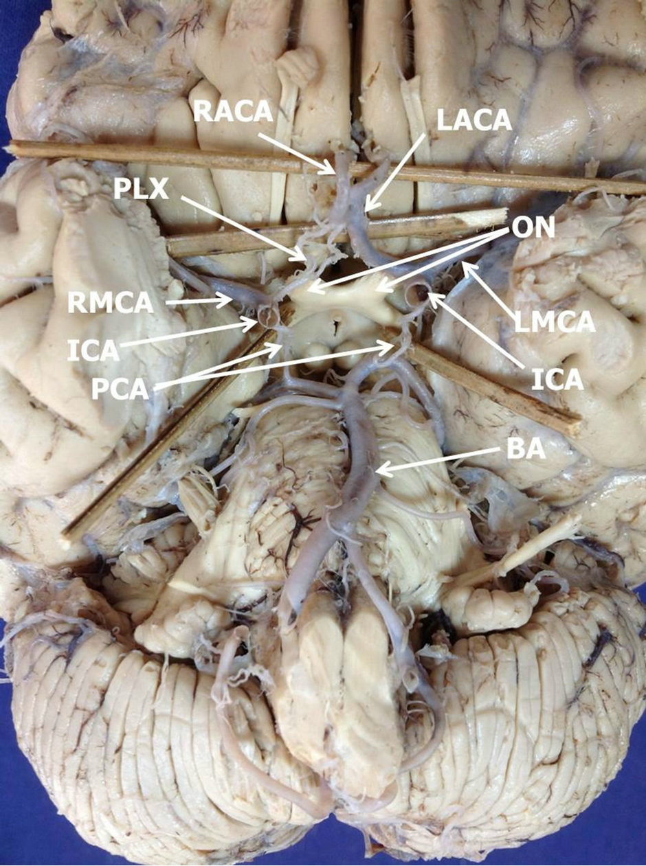
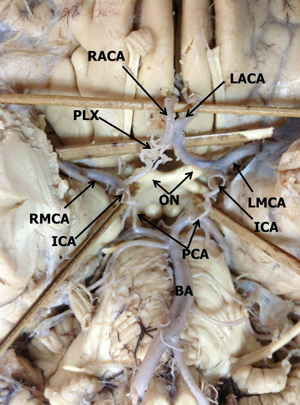
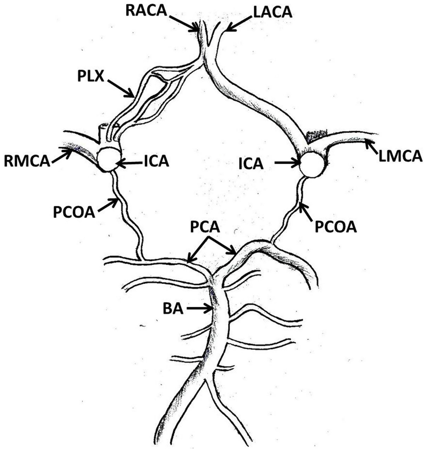
During the dissection of the brain for undergraduate Medical students, we found a rare variation of the right anterior cerebral artery at the base of the brain. The brain belonged to a South Indian male cadaver, approximately aged 55 years. The body was donated to the college for medical undergraduate dissection classes. The right internal carotid artery was smaller (about 4 mm) than its left counterpart (about 5 mm). It divided into anterior cerebral and middle cerebral arteries near the anterior perforated substance. The anterior cerebral artery was very small in size (about 2 mm) and it further divided into a few small branches which formed a plexus near

Figure 1. Base of the brain showing circle of Willis. Note the plexiform nature of the proximal part of right anterior cerebral artery. (ICA—internal carotid artery; ON—optic nerve; PLX— plexiform part of the anterior cerebral artery; RACA—right anterior cerebral artery; LACA—left anterior cerebral artery; RMCA—right middle cerebral artery; LMCA—left middle cerebral artery; PCA—posterior communicating arteries; BA—basilar artery).

Figure 2. Closer view of the circle of Willis. Note the plexiform nature of the proximal part of right anterior cerebral artery. (ICA—internal carotid artery; ON—optic nerve; PLX—plexiform part of the anterior cerebral artery; RACA–right anterior cerebral artery; LACA—left anterior cerebral artery; RMCA—right middle cerebral artery; LMCA—left middle cerebral artery; PCA—posterior communicating arteries; BA—basilar artery).

Figure 3. A Schematic diagram of the arteries at the base of the brain. (ICA—internal carotid artery; PLX—plexiform part of the anterior cerebral artery; RACA—right anterior cerebral artery; LACA—left anterior cerebral artery; RMCA—right middle cerebral artery; LMCA—left middle cerebral artery; PCOA—posterior communicating arteries; PCA—posterior cerebral artery; BA—basilar artery).
the anterior perforated substance (Figures 1-3). This plexus continued medially above the optic nerve and reunited to form a single artery. This single artery was almost of the same size as the anterior cerebral artery ofthe opposite side (about 3 mm). The anterior communicating artery was absent. The right and left anterior cerebral arteries fused with each other for a distance of about one centimeter (Figures 1-3). The arteries contributing to the left half of the circle of Willis were slightly larger than their counterparts.
3. DISCUSSION
Variations of the origin and distribution of the arteries at the base of the brain are common. Most of these variations do not alter the brain function due to the collateral circulation and compensation from the arteries of the other side [6]. Anterior cerebral artery is one of the arteries that show frequent variations. Bergman et al. have reported some variations of the same [7]. According to their findings, both anterior cerebral arteries can start as a single vessel and then divide into two distally. Frequently the two arteries differ in size. The larger artery then will send branches to supplement the weaker artery. They have also found a third anterior cerebral artery arising from the anterior communicating artery. Very recently, a hypoplastic forked right anterior cerebral artery has been reported [8]. The term hypoplastic is used when the external diameter of the artery is less than 1 mm [9]. Different types of hypoplastic anterior cerebral arteries have also been reported by different workers in the past [10-12]. In an angiographic study conducted by Akira et al. there was aplasia of A1 segment in 5.6% of cases, three A2 segments in 3% of cases and unpaired A2 segment in 2% of cases [13]. The hypoplasia of A1 segment of the anterior cerebral artery might lead to ischemic stroke [14]. Plexiform anterior cerebral artery is a very rare occurrence. In our literature survey, we could not find a report on occurrence of such a variation. Anterior communicating and posterior communicating arteries may be absent and their absence is frequently associated with defective perfusion of the blood into one of the hemispheres [15].
The primitive internal carotid arteries develop from the third aortic arches, dorsal aortae, and a plexiform primordial vascular network surrounding the developing forebrain and mid-brain. Later, the primitive internal carotid arteries divide into cranial and caudal divisions. Then, the cranial divisions terminate as the primitive olfactory arteries which eventually give rise to definitive anterior cerebral arteries, anterior choroidal and middle cerebral arteries. The plexiform nature of the anterior cerebral artery observed in the present study may be due to the incomplete fusion of the primitive plexiform anterior cerebral artery to form a single vessel [16]. The current case is unique in having a hypoplastic, plexiform initial segment of the right anterior cerebral artery. Since the distal part of the anterior cerebral artery was large and had a fusion with the left anterior cerebral artery, this variation might not cause any functional disturbances. But it might cause serious infarct of both hemispheres in case of thrombosis or rupture of the initial segment of the left anterior cerebral artery because of the poor collateral circulation provided by the artery of the right side [5,6]. Knowledge of variant arteries of the brain such as the one reported here is important for radiologist and neurosurgeons in diagnosing the reasons for the stroke and in planning the surgical procedures.
REFERENCES
- Williams, P.L. (1995) Gray’s anatomy (The anatomical basis of medicine and surgery). 38th Edition, Churchill Livingstone, Edinburgh, 1523-1530.
- Armand, J.P., Dousset, V., Viard, B., Huot, P., Chehab, Z., Dos Santos, E., Berge, J. and Caille, J.M. (1996) Agenesis of the internal carotid artery associated with an aneurysm of the anterior communicating artery. Journal of Neuroradiology, 23, 164-167.
- Nakamura, H., Yamada, H., Nagao, T., Fujita, K. and Tamaki, N. (1993) A case of hypoplasia of the left internal carotid manifested as convulsion attack. No Shinkei Geka: Neurological Surgery, 21, 843-848.
- Stefani, M.A., Schneider, F.L., Marrone, A.C., Severino, A.G., Jackowski, A.P. and Wallace, M.C. (2000) Anatomic variations of the anterior cerebral cortical branches. Clinical Anatomy, 13, 231-236. doi:10.1002/1098-2353(2000)13:4<231::AID-CA1>3.0.CO;2-T
- Puchades-Orts, A., Nombela-Gomez, M. and OrtunoPacheco, G. (1975) Variation in form of circle of Willis: Some anatomical and embryological considerations. Anatomical Record., 185, 119-124. doi:10.1002/ar.1091850112
- Rhoton Jr., A.L. (2002) The supratentorial arteries. Neurosurgery, 51, 53-120. doi:10.1097/00006123-200210001-00003
- Bergman, R.A., Afifi, A.K. and Miyauchi, R. (2013) Anterior cerebral artery. Illustrated encyclopedia of human anatomic variation: Opus II: Cardiovascular system: Arteries: Head, neck, and thorax. http://www.anatomyatlases.org/AnatomicVariants/Cardiovascular/Text/Arteries/CerebralAnterior.shtml
- Baburao, K.P., Manohar, U.J., Namdeo, U.M. and Pandit, S.V. (2012) Anomalous anterior cerebral artery. International Journal of Recent Trends in Science and Technology, 5, 53-54.
- Padget, D.H. (1948) The circle of Willis: Its embryology and anatomy. In: Dandy, W.E., Ed., Intracranial Arterial Aneurysms, Chapter III, Comstock Publishing, New York, 67-90.
- Alper, B.J., Berry, R.G. and Paddison, R.M. (1959) Anatomical studies of the circle of Willis in normal brain. Archives of Neurology and Psychiatry, 81, 409-418. doi:10.1001/archneurpsyc.1959.02340160007002
- Battacharji, S.K., Hutchinson, E.C. and McCall, A.J. (1967) The circle of Willis—The incidence of developmental abnormalities in normal and infracted brains. Brain, 90, 747-758. doi:10.1093/brain/90.4.747
- Puchades-Orts, A., Nombela-Gomez, M. and Ortuno, P.G. (1976) Variations in the form of circle of Willis: Some anatomical and embryological considerations. Anatomical Record, 185, 119-123. doi:10.1002/ar.1091850112
- Uchino, A., Nomiyama, K., Takase Y. and Kudo, S. (2006) Anterior cerebral artery variations detected by MR angiography. Neuroradiology, 48, 647-652. doi:10.1007/s00234-006-0110-3
- Chuang, Y.-M., Liu, C.-Y., Pan, P.-J. and Lin, C.-P. (2007) Anterior cerebral artery A1 segment hypoplasia may contribute to A1 hypoplasia syndrome. European Neurology, 57, 208- 211. doi:10.1159/000099160
- Merkkola, P., Tulla, H., Ronkainen, A., Soppi, V., Oksala, A., Koivisto, T. and Hippeläinen, M. (2006) Incomplete circle of Willis and right axillary artery perfusion. The Annals of Thoracic Surgery, 82, 74-79. doi:10.1016/j.athoracsur.2006.02.034
- Padget, D.H. (1948) The development of the cranial arteries in the human embryo. Contribution to embryology. Carnegie Institution, 32, 205-261.

