International Journal of Medical Physics, Clinical Engineering and Radiation Oncology, 2013, 2, 15-18
Published Online February 2013 (http://www.scirp.org/journal/ijmpcero)
http://dx.doi.org/10.4236/ijmpcero.2013.21003
Copyright © 2013 SciRes. IJMPCERO
Technical Note: The Uses of I’mRT MatriXX in Electron
Beams
Mutian Zhang, Sicong Li, Hua Deng, Sumin Zhou
Department of Radiation Oncology, University of Nebraska Medical Center, Omaha, USA
Email: mutianzhang@unmc.edu
Received Ocotber 20, 2012; revised November 22, 2012; accepted November 30, 2012
ABSTRACT
Purpose: The objective of this study is to investigate the properties of I’mRT MatriXX device in electron beams, and to
validate MatriXX in electron dosimetry and quality assurance (QA). Methods: The measurements were conducted us-
ing MatriXX in electron and photon beams from Siemens linacs. The MatriXX was placed horizontally on the linac
tabletop. Solid Water layers were used for buildup. For all the measurements, the linac gantry angle was 0˚, and the
source-to-surface distance was 100 cm from the Solid Water surface. The electron cone factors, cutout factors, and
beam profiles were measured and compared with thimble ionization chamber results. Results: The effective water
equivalent depth of MatriXX measurement point is larger than 4 mm. When measuring at the respective depths of
maximum dose, MatriXX has different responses to different beam energies. The cone factors measured by MatriXX
are nearly identical or close to those derived by ionization chambers. Beam profiles (flatness and symmetry) can be eas-
ily determined using MatriXX and are comparable to water tank results. The planar dose map of electron cutout blocks
can be visually observed, and the cutout factors can be conveniently measured. Conclusions: The MatriXX needs
separate dose calibration factors for electron and photon beams. MatriXX can be used to measure electron cutout factors
and beam profiles, thus has the potentials in electron beam dosimetry and routine linac and patient-specific QA tests.
Keywords: Electron Beam; MatriXX; Dosimetry; Quality Assurance
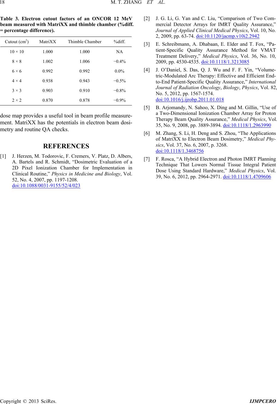
1. Introduction
The I’mRT MatriXX (IBA Dosimetry GmbH, Germany)
device consists of a two-dimensional (2D) array of ioni-
zation chambers. There are 1,020 vented parallel plate
ion chambers on the array detector, arranged in 32 × 32
grid. The chamber center-to-center distance is 7.62 mm,
and the active area is 24.4 × 24.4 cm2. MatriXX has been
validated for 2D dose measurements [1], and is increas-
ingly used in photon beam dosimetry and patient-specific
quality assurance (QA) [2-4]. The application of Ma-
triXX is also extended to QA checks for proton therapy
[5].
In this work, we report our investigation on the feasi-
bility of using MatriXX in electron beam dosimetry and
routine linac QA or patient-specific treatment QA. This
note is the expansion of an abstract submitted to the 2010
American Association of Physicists in Medicine annual
meeting [6].
2. Methods
The measurements were conducted using electron and
photon beams from Siemens ONCOR and PRIMUSTM
linacs (Siemens Healthcare, Germany). The linacs could
produce 6 MV and 23 MV photons, and electron beams
with multiple energies between 5 MeV and 21 MeV. The
MatriXX was placed horizontally on the linac treatment
couch, supported by 5 or 6 cm Solid Water (Gammex
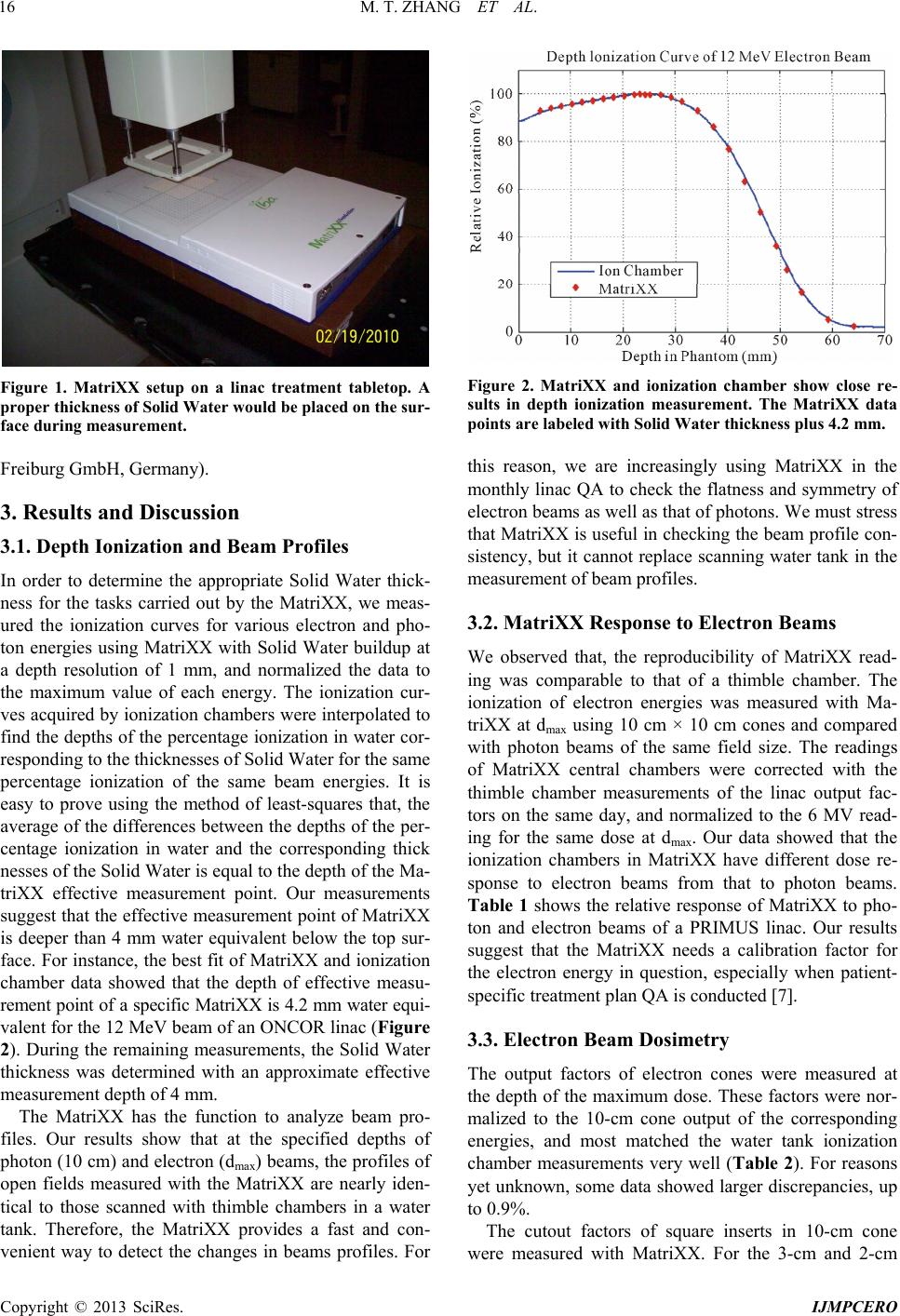
Inc., USA) blocks (Figure 1). The MatriXX was posi-
tioned using the linac light field. On the MatriXX surface,
30 cm × 30 cm Solid Water layers served as beam
buildup with 1 mm thickness resolution. The linac gantry
angle was 0˚, and the source-to-surface distance was set
at 100 cm from the Solid Water surface.
The MatriXX was previously calibrated for the photon
beams. Before each use, the MatriXX was powered on
for 30 minutes, and irradiated with at least 500 cGy until
stable readings were achieved. For each reading, 100
monitor units were delivered. The measurements of each
data point were repeated three times. When taking the
readings, the calibration factor of 6 MV photon beam
was used, so that the MatriXX responses to different
beam energies could be compared. The chamber array
was placed at the depth of maximum dose (dmax) of the
corresponding beam energy for the measurements of
dose response, cone factor or cutout factor. The MatriXX
measurements were compared with beam data acquired
using calibrated PTW semiflex thimble chambers (PTW
M. T. ZHANG ET AL.
16
Figure 1. MatriXX setup on a linac treatment tabletop. A
proper thickness of Solid Water would be placed on the sur-
face during measurement.
Freiburg GmbH, Germany).
3. Results and Discussion
3.1. Depth Ionization and Beam Profiles
In order to determine the appropriate Solid Water thick-
ness for the tasks carried out by the MatriXX, we meas-
ured the ionization curves for various electron and pho-
ton energies using MatriXX with Solid Water buildup at
a depth resolution of 1 mm, and normalized the data to
the maximum value of each energy. The ionization cur-
ves acquired by ionization chambers were interpolated to
find the depths of the percentage ionization in water cor-
responding to the thicknesses of Solid Water for the same
percentage ionization of the same beam energies. It is
easy to prove using the method of least-squares that, the
average of the differences between the depths of the per-
centage ionization in water and the corresponding thick
nesses of the Solid Water is equal to the depth of the Ma-
triXX effective measurement point. Our measurements
suggest that the effective measurement point of MatriXX
is deeper than 4 mm water equivalent below the top sur-
face. For instance, the best fit of MatriXX and ionization
chamber data showed that the depth of effective measu-
rement point of a specific MatriXX is 4.2 mm water equi-
valent for the 12 MeV beam of an ONCOR linac (Figure
2). During the remaining measurements, the Solid Water
thickness was determined with an approximate effective
measurement depth of 4 mm.
The MatriXX has the function to analyze beam pro-
files. Our results show that at the specified depths of
photon (10 cm) and electron (dmax) beams, the profiles of
open fields measured with the MatriXX are nearly iden-
tical to those scanned with thimble chambers in a water
tank. Therefore, the MatriXX provides a fast and con-
venient way to detect the changes in beams profiles. For
Figure 2. MatriXX and ionization chamber show close re-
sults in depth ionization measurement. The MatriXX data
points are labeled with Solid Water thickne ss plus 4.2 mm.
this reason, we are increasingly using MatriXX in the
monthly linac QA to check the flatness and symmetry of
electron beams as well as that of photons. We must stress
that MatriXX is useful in checking the beam profile con-
sistency, but it cannot replace scanning water tank in the
measurement of beam profiles.
3.2. MatriXX Response to Electron Beams
We observed that, the reproducibility of MatriXX read-
ing was comparable to that of a thimble chamber. The
ionization of electron energies was measured with Ma-
triXX at dmax using 10 cm × 10 cm cones and compared
with photon beams of the same field size. The readings
of MatriXX central chambers were corrected with the
thimble chamber measurements of the linac output fac-
tors on the same day, and normalized to the 6 MV read-
ing for the same dose at dmax. Our data showed that the
ionization chambers in MatriXX have different dose re-
sponse to electron beams from that to photon beams.
Table 1 shows the relative response of MatriXX to pho-
ton and electron beams of a PRIMUS linac. Our results
suggest that the MatriXX needs a calibration factor for
the electron energy in question, especially when patient-
specific treatment plan QA is conducted [7].
3.3. Electron Beam Dosimetry
The output factors of electron cones were measured at
the depth of the maximum dose. These factors were nor-
malized to the 10-cm cone output of the corresponding
energies, and most matched the water tank ionization
chamber measurements very well (Table 2). For reasons
yet unknown, some data showed larger discrepancies, up
to 0.9%.
The cutout factors of square inserts in 10-cm cone
were measured with MatriXX. For the 3-cm and 2-cm
Copyright © 2013 SciRes. IJMPCERO
M. T. ZHANG ET AL.
IJMPCERO
17
Table 1. The relative response of a MatriXX to clinical photon and electron beams of a PRIMUS linac. The readings were
normalized to 6 MV photon energy using the actual linac output of each modality.
Beam Energy 6 X 23 X 6 MeV 9 MeV 12 MeV 15 MeV 18 MeV
Normalized Reading 1.000 0.993 1.070 1.069 1.068 1.077 1.101
Table 2. The electron cone factors measured with MatriXX (MX) and ionization chamber (IC) on a PRIMUS linac. The 5-cm
cone is circular while the rest cones are square (%diff. = percentage difference).
Energy 6 MeV 9 MeV 12 MeV 15 MeV 18 MeV
Cone size MX IC %diff.MX IC %diff.MX IC %diff.MX IC %diff. MX IC %diff.
5 cm (cir) 0.787 0.789 0.3%0.879 0.886 0.8%0.9200.920 0.0% 0.9420.9420.0% 0.968 0.968 0.0%
10 cm 1.000 1.000 NA 1.000 1.000 NA 1.0001.000 NA 1.0001.000 NA 1.000 1.000 NA
15 cm 1.018 1.016 0.2% 0.993 0.992 0.1% 0.9950.993 0.2% 1.0011.001 0.0% 1.001 1.001 0.0%
20 cm 1.029 1.026 0.3% 0.973 0.973 0.0% 0.9640.961 0.3% 0.9690.967 0.2% 0.962 0.9660.4%
25 cm 1.018 1.009 0.9% 0.965 0.964 0.1% 0.9600.956 0.4% 0.9730.968 0.5% 0.963 0.9670.4%
cutout, a MatriXX central chamber was placed at the
center of the field for accurate readings. These inserts
were made for electron beam commissioning and their
cutout factors had been measured with a thimble cham-
ber in water phantom. Table 3 compares the cutout fac-
tors for the ONCOR 12 MeV beam from MatriXX and
thimble chamber. The data suggest that the MatriXX may
be a useful tool for the measurement of electron cutout
factor. In most cases, the MatriXX can provide clinically
acceptable cutout factors, and we routinely use the Ma-
triXX to measure electron cutout factors. However, the
MatriXX seems to underestimate the cutout factors of
very small inserts (nearly 1%). This tendency could be
caused by the MatriXX air cavities, which make the dif-
ferences in lateral scattering and attenuation of electrons.
The accuracy of MatriXX for very small electron cutouts
may need further investigation.
With Solid Water buildup of appropriate thickness, the
MatriXX can provide a 2D dose map at the desired depth.
This ability provides a convenient way to visualize elec-
tron dose distribution within a phantom. The 2D dose
profile can help the clinician to judge whether a custom
cutout block can provide enough coverage of the lesion
to be treated (Figure 3). This is a rather useful feature
when the electron treatment plan is based on clinical se-
tup and without a 3D image.
4. Conclusion
The MatriXX responds differently to electron beams and
photon beams, thus separate dose calibration factors
should be established for electron dosimetry. It has been
shown that MatriXX can be used to obtain electron cut-
out factors. The ability of MatriXX to display planar
Figure 3. The MatriXX 2D electron beam profiles of 10 cm
open field (top) and a custom cutout (bottom) at dmax with
Solid Water buildup. The beam energy is 18 MeV, and the
profiles are normalized to the same nominal dose.
Copyright © 2013 SciRes.
M. T. ZHANG ET AL.
18
Table 3. Electron cutout factors of an ONCOR 12 MeV
beam measured with MatriXX and thimble chamber (%diff.
= percentage difference).
Cutout (cm2) MatriXX Thimble Chamber %diff.
10 × 10 1.000 1.000 NA
8 × 8 1.002 1.006 0.4%
6 × 6 0.992 0.992 0.0%
4 × 4 0.938 0.943 0.5%
3 × 3 0.903 0.910 0.8%
2 × 2 0.870 0.878 0.9%
dose map provides a useful tool in beam profile measure-
ment. MatriXX has the potentials in electron beam dosi-
metry and routine QA checks.
REFERENCES
[1] J. Herzen, M. Todorovic, F. Cremers, V. Platz, D. Albers,
A. Bartels and R. Schmidt, “Dosimetric Evaluation of a
2D Pixel Ionization Chamber for Implementation in
Clinical Routine,” Physics in Medicine and Biology, Vol.
52, No. 4, 2007, pp. 1197-1208.
doi:10.1088/0031-9155/52/4/023
[2] J. G. Li, G. Yan and C. Liu, “Comparison of Two Com-
mercial Detector Arrays for IMRT Quality Assurance,”
Journal of Applied Clinical Medical Physics, Vol. 10, No.
2, 2009, pp. 63-74. doi:10.1120/jacmp.v10i2.2942
[3] E. Schreibmann, A. Dhabaan, E. Elder and T. Fox, “Pa-
tient-Specific Quality Assurance Method for VMAT
Treatment Delivery,” Medical Physics, Vol. 36, No. 10,
2009, pp. 4530-4535. doi:10.1118/1.3213085
[4] J. O’Daniel, S. Das, Q. J. Wu and F. F. Yin, “Volume-
tric-Modulated Arc Therapy: Effective and Efficient End-
to-End Patient-Specific Quality Assurance,” International
Journal of Radiation Oncology, Biology, Physics, Vol. 82,
No. 5, 2012, pp. 1567-1574.
doi:10.1016/j.ijrobp.2011.01.018
[5] B. Arjomandy, N. Sahoo, X. Ding and M. Gillin, “Use of
a Two-Dimensional Ionization Chamber Array for Proton
Therapy Beam Quality Assurance,” Medical Physics, Vol.
35, No. 9, 2008, pp. 3889-3894. doi:10.1118/1.2963990
[6] M. Zhang, S. Li, H. Deng and S. Zhou, “The Applications
of MatriXX to Electron Beam Dosimetry,” Medical Phy-
sics, Vol. 37, No. 6, 2007, p. 3268.
doi:10.1118/1.3468756
[7] F. Rosca, “A Hybrid Electron and Photon IMRT Planning
Technique That Lowers Normal Tissue Integral Patient
Dose Using Standard Hardware,” Medical Physics, Vol.
39, No. 6, 2012, pp. 2964-2971. doi:10.1118/1.4709606
Copyright © 2013 SciRes. IJMPCERO