K.J. Cross et al. / Open Journal of Genetics 2 (2012) 18-22
Copyright © 2012 SciRes. OJGen
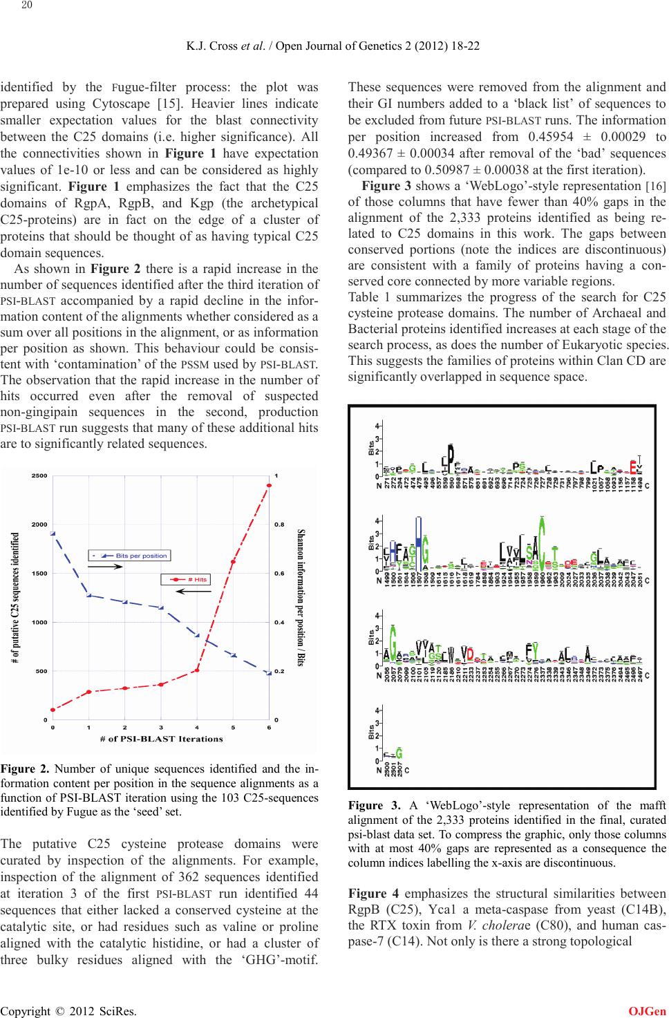
gested by Figure 5.
Figure 5. A plot of the frequency of occurrence of various re-
sidues that align with the ‘S1’-pocket residues of RgpB in the
‘seed’ set of 103 C25 cysteine protease sequences. As shown in
Table 1, these are Bacterial or Archaeal proteins. These resi-
dues determine the substrate specificity of the various C25
proteases. The low overall conservation suggests a broad range
of C25 substrate preferences.
4. CONCLUSION
The bacterial C25 cysteine proteases share significant
sequential and structural similarity with other Clan CD
cysteine proteases. The number of identified bacterial
and archaeal sequences increases at each stage of the
search procedure as seen in Table 1, while the number of
sequences associated with other phyla increases dramat-
ically in the final round of the search.
The lack of sequence conservation in the ‘S1’-binding
site argues for a wide-range of substrate specificities
among the C25 cysteine proteases further blurring the
distinctions between the various protease families within
the Clan CD proteases.
5. ACKNOWLEDGEMENTS
We acknowledge funding from the Oral Health CRC and NH&MRC.
REFERENCES
[1] Carlsson, J., B.F. Herrmann, J.F. Hofling, and G.K.
Sundqvist. (1984) Degradation of the human proteinase
inhibitors alpha-1-antitrypsin and alpha-2-macroglobulin
by Bacteroides gingivalis. Infection and Immunity. 43,
644-648.
[2] Rawlings, N.D., A.J. Barrett, and A. Bateman. (2010)
MEROPS: the peptidase database. Nucleic Acids
Research. 38, D227-D233.
[3] Walker, N.P.C., R.V. Talanian, K.D. Brady, L.C. Dang,
N.J. Bump, et al. (1994) Crystal structure of the cysteine
protease interleukin-1 beta-converting enzyme: a
(p20/p10)2 homodimer. Cell. 78,
doi:10.1093/nar/gkp971
343-352.
[4] Eichinger, A., H.G. Beisel, U. Jacob, R. Huber, F.J.
Medrano, et al. (1999) Crystal structure of gingipain R:
an Arg-specific bacterial cysteine proteinase with a
caspase-like fold. EMBO Journal. 18,
5453-5462.
doi:10.1016/0092-8674(94)90303-4
[5] Lupardus, P.J., A. Shen, M. Bogyo, and K.C. Garcia.
(2008) Small molecule-induced allosteric activation of
the Vibrio cholerae RTX cysteine protease domain.
Science. 322, 265-8.
doi:10.1093/emboj/18.20.5453
[6] Chen, J.M., N.D. Rawlings, R.A. Stevens, and A.J.
Barrett. (1998) Identification of the active site of
legumain links it to caspases, clostripain and gingipains
in a new clan of cysteine endopeptidases. FEBS Letters.
441, 361-365.
doi:10.1126/science.1162403
[7] nr database. [cited 2012 September 3]; Available
from:
doi:10.1016/S0014-5793(98)01574-9
ftp://ftp.ncbi.nih.gov/blast/db.
[8] Shi, J., T.L. Blundell, and K. Mizuguchi. (2001) FUGUE:
sequence-structure homology recognition using
environment-specific substitution tables and
structure-dependent gap penalties. Journal of Molecular
Biology. 310, 243-257.
[9] Mizuguchi, K., C.M. Deane, T.L. Blundell, and J.P.
Overington. (1998) HOMSTRAD: a database of protein
structure alignments for homologous families. Protein
Science. 7, 2469-2471.
doi:10.1006/jmbi.2001.4762
[10] Notredame, C., D.G. Higgins, and J. Heringa. (2000)
T-Coffee: A novel method for multiple sequence
alignments. Journal of Molecular Biology. 302, 205-217.
doi:10.1002/pro.5560071126
[11] Katoh, K., K. Misawa, K. Kuma, and T. Miyata. (2002)
MAFFT: a novel method for rapid multiple sequence
alignment based on fast Fourier transform. Nucleic Acid
Research. 30, 3059-3066
[12] Camacho, C., G. Coulouris, V. Avagyan, N. Ma, J.
Papadopoulos, et al. (2009) BLAST+: architecture and
applications. BMC Bioinformatics. 10,
421.
[13] Shannon, C.E. (1948) A Mathematical Theory of
Communication. Bell System Technical Journal. 27,
379–423.
doi:10.1186/1471-2105-10-421
[14] [14] Côté, R.G., P. Jones, L. Martens, S. Kerrien, F.
Reisinger, et al. (2007) The Protein Identifier
Cross-Referencing (PICR) service: reconciling protein
identifiers across multiple source databases. BMC
Bioinformatics. 8, 401.
[15] [15] Smoot, M.E., K. Ono, J. Ruscheinski, P.L. Wang,
and T. Ideker. (2010) Cytoscape 2.8: new features for
data integration and network visualization.
Bioinformatics. 27,
431-2.
doi:10.1186/1471-2105-8-401
[16] Crooks, G.E., G. Hon, J.M. Chandonia, and S.E. Brenner.
(2004) WebLogo: a sequence logo generator. Genome
Research. 14, 1188-1190.
doi:10.1093/bioinformatics/btq675
doi:10.1101/gr.849004