Paper Menu >>
Journal Menu >>
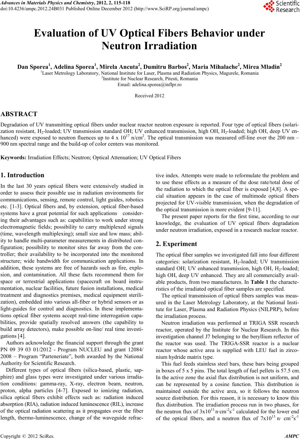 Advances in Ma terials Physics and Che mist ry, 2012, 2, 115-118 doi:10.4236/ampc.2012.24B031 Published Online December 2012 (htt p://www.SciRP.org/journal/ampc) Copyright © 2012 SciRes. AMPC Evaluation of UV Optical Fibers Behavior under Neutron Irradiation Dan Spore a1, Adelina Sporea1, Mirela Ancuta2, Du mitru Barbos2, Maria Mihalach e2, Mirea Mladin2 1Laser Metrology Laboratory, National Institute for Laser, Plasma and Radiation Physics, Magu rele, Roma nia 2Institute for N uclear Researc h, Pitesti, Romania Email: adelina.sporea@inflpr.ro Received 2012 ABSTRACT Degradation of UV transmitting optical fibers under nuclear reactor neutron exposure is reported. Four type of optical fibers (solari- zation resistant, H2-loaded; UV transmission standard OH; UV enhanced transmission, high OH, H2-loaded; high OH, deep UV en- hanced) were exposed to neutron fluences up to 4 x 10¹⁷ n/cm2. The optical transmission was measured off-line over the 200 nm – 900 nm spectral range and the build-up of color centers was monitored. Keywords: Irradiation Effects; Neutron; Optical Attenuation; UV Optical Fibers 1. Introduction In the last 30 years optical fibers were extensively studied in order to assess their possible use in radiation environments for communications, sensing, remote control, light guides, robotics etc. [1-3]. Optical fibers and, by extension, optical fiber-based systems h ave a great p otential for such applications consider- ing their advantages such as: capabilities to work under strong electromagnetic fields; possibility to carry multiplexed signals (time, wavelength multiplexing); small size and low mass; abil- ity to handle multi-parameter measu rements in di stributed con- figuration; possibility to monitor sites far away from the con- troller; their availability to be incorporated into the monitored structure; wide bandwidth for communication applications. In addition, these systems are free of hazards such as fire, explo- sion, and contamination. All these facts recommend them for space or terrestrial applications (spacecraft on board instru- mentation, nuclear facilities, future fusion installations, medical treatment and diagnostics premises, medical equipment sterili- zation), embedded into various all-fiber or hybrid sensors or as light-guides for control and diagnostics. In these implementa- tions optical fiber systems accept real-time interrogation capa- bilities, provide spatially resolved answers (the capability to build array detectors), make possible on-line/ real time investi- gations [4]. Authors acknowledge the financial support through the grant PN 09 39 03 01/2012 - Program NUCLEU and grant 12084/ 2008 – Program “Parteneriate”, both awarded by the National Autho r ity for Scient ific Research. Different types of optical fibers (silica-based, plastic, sap- phire) and glass types were investigated under various irradia- tion conditions: gamma-ray, X-ray, electron beam, neutron, proton, alpha particles [4-7]. Exposed to ionizing radiation, silica optical fibers exhibit effects such as: radiation induced absorption (RIA), radiation induced luminescence (RIL), increase of the optical radiation scattering as it propagates over the fiber length, thermo-luminescence, change of the waveguide refrac- tive ind ex. Attempt s were made to reformulate the problem and to use these effects as a measure of the dose rate/total dose of the radiation to which the optical fiber is exposed [4,8]. A spe- cial situation appears in the case of multimode optical fibers proj ected for UV-visible transmission, when the degradation of the optical transmission is more evident [9-11]. The present paper reports for the first time, according to our knowledge, the evaluation of UV optical fibers degradation under neutron irradiation, exposed in a research nuclear reactor . 2. Experiment The optical fiber samples we investigated fall into four different categories: solarization resistant, H2-loaded; UV transmission standard OH; UV enhanced transmission, high OH, H2-loaded; high OH, deep UV enhanced. Th ey ar e all commerciall y avail- able products, from two manufactures. In Table 1 the characte- ristics of the irr adiated optical fiber s amples are specified . The optical transmission of optical fibers samples was meas- ured in the Laser Metrology Laboratory, at the National Insti- tute for Laser, Plasma an d Radiatio n Physics (NILP RP), before the irra di a tion proce s s. Neutron irradiation was performed at TRIGA SSR research reactor, operated by the Institute for Nuclear Research. In this investigation channel J7 belonging to the beryllium reflector of the reactor was used. The TRIGA-SSR reactor is a nuclear reactor whose active area is supplied with LEU fuel in zirco- nium hydride matrix type. This fuel feeds stainless steel bars, these bars being grouped in boxes of 5 x 5 pins. The total length of fuel pellets is 57.5 cm. In the active zone the axial flux distribution is not uniform, and can be represented by a cosine function. This distribution is maintained outside the active area, so it follows the neutron source distribution. For this reason, it is necessary to know this flux distribution. The irradiation process run in two phases, for the neutron flux of 3x10¹³ n∙cm-2s-1 calculated for th e lower end of the optical fibers, and a neutron flux of 7x10¹³ n∙ cm-2s-1 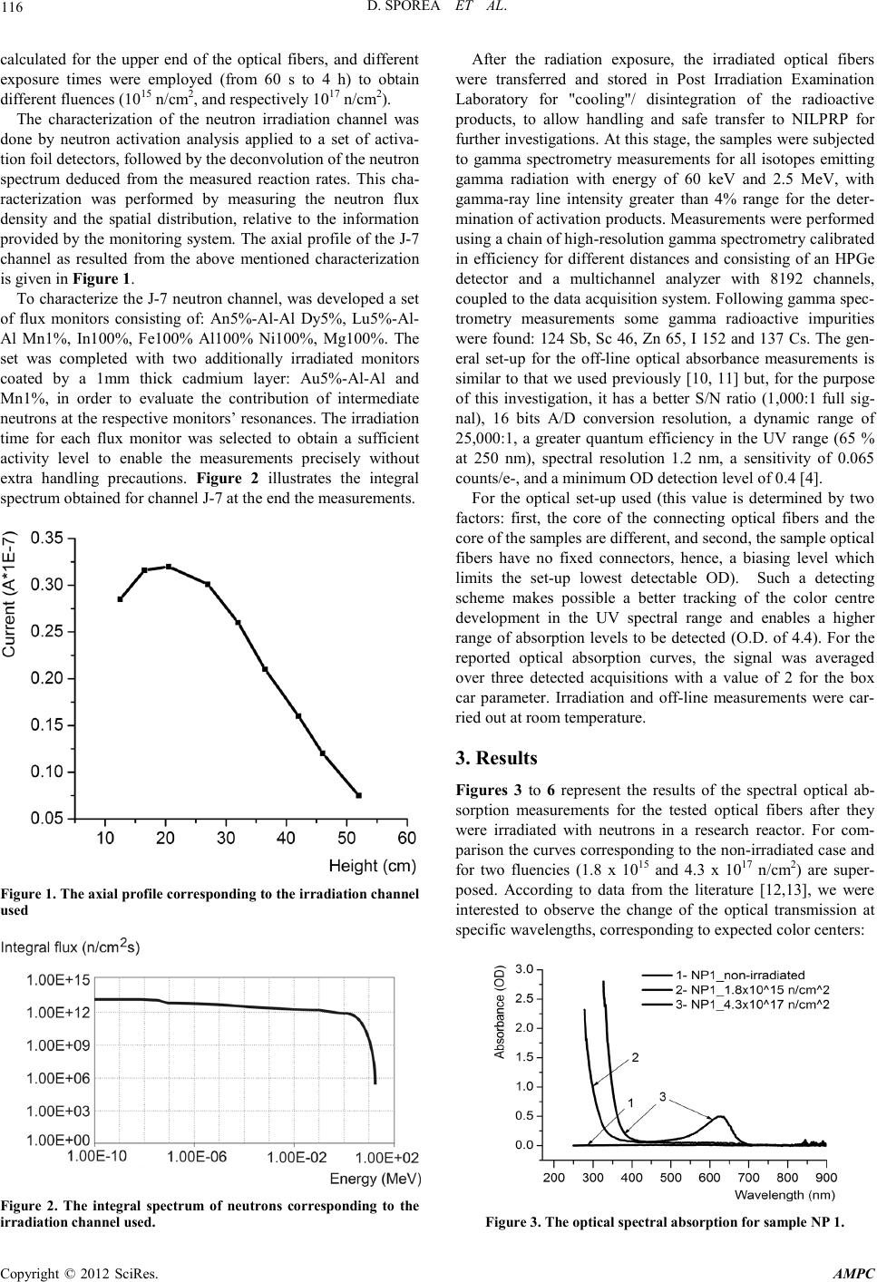 D. SPOREA ET AL. Copyright © 2012 SciRes. AMPC 116 calculated for the upper end of the optical fibers, and different exposure times were employed (from 60 s to 4 h) to obtain different fluences ( 1015 n /cm2, and respectively 1017 n/cm2). The characterization of the neutron irradiation channel was done by neutron activation analysis applied to a set of activa- tion foil detectors, followed by the deconvolution of the neutron spectrum deduced from the measured reaction rates. This cha- racterization was performed by measuring the neutron flux density and the spatial distribution, relative to the information provided by the monitoring system. The axial profile of the J-7 channel as resulted from the above mentioned characterization is given in Figure 1. To character ize the J-7 neutron channel, was developed a set of flux monitors consisting of: An5%-Al-Al Dy5%, Lu5%-Al- Al Mn1%, In100%, Fe100% Al100% Ni100%, Mg100%. The set was completed with two additionally irradiated monitors coated by a 1mm thick cadmium layer: Au5%-Al-Al and Mn1%, in order to evaluate the contribution of intermediate neut rons at th e respect ive moni tors’ reso nances. The irrad iation time for each flux monitor was selected to obtain a sufficient activity level to enable the measurements precisely without extra handling precautions. Figure 2 illustrates the integral spectrum obtained for channel J-7 at the end the measurements. Figure 1. T he axial profile corresponding to the irradiation c hannel used Figure 2. The integral spectrum of neutrons corresponding to the irradiation channel used. After the radiation exposure, the irradiated optical fibers were transferred and stored in Post Irradiation Examination Laboratory for "cooling"/ disintegration of the radioactive products, to allow handling and safe transfer to NILPRP for further investigations. At this stage, the samples were subjected to gamma spectrometry measurements for all isotopes emitting gamma radiation with energy of 60 keV and 2.5 MeV, with gamma-ray line intensity greater than 4% range for the deter- mination of activation products. Measurements were per formed using a chain of high-resolu tion ga mma spectro metr y cali brat ed in efficiency for different distances and consisting of an HPGe detector and a multichannel analyzer with 8192 channels, coupled to the data acquisition system. Following gamma spec- trometry measurements some gamma radioactive impurities were found: 124 Sb, Sc 46, Zn 65, I 152 and 137 Cs. The gen- eral set-up for the off-line optical absorbance measurements is similar to that we used previously [10, 11] but, for the purpose of this investigation, it has a better S/N ratio (1,000:1 full sig- nal), 16 bits A/D conversion resolution, a dynamic range of 25,000:1, a greater quantum efficiency in the UV range (65 % at 250 nm), spectral resolution 1.2 nm, a sensitivity of 0.065 counts/e-, and a minimum OD detection level of 0.4 [4]. For the optical set-up used (this value is determined by two factors: first, the core of the connecting optical fibers and the core of the samples ar e different, and second, the sample optical fibers have no fixed connectors, hence, a biasing level which limits the set-up lowest detectable OD). Such a detecting scheme makes possible a better tracking of the color centre development in the UV spectral range and enables a higher range of absorption levels to be detected (O.D. of 4.4). For the reported optical absorption curves, the signal was averaged over three detected acquisitions with a value of 2 for the box car parameter. Irradiation and off-line measurements were car- ried ou t at room temperature. 3. Results Figures 3 to 6 represent the results of the spectral optical ab- sorption measurements for the tested optical fibers after they were irradiated with neutrons in a research reactor. For com- parison the curves corresponding to the non-irradi ated case and for two fluencies (1.8 x 10¹⁵ and 4.3 x 10¹⁷ n/cm2) are super- posed. According to data from the literature [12,13], we were interested to observe the change of the optical transmission at specific wa vel engths, correspon ding t o expected color centers: Figure 3. Th e optical spectral absorp t i on f or s ample NP 1. 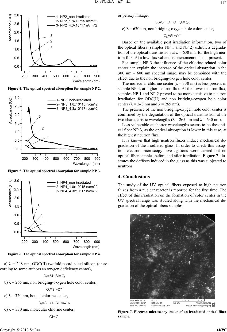 D. SPOREA ET AL. Copyright © 2012 SciRes. AMPC 117 Figure 4. Th e optical spectral absorp t i on f or s ample NP 2. Figure 5. The op t ic al spe ct ra l absorp t ion for sample NP 3. Figure 6. Th e optical spectral absorp t i on f or s ample NP 4. a) λ = 248 nm, ODC(II) twofold coordinated silicon (or ac- cording to some authors an oxygen deficiency center), b) λ = 265 nm, non bridging-oxygen hole color center, c) λ = 320 nm, bound chlorine center, d) λ = 330 nm, molecular chlorine center, or peroxy linkage, e) λ = 630 nm, non bridging-oxygen hole color center, Based on the available post irradiation information, two of the optical fibers (samples NP 1 and NP 2) exhibit a degrada- tion of the optical transmission at λ = 630 nm, for the high neu- tron flux. At a low flux value this phenomenon is not present. For sample NP 3 the influence of the chlorine related color center can explain the increase of the optical absorption in the 300 nm – 600 nm spectral range, may be combined with the effect due to the non bridging-oxygen hole color center. The molecular chlorine center (λ = 330 nm) is less present in sample NP 4, at higher neutron flux. At the lower neutron flu x, samples NP 1 and NP 2 proved to be more sensitive to neutron irradiation for ODC(II) and non bridging-oxygen hole color center (λ = 248 nm and λ = 265 nm). The presence of the non bridging-oxygen hole color center is confirmed by the degradation of the optical transmission at the two characteristic wavelengths (λ = 265 nm and λ = 630 nm). Less vulnerable at shorter wavelengths seems to be the opti- cal fiber NP 3, as the optical ab sorption is lower in this case, at the highest neutron flux. It is known that high neutron fluxes induce mechanical de- gradation of the irradiated glass. In order to check this assup- tion electron microscopy investigations were carried out on opt ical fiber samples befo re and after irardi ation. Figure 7 illu- strates th e deffects induced in the glass as this was subjected to neutrons. 4. Conclusions The study of the UV optical fibers exposed to high neutron fluxes from a nuclear reactor is reported for the first time. The effect of this irradiation on the formation of color center in the UV spectral range was studied along with the mechanical de- gradation of the optical fiber s sampl es . Figure 7. Electron microscopy image of an irradiated optical fiber sample. 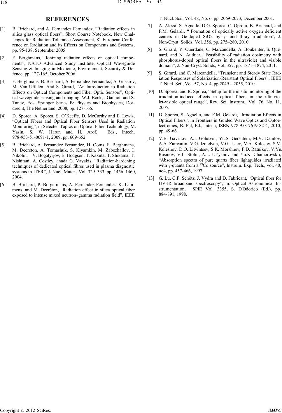 D. SPOREA ET AL. Copyright © 2012 SciRes. AMPC 118 REFERENCES [1] B. Brichard, and A. Fernandez Fernandez, “Radiation effects in silica glass optical fibers”, Short Course Notebook, New Chal- lenges for Ra diati on Tolera nce Ass essmen t, 8th European Confe- rence on Radiation and its Effects on Components and Systems, pp. 95-138, Se ptember 2005 [2] F. Berghmans, “Ionizing radiation effects on optical compo- nents”, NATO Advanced Study Institute, Optical Waveguide Sensing & Imaging in Medicine, Environment, Security & De- fence , pp. 127-165, October 2006 [3] F. Berghmans, B. Brichard, A. Fernandez Fernandez, A. Gusarov, M. Van Uffelen. And S. Girard, “An Introduction to Radiation Effects on Optical Components and Fiber Optic Sensors”, Opt i- cal waveguide sensing and imaging, W.J. Bock, I.Gannot, and S. Tanev, Eds. Springer Series B: Physics and Biophysics, Dor- drecht, The Netherland, 2008, pp. 127-166. [4] D. Sporea, A. Sporea, S. O’Keeffe, D. McCarthy and E. Lewis, “Optical Fibers and Optical Fiber Sensors Used in Radiation Monitoring”, in Selected Topics on Optical Fiber Technology, M. Yasin, S. W. Harun and H. Arof, Eds., Intech, 978-953-51-0091-1, 2 00 9, pp. 609-652. [5] B. Brichard, A. Fernandez Fernandez, H. Ooms, F. Berghmans, M. Decréton, A. Tomashuk, S. Klyamkin, M. Zabezhailov, I. Nikolin, V. Bogatyrjov, E. Hodgson, T. Kakuta, T. Shikama, T. Nishitani, A. Costley, anada G. Vayakis, “Radiation-hardening techniques of dedicated optical fibres used in plasma diagnostic systems in ITER”, J. Nucl. Mater., Vol. 329 –333, pp . 1456–1460, 2004. [6] B. Brichard, P. Borgermans, A. Fernandez Fernandez, K. Lam- mens, and M. Decréton, “Radiation effect in silica optical fiber expos ed to i nten se mix ed neut ron–gamma radiation field”, IEEE T. Nucl. Sci., Vol. 48, No. 6, pp. 2069-2 073, December 2001. [7] A. Alessi, S. Agnello, D.G. Sporea, C. Oproiu, B. Brichard, and F.M. Gelardi, “ Formation of optically active oxygen deficient centers in Ge-doped SiO2 by γ- and β-ray irradiation”, J. Non-Cryst. Solids, Vol. 356, pp. 275–280, 2010. [8] S. Girard, Y. Ouerdane, C. Marcandella, A. Boukenter, S. Que- nard, and N. Authier, “Feasibility of radiation dosimetry with phosphorus-doped optical fibers in the ultraviolet and visible domain”, J. Non-Cryst. Solids, Vol. 357, pp. 1871–1874, 2011. [9] S. Girard , and C. M arcand ella, “Tra nsi ent and S teady St ate Rad- iation Responses of Solarization-Resist ant Optical Fi bers” , IEEE T. Nucl. Sci., Vol. 57, No. 4, pp.2049 – 2055, 2010. [10] D. S porea, and R. Sp orea, “Setup for the in s itu monitorin g of the irradiation-induced effects in optical fibers in the ultravio- let-visible optical range”, Rev. Sci. Instrum., Vol. 76, No. 11, 2005. [11] D. Sporea, S. Agnello, and F.M. Gelardi, “Irradiation Effects in Optical Fibers”, in Frontiers in Guided Wave Optics and Optoe- lectronics, B. Pal, Ed., Intech, ISBN 978-953-7619-82-4, 2010, pp. 49-66. [12] V.B. Gavrilov, A.I. Golutvin, Yu.S. Gershtein, M.V. Danilov, A.A. Zamyatin, V.G. Izraelyan, V.G. Isaev, V.A. Kolosov, S.V. Koleshov, D.O. Lit vintsev, S.K. Morsh nev, F.D. Rat nikov, V. Yu. Rasinov, V.L. Stolin, A.L. Ul’yanov and Yu.K. Chamorovskii, “Absorption spectra of pure quartz fiber lightguides irradiated with γ-qu anta from a 60Co sou rce”, Inst rum. E xp. Tech., vol. 40, no4, pp. 457-466, 1997. [13] G. Lu, G.F. Schötz, J. Vydra and D. Fabricant, “Optical fiber for UV-IR broadband spectroscopy”, in: Optical Astronomical In- strumentation, SPIE Vol. 3355, S. D'Odorico (Ed.), pp. 884-891, 1998. |