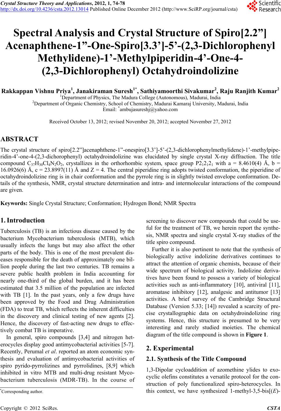
 Crystal Structure Theory and Applications, 2012, 1, 74-78 http://dx.doi.org/10.4236/csta.2012.13014 Published Online December 2012 (http://www.SciRP.org/journal/csta) Spectral Analysis and Crystal Structure of Spiro[2.2”] Acenaphthene-1”-One-Spiro[3.3’]-5’-(2,3-Dichlorophenyl Methylidene)-1’-Methylpiperidin-4’-One-4- (2,3-Dichlorophenyl) Octahydroindolizine Rakkappan Vishnu Priya1, Janakiraman Suresh1*, Sathiyamoorthi Sivakumar2, Raju Ranjith Kumar2 1Department of Physics, The Madura College (Autonomous), Madurai, India 2Department of Organic Chemistry, School of Chemistry, Madurai Kamaraj University, Madurai, India Email: *ambujasureshj@yahoo.com Received October 13, 2012; revised November 20, 2012; accepted November 27, 2012 ABSTRACT The crystal structure of spiro[2.2”]acenaphthene-1”-onespiro[3.3’]-5’-(2,3-dichlorophenylmethylidene)-1’-methylpipe- ridin-4’-one-4-(2,3-dich orophenyl) octahydroindolizine was elucidated by single crystal X-ray diffraction. The title compound C37H30Cl4N2O2, crystallizes in the orthorhombic system, space group P212121 with a = 8.4610(4) Å, b = 16.0926(6) Å, c = 23.8997(11) Å and Z = 4. The central piperidine ring adopts twisted conformation, the piperidine of octahydroindolizine ring is in chair conformation and the pyrrole ring is in slightly twisted envelope conformation. De- tails of the synthesis, NMR, crystal structure determination and intra- and intermolecular interactions of the compound are given. Keywords: Single Crystal Structure; Conformation; Hydrogen Bond; NMR Spectra 1. Introduction Tuberculosis (TB) is an infectious disease caused by the bacterium Mycobacterium tuberculosis (MTB), which usually infects the lungs but may also affect the other parts of the body. This is one of the most prevalent dis- eases responsible for the death of approximately one bil- lion people during the last two centuries. TB remains a severe public health problem in India accounting for nearly one-third of the global burden, and it has been estimated that 3.5 million of the population are infected with TB [1]. In the past years, only a few drugs have been approved by the Food and Drug Administration (FDA) to treat TB, which reflects the inherent difficulties in the discovery and clinical testing of new agents [2]. Hence, the discovery of fast-acting new drugs to effec- tively combat TB is imperative. In general, spiro compounds [3,4] and nitrogen het- erocycles display good antimycobacterial activities [5-7]. Recently, Perumal et al. reported an atom economic syn- thesis and evaluation of antimycobacterial activities of spiro pyrido-pyrrolizines and pyrrolidines, [8,9] which inhibited in vitro MTB and multi-drug resistant Myco- bacterium tuberculosis (MDR-TB). In the course of screening to discover new compounds that could be use- ful for the treatment of TB, we herein report the synthe- sis, NMR spectra and single crystal X-ray studies of the title spiro compound. Further it is also pertinent to note that the synthesis of biologically active indolizine derivatives continues to attract the attention of organic chemists, because of their wide spectrum of biological activity. Indolizine deriva- tives have been found to possess a variety of biological activities such as anti-inflammatory [10], antiviral [11], aromatase inhibitory [12], analgesic and antitumor [13] activities. A brief survey of the Cambridge Structural Database (Version 5.33; [14]) revealed a scarcity of pre- cise crystallographic data on octahydroindolizine ring systems. Hence, this structure is presumed to be very interesting and rarely studied moieties. The chemical diagram of the title compound is shown in Figure 1. 2. Experimental 2.1. Synthesis of the Title Compound 1,3-Dipolar cycloaddition of azomethine ylides to exo- cyclic olefins constitutes a versatile protocol for the con- struction of poly functionalized spiro-heterocycles. In this context, we have synthesized 1-methyl-3,5-bis[(E)- *Corresponding author. C opyright © 2012 SciRes. CSTA  R. V. PRIYA ET AL. 75 Figure 1. Chemical diagram of the molecule. 2,3-dichlorophenylmethylidene]tetrahydro-4(1H)-pyridi none, an exocyclic dipolarophile, from the reaction of 1-methyl-4-piperidone and 2,3-dichlorobenzaldehyde in ethanol in the presence of NaOH. The cycloaddition of the above dipolarophile with azomethine ylide generated in situ from the decarboxylative condensation of ace- naphthenequinone and piperidine-2-carboxylic acid led to the formation of the title compound in excellent yield. A mixture of 1-methyl-3,5-bis[(E)-2,3-dichlorophenyl- methylidene]tetrahydro-4(1H)-pyridinone (1 mmol), acena- phthenequinone (1 mmol) and piperidine-2-carboxylic acid (1 mmol) was dissolved in isopropyl alcohol (15 mL) and heated to reflux for 60 min. After completion of the reac- tion as evident from TLC, the mixture was poured into water (50 mL), the precipitated solid was filtered and washed with water (100 mL) to obtain the product as pure yellow solid. The product was recrystallized from ethyl acetate to obtain suitable crystals for the X-ray ana- lysis. Melting point: 494 (2) K, Yield: 94%. 2.2. Structure Determination and Refinement For the crystal structure determination, the intensity data of the single-crystal of the compound C37H30Cl4N2O2 was collected using a Bruker AXS Kappa APEX II single crystal CCD Diffractometer equipped with graphite- monochromated MoKα radiation (λ = 0.71073 Å) at room temperature with a crystal dimension of 0.35 × 0.25 × 0.20 mm3. Accurate unit cell parameters were determined from the reflections of 36 frames measured in three dif- ferent crystallographic zones. The data collection, data reduction and absorption correction were performed by APEX2, SAINT-plus and SADABS program [15]. The structure was solved by the direct method procedure and the non-hydrogen atoms were subjected to anisotropic refinement by full-matrix least squares on F2 using SHELXL-97 program [16]. The positions of all the hy- drogen atoms were identified from difference electron density map, and they were constrained to ride on the corresponding non-hydrogen atoms. Molecular graphics were drawn using PLATON [17]. The crystal data, ex- perimental conditions and structure refinement parame- ters for the title compound are presented in Table 1. Table 1. The crystal data, experimental conditions and stru- cture refinement parameters of the title compound. Empirical formula C37H30Cl4N2O2 Formula weight 676.43 Temperature 293(2) K Wavelength 0.71073 Å Crystal system, space group P212121, orthorhombic Unit cell dimensions a = 8.4610(4) Å b = 16.0926(6) Å c = 23.8997(11) Å Volume 3254.2(2) Å3 Z, Calculated density 4, 1.38 mg/m3 Absorption coefficient 0.401 mm–1 F(000) 1400 Crystal size 0.23 × 0.21 × 0.19 mm3 Theta range for data collection2.12 to 24.91 deg Limiting indices –8< = h < = 10, –18, = k < = 19 –22< = l < = 28 Reflections collected/unique 16629/5642 [R(int) = 0.032] Completeness to theta 99.6% Absorption correction ω-scan Refinement method Full-matrix least-squares on F2 Data/restraints/parameters 5642/0/407 Goodness-of-fit on F2 1.024 Final R indices [I > 2sigma(I)]R1 = 0.038, wR2 = 0.077 R indices (all data) R1 = 0.058, wR2 = 0.085 Largest diff. peak and hole 0.194 and −0.203 e·A–3 3. Results and Discussion 3.1. Spectral Data The structure of title compound has been elucidated with the help of 1H, 13C and two dimensional NMR spectro- scopic studies. The H,H-COSY spectrum of the com- pound assigns a doublet and multiplet at 4.81 ppm (J = 9.9 Hz) and 4.00 ppm to H-4 and H-4a respectively. Further, H-4 shows HMBC correlations with C-4’, C-3, C-4a and C-5 at 195.7, 64.4, 64.5 and 25.5 ppm respec- tively. The signals of 5, 6, 7 and 8-CH2 protons overlap and appear as a multiplets from 1.26 to 2.15 ppm whereas the carbon signals appear at 25.5, 24.1, 30.8 and 45.6 respectively. The multiplet in a range 1.26 - 1.30 ppm and the doublet at 2.95 ppm (J = 12.6 Hz) are as- signed to 2’-CH2 protons. The 6’-CH2 protons appears as doublets at 2.71 and 2.48 ppm (J = 15.3 Hz). The C,H- COSY correlations assign the carbon signals at 56.9 and 55.3 to C-2’ and C-6’ carbons respectively. The singlet at Copyright © 2012 SciRes. CSTA  R. V. PRIYA ET AL. 76 1.69 ppm is due to the N-CH3 protons. The aromatic protons appear as a multiplet in a range 6.58 - 7.97 ppm. The 1H and 13C NMR chemical shifts of the title com- pound are shown in Figure 2. It is pertinent to observe that the chemical shifts of 2’-CH2 of the compound (2.95 ppm ~1.30 ppm) differ very much by 1.65 ppm. This suggests that probably the H-2eq is spatially proximate to the carbonyl of ace- naphthylen-1 (2H)-one shifting it downfield, while H-2ax lies in the shielding zone of the acenaphthylen-1(2H)- one ring shifting it downfield suggesting relative con- figuration at C-2 for the compound. The alternative ste- reochemistry with inversion of configuration at C-2 rela- tive to that shown on the compound bringing both the carbonyls of the piperidone and acenapthene rings to- wards each other in spatial proximity, probably renders the transition state of the cycloaddition unstable by elec- trostatic repulsion. This could raise the free energy of activation (transition state B in Figure 3), relative to the transition state leading to the formation of the compound with both carbonyls placed far off (transition state A in Figure 3). Figure 2. 1H and 13C NMR chemical shifts of the com- pound. Figure 3. Stereochemistry of formation of cycloadducts dif- fering in their configurations at C-2. 3.2. Crystal Structure Figure 4 shows the ORTEP plot drawn at 50% probabi- lity displacement ellipsoids of title compound and the atom-numbering scheme. The six membered piperidine ring in the title compound adopts the half chair confor- mation as evident from puckering parameters Q = 0.561 (3) Å, θ = 136.9(3)˚ and Φ = 140.6(4)˚ [18]. The olefinic double bond in the structure has an E configuration. The piperidine of octahydroindolizine ring is in the chair con- formation as evident from the puckering parameters Q = 0.574(2) Å, θ = 180(3)˚ and Φ = 69(11)˚ [18]. The pyrrole ring is in the twisted envelope conformation with atom N2 at the flap as the puckering parameters are, Q = 0.424(3) Å, and Φ = 12.2(4)˚ [18,19]. The dihedral angles between the mean plane of the piperidone ring and the aryl rings are 38.69(1)˚, 82.28(1)˚ which indicate that the aryl rings in the structure are not coplanar with the mean plane of the piperidone ring. As a result, the torsion angle C3C31C32C33 is 41.24(3)˚. This lack of coplanarity is caused by non- bonded interactions between one of the ortho H atoms in the aryl ring and the equatorial H atoms at the 2-position of the piperidone ring (H33/H2A or H2B). As a result of these steric repulsions, the bond angle C3C31C32 expands to 128.69 (19)˚ instead of 120˚. The dichloro- phenyl rings are planar as confirmed by the values of the r.m.s. deviations 0.0186 and 0.0127 Å. The dihedral an- gle between the dichlorophenyl rings is 74.22 (1)˚. The di- hedral angles of these dichlorophenyl rings with ace- naphthene group are 30.84 (1)˚ and 78.99 (1)˚. The Csp2-Csp2 distances in the acenaphthene group range from 1.346(3) (C19-C20) to 1.574(2) Å (C13-C14) and the C-C-C bond angles from 101.54(16)˚ (C12-C11- Figure 4. The molecular structure of the tittle compound showing the atom numbering scheme. Displacement ellip- soids are drawn at 50% probability level, using ORTEP 3. Hydrogen atoms are omitted for clarity. Copyright © 2012 SciRes. CSTA  R. V. PRIYA ET AL. 77 C19) to 123.32(2)˚ (C16-C17-C21). These values are in agreement with related structures [20-24]. The C8-N2 bond distance being 1.450 (3) Å is comparable to the Csp2-Nsp2 distances found in similar structures [25,26]. In the crystal structure some weak C—H···O intramolecular interactions have been observed (Table 2). A C---H...π interaction (Ta bl e 2) C10---H10B…Cg1, (Cg1 is the centroid of the ring C32-C37) forms, a two dimensional linear zig zag chain running parallel to the b-axis as shown in Figure 5. 4. Conclusion Spiro[2.2”]acenaphthene-1”-onespiro[3.3’]-5’-(2,3-dich- lorophenylmethylidene)-1’-methylpiperidin-4’- one-4-(2,3-dichlorophenyl) octahydroindolizine was syn- thesized through 1,3-dipolar cycloaddition reaction. The single crystal of the title compound is obtained by slow evaporation method (solvent: 1:1 ethyl acetate-ethanol). The conformational features of the compound are deter- mined in the solid phase by X-ray method and in liquid phase by NMR method. The replacements of the equato- rial H atoms at the 2- and 6-positions, and the attachment of different atoms to one or two of the atoms C33, C37, C72 and C76, are likely to alter the C3-C31-C32 and C8-C7-C71 bond angles. Correlations have been established Table 2. Hydrogen bonds [Å and ] of the compound. D-H...A d(D-H)d(H...A) d(D...A)DHA C(6)-H(6A)...O(2) 0.97 2.37 3.003(3)122 C(7)-H(7)...Cl1(1) 0.98 2.53 3.062(3)114 C(8)-H(8)...O(2) 0.98 2.45 3.091(3)123 C(18)-H(18)...O(1) 0.93 2.55 3.192(4)126 C(7)-H(7)...O(1) 0.98 2.23 2.774(3)113 C(10)-H(10B)...Cg1 (i) 0.93 2.88 3.675(4)140 Symmetry transformations used to generate equivalent atoms: (i) 1 – x, 1/2 + y, 1/2 – z. Figure 5. Partial packing diagram showing the C---H…π interaction along “b” axis. between the bond angle values and bio-activity [27]. In addition, the orientation of aryl rings will affect the alignment of these rings at a binding site and hence in- fluence bioactivity [28]. Further studies on structure- bioactivity relationship of this compound are in progress in our research group. 5. Acknowledgements JS thanks the UGC for the FIST support. JS and RV thank the management of Madura College for their en- couragement and support. RRK thanks DST, New Delhi for funds under fast track scheme (No.SR/FT/CS-073/ 2009). REFERENCES [1] R. Granich, F. Wares, S. Suvanand and L. S. Chauhan, “Lancet Infectious Disease,” Tuberculosis Control in In- dia, Vol. 3, No. 9, 2003, p. 535. doi:10.1016/S1473-309 9(03)00744-8 [2] R. J. O’Brien and P. P. Nunn, “The Need for New Drugs against Tuberculosis. Obstacles, Opportunities, and Next Steps,” American Journal of Respiratory and Critical Care Medicine, Vol. 163, No. 5, 2001, pp. 1055-1058. [3] M. S. Chande, R. S. Verma, P. A. Barve, R. R. Khanwel- kar, R. B. Vaidya and K. B. Ajaikumar, “Facile Synthesis of Active Antitubercular, Cytotoxic and Antibacterial Agents: A Michael Addition Approach,” Journal of Me- dicinal Chemistry, Vol. 40, No. 11, 2005, p. 1143. [4] A. Dandia, M. Sati, K. Arya, R. Sharma and A. Loupy, “Facile One Pot Microwave Induced Solvent-Free Syn- thesis and Antifungal, Antitubercular Screening of Spiro [1,5]-Benzothiazepin-2,3’[3’H]indol-2[1’H]-ones,” Che- mical & Pharmaceutical Bulletin, Vol. 51, No. 10, 2003, p. 1137. [5] D. Sriram, P. Yogeeswari and K. Madhu, “Synthesis and in Vitro Antitubercular Activity of Some 1-[(4-sub) Phenyl]-3-(4-{1-[(Pyridine-4-Carbonyl)Hydrazono]Ethyl} Phenyl)Thiourea,” Bioorganic & Medicinal Chemistry Letters, Vol. 16, No. 4, 2006, pp. 876-878. doi:10.1016/j.bmcl.2005.11.004 [6] M. Biava, G. C. Porretta, G. Poc, S. Supino, D. Deidda, R. Pompei, P. Molicotti, F. Manetti and M. J. Botta, “Anti- mycobacterial Agents. Novel Diarylpyrrole Derivatives of BM212 Endowed with High Activity toward Myco- bacterium Tuberculosis and Low Cytotoxicity,” Journal of Medicinal Chemistry, Vol. 49, No. 16, 2006, pp. 4946- 4952. doi:10.1021/jm0602662 [7] M. Shaharyar, A. A. Siddiqui, M. A. Ali, D. Sriram and P. Yogeeswari, “Synthesis and in Vitro Antimycobacterial Activity of N1-Nicotinoyl-3-(4’-Hydroxy-3’-Methyl Phenyl)- 5-[(sub)Phenyl]-2-Pyrazolines,” Bioorganic & Medicinal Chemistry Letters, Vol. 16, No. 15, 2006, pp. 3947-3949. doi:10.1016/j.bmcl.2006.05.024 [8] R. R. Kumar, S. Perumal, P. Senthilkumar, P. Yogeeswari and D. Sriram, “A Highly Atom Economic, Chemo-, Regio- and Stereoselective Synthesis, and Discovery of Copyright © 2012 SciRes. CSTA  R. V. PRIYA ET AL. Copyright © 2012 SciRes. CSTA 78 Spiro-Pyrido-Pyrrolizines and Pyrrolidines as Antimyco- bacterial Agents,” Tetrahedron, Vol. 64, No. 13, 2008, pp. 2962-2971. doi:10.1016/j.tet.2008.01.072 [9] R. R. Kumar, S. Perumal, P. Senthilkumar, P. Yogeeswari and D. Sriram, “Discovery of Antimycobacterial Spiro- piperidin-4-Ones: An Atom Economic, Stereoselective Syn- thesis, and Biological Intervention,” Journal of Medicinal Chemistry, Vol. 51, No. 18, 2008, pp. 5731-5735. doi:10.1021/jm800545k [10] H. Malonne, J. Hanuise and J. Fontaine, “Topical Anti- Inflammatory Activity of New 2-(1-Indolizinyl)Propionic Acid Derivatives in mice,” Pharmacy and Pharmacology Communications, Vol. 4, No. 5, 1998, pp. 241-243. [11] S. Medda, P. Jaisankar, R. K. Manna, B. Pal, V. S. Giri and M. K. Basu, “Phospholipid Microspheres: A Novel Delivery Mode for Targeting Antileishmanial Agent in Experimental Leishmaniasis,” Journal of Drug Targeting, Vol. 11, No. 2, 2003, pp. 123-128. doi:10.1080/1061186031000119101 [12] P. Sonnet, P. Dallemagne, J. Guillom, C. Engueard, S. Stiebing, J. Tangue, B. Bureau, S. Rault, P. Auvray, S. Moslemi, P. Sourdaine and G. E. Seralini, “New Aro- matase Inhibitors. Synthesis and Biological Activity of Aryl-Substituted Pyrrolizine and Indolizine Derivatives,” Bioorganic & Medicinal Chemistry, Vol. 8, No. 5, 2000, pp. 945-955. doi:10.1016/S0968-0896(00)00024-9 [13] W. H. Pearson and L. Guo, “Synthesis and Mannosidase Inhibitory Activity of 3-Benzyloxymethyl Analogs of Swainsonine,” Tetrahedron Letters, Vol. 42, No. 47, 2001, pp. 8267-8271. doi:10.1016/S0040-4039(01)01777-4 [14] F. H. Allen, “The Cambridge Structural Database: A Quar- ter of a Million Crystal Structures and Rising,” Acta Cry- stallographica, Vol. B58, No. 1, 2002, pp. 380-388. [15] Bruker, “APEX2, SAINT-Plus and XPREP,” Bruker AXS Inc., Madison, 2004. [16] G. M. Sheldrick, “A Short History of SHELX,” Acta Cry- stallographica, Vol. A64, Part 1, 2008, pp. 112-122. [17] A. L. Spek, “Structure Validation in Chemical Crystallo- graphy,” Acta Crystallographica, Vol. D65, Part 2, 2009, pp. 148-155. [18] D. Cremer and A. J. Pople, “General Definition of Ring Puckering Coordinates,” Journal of the American Che- mical Society, Vol. 97, No. 6, 1975, pp. 1354-1358. doi:10.1021/ja00839a011 [19] M. Nardelli, “Ring Asymmetry Parameters from Out-of- Plane Atomic Displacements,” Acta Crystallographica, Vol. C39, Part 8, 1983, pp. 1141-1142. [20] A. C. Hazell, “The Crystal Structure of 6b,8a-Dihydro- cyclobut[a]Acenaphthylene, C14H10,” Acta Crystallo- graphica, Vol. B32, Part 7, 1976, pp. 2010-2013. [21] A. C. Hazell and R. G. Hazell, “The Crystal Structure of 6b, 10b-Dihydrobenzo[j]Cyclobut[a]Acenaphthylene, C18H12,” Acta Crystallographica, Vol. B33, Part 2, 1977, pp. 360- 365. [22] A. C. Hazell and Weigelt, “6b,12b-Dihydronaphtho[2,3- j]Cyclobut[a]Acenaphthylene,” Acta Crystallographica, Vol. B32, Part 1, 1976, pp. 306-308. [23] P. G. Jones, P. Bubenitschek, G. M. Sheldrick and G. Dyker, “Acenaphtho[1,2a]acenaphthylene at 178 K,” Acta Crystallographica, Vol. C48, Part 9, 1992, pp. 1633-1635. [24] T. V. Sundar, V. Parthasarathi, A’lvarez-Ruá, C. S. Gar- cıá-Granda, A. Saxena, P. Pardasani and R. T. Par- dasani, “(rac-5RS,7RS,8SR)-Spiro[7-Methoxycar-bonyl-1- aza-3-tHiabicyclo[3.3.0]octane-8,1’-acenaphthylen]-2’-one,” Acta Crystallographica, Vol. E58, Part 12, 2002, pp. o1405-o1407. [25] J. L. Sussman and S. J. Wodak, “The Crystal Structure of Fulvine: A Pyrrolizidine Alkaloid,” Acta Crystallogra- phica, Vol. B29, Part 12, 1973, pp. 2918-2926. [26] S. J. Wodak, “The Crystal Structure of Heliotrine: A Pyr- Rolizidine Alkaloid Monoester,” Acta Crystallographica, Vol. B31, Part 2, 1975, pp. 569-573. [27] S. N. Pandeya and J. R. Dimmock, “An Introduction to Drug Design,” New Age International Publishers, New Delhi, 1997. [28] J. W. Quail, A. Doroud, H. N. Pati, U. Das and J. R. Dim- mock, “(E,E)-2-(4-Fluorophenylmethylene)-6-(4-Nitrophen- ylmeThylene)Cyclohexanone,” Acta Crystallographica, Vol. E61, Part 6, 2005, pp. o1774-o1776.
|