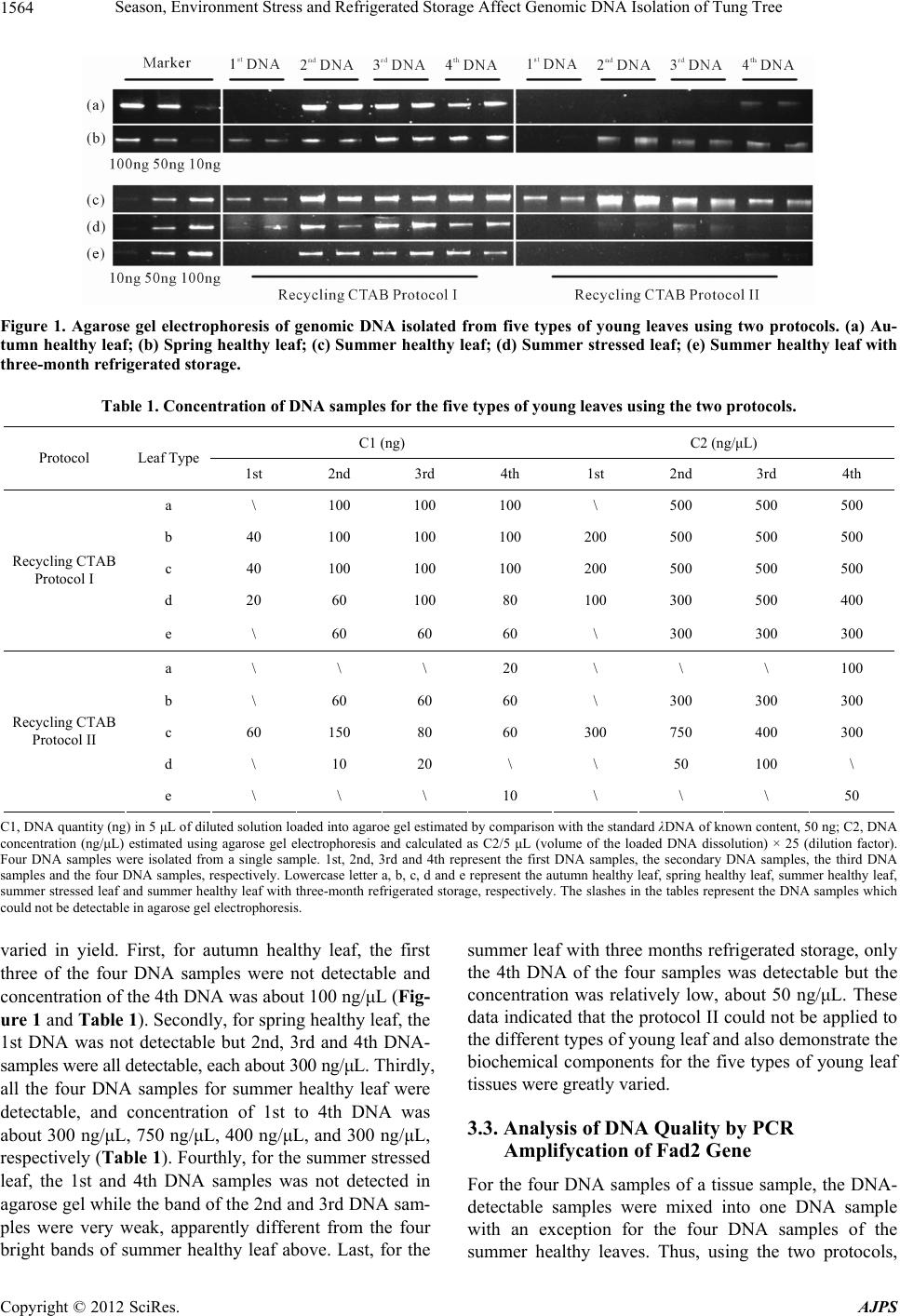
Season, Environment Stress and Refrigerated Storage Affect Genomic DNA Isolation of Tung Tree
1566
lar Biology, Vol. 3, No, 2, 1961, pp. 208-218.
[8] M. G. Murray and W. F. Thompson, “Rapid Isolation of
High Molecular Weight Plant DNA,” Nucleic Acids Re-
search, Vol. 8, No. 19, 1980, pp. 4321-4325.
doi:10.1093/nar/8.19.4321
[9] J. J. Doyle and J. L. Doyle, “Isolation of Plant DNA from
Fresh Tissue,” Focus, Vol. 12, No. 1, 1990, pp. 13-15.
[10] D. Goldenberger, I. Perschil, M. Ritzler and M. Altwegg,
“A Simple Universal DNA Extraction Procedure Using
SDS and Proteinase K Is Compatible with Directly PCR
Amplification,” Genome Research, Vol. 4, 1995, pp. 368-
370.
[11] S. M. Aljanabi and I. Martinez, “Universal and Rapid
Salt-Extraction of High Quality Genomic DNA for PCR-
Based Techniques,” Nucleic Acids Research, Vol. 25, No.
22, 1997, pp. 4692-4693. doi:10.1093/nar/25.22.4692
[12] N. Stein, G. Herren and B. Keller, “A New DNA Extrac-
tion Method for High-Throughput Marker Analysis in a
Large-Gemone such as Triticum aestivum,” Plant Breed-
ing, Vol. 120, No. 4, 2001, pp. 354-356.
doi:10.1046/j.1439-0523.2001.00615.x
[13] K. L. Hill-Ambroz, G. L. Brown-Guedira and J. P. Fellers,
“Modified Rapid DNA Extraction Protocol for High
throught Microsatellite Analysis in Wheat,” Crop Science,
Vol. 42, No. 6, 2002, pp. 2088-2091.
doi:10. 2135/cropsci2002.2088
[14] R. J. Mogg and J. M. Bond, “A Cheap, Reliable and
Rapid Method of Extracting High-Quality DNA from
Plants,” Molecular Ecology Notes, Vol. 3, No. 4, 2003,
pp. 666-668. doi:10.1046/j.1471-8286.2003.00548.x
[15] G. C. Allen, M. A. Flores-Vergara, S. Krasynanski, S.
Kumar and W. F. Thompson, “A Modified Protocol for
Rapid DNA Isolation from Plant Tissues Using Cetyl-
trimethylammonium Bromide,” Nature Protocol, Vol. 1,
No. 5, 2006, pp. 2320-2325.
[16] J. Amani, R. Kazemi, A. R. Abbasi and A. H. Salmanian,
“A Simple and Rapid Leaf Genomic DNA Extraction
Method for Polymerase Chain Reaction Analysis,” Ira-
nian Journal of Biotechnology, Vol. 9, No. 1, 2011, pp.
69-71.
[17] H. A. Souza, L. A. Muller, R. L. Brandao and M. B.
Lovato, “Isolation of High Quality and Polysaccharide-
Free DNA from Leaves of Dimorphandra mollis (Legu-
minosae), a Tree from the Brazilian Cerrado,” Genetic
Molecular Research, Vol. 11, No. 1, 2012, pp. 756-764.
[18] F. Lefort and G. C. Douglas, “An Efficient Micro-Method
of DNA Isolation from Mature Leaves of Four Hardwood
Tree Species Acer, Fraxinus, Prunus and Quercu,” An-
nual Forest Science, Vol. 56, No. 3, 1999, pp. 259-263.
doi:10.1051/forest: 19990308
[19] P. A. Moreira and D. A. Oliveira, “Leaf Age Affects the
Quality of DNA Extracted from Dimorphandra mollis
(Fabaceae), a Tropical Tree Species from the Cerrado
Region of Brazil,” Genetic Molecular Research, Vol. 10,
No. 1, 2011, pp. 353-358. doi:10.4238/vol10-1gmr1030
[20] N. M. Van Dam, R. Verpoorte and E. Van der Meljden,
“Extreme Difference in Pyrrolizidine Alkaloid Levels
between Leaves of cynoglossum officinale,” Phytochem-
istry, Vol. 37, No. 4, 1994, pp. 1013-1016.
doi:10.1016/S0031-9422(00)89519-9
[21] M. A. Azmat, I. A. Khan and H. M. N. Cheema, “Extrac-
tion of DNA Suitable for PCR Applications from Mature
Leaves of Mangifera indica L.,” Journal of Zhejiang Uni-
versity Science B., Vol. 13, No. 4, 2012, pp. 239-243.
doi:10.1631/jzus.B1199194
[22] Z. Liu, S. B. Carpenter, W. J. Bourgeois, Y. Yu, R. J.
Constantin, M. J. Falcon and J. C. Adams, “Variations in
the Secondary Metabolite Camptothecin in Relation to
Tissue Age and Season in Camptotheca acuminate,” Tree
Physiology, Vol. 18, No. 4, 1998, pp. 265-270.
[23] F. Vallejo, F. A. Tomas-Barberan and C. Garcia-Viguera,
“Effect of Climatic and Sulphur Fertilization Conditions,
on Phenolic Compounds and Vitamin C, in the Inflores-
cences of Eight Broccoli Cultivars,” European Food Re-
search and Technology, Vol. 216, No. 5, 2003, pp. 395-
401. doi:10.1007/s00217-003-0664-9
[24] M. C. Shih, C. M Chang, S. M. Kang and M. L. Tsai,
“Effect of Different Parts (Leaf, Stem and Stalk) and
Season (Summer and Winter) on the Chemical Composi-
tions and Antioxidant Activity of Moringa oleifera,” In-
ternational Journal of Molecular Sciences, Vol. 12, No. 9,
2011, pp. 6077-6088. doi:10.3390/ijms12096077
[25] Z. Yang, L. L. Geng, W. Wei and J. Zhang, “Combined
Effects of Temperature, Light Intensity, and Nitrogen
Concentration on the Growth and Polysaccharide Content
of Microcystis aeruginosa in Batch Culture,” Biochemical
Systematics and Ecology, Vol. 41, 2012, pp. 130-135.
doi:10.1016/j.bse.2011.12.015
[26] N. Benhamou, “Elicitor-Induced Plant Defence Path-
ways,” Trends Plant Science, Vol. 1, No. 7, 1996, pp.
233-240. doi:10.1016/1360-1385(96)86901-9
[27] F. Bourgaud, A. Gravot, S. Milesi and E. Gontier, “Pro-
duction of Plant Secondary Metabolites: A Historial Per-
spective,” Plant Science, Vol. 161, No. 5, 2001, pp. 839-
851. doi:10.1016/S0168-9452(01)00490-3
[28] T. Siatka and M. Kasparova, “Seasonal Variation in Total
Phenolic and Flavonoid Content and DPPH Scavenging
Activity of Bellis perennis L. Flowers,” Molecular, Vol.
15, No. 12, 2010, pp. 9450-9461.
[29] A. S. Rodrigues, M. R. Perez-Gregorio, M. S. Garcia-
Falcon, J. Simal-Gandara and D. P. F. Almeida, “Effect
of Meteorological Conditions on Antioxidant Flavonoids
in Portuguese Cultivars of White and Red Onions,” Food
Chemistry, Vol. 124, No. 1, 2011, pp. 303-308.
doi:10.1016/j.foodchem.2010.06.037
[30] J. Y. Park, D. K. Kim, Z. M. Wang, P. Lu, S. C. Park and
J. S. Lee, “Production and Characterization of Biodiesel
from Tung Oil,” Applied Biochemistry and Biotechnology,
Vol. 148, No. 6, 2008, pp. 109-117.
doi:10.1007/s12010-007-8082-2
[31] Q. Shang, W. Jiang, H. F. Lu and B. Liang, “Properties of
Tung Oil Biodiesel and Its Blends with 0# Diesel,” Bio-
resource Technology, Vol. 101, No. 2, 2010, pp. 826-828.
doi:10.1016/j.biortech.2009.08.047
[32] Y. H. Chen, J. H. Chen and Y. M. Luo, “Complementary
Biodiesel Combination from Tung and Medium-Chain
Copyright © 2012 SciRes. AJPS