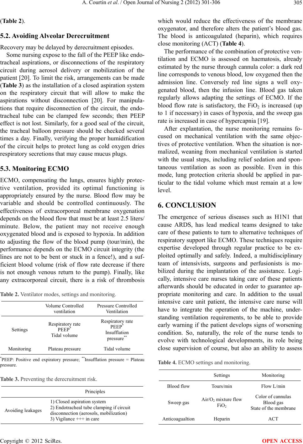
A. Courtin et al. / Open Journal of Nursing 2 (2012) 301-306
Copyright © 2012 SciRes.
306
the risks to which expert knowledge and a good under-
standing of the treatment plan are necessary.
OPEN ACCESS
REFERENCES
[1] Bernard, G.R., Arti gas, A., Brigham, K. L., Carlet, J., Fa l-
ke, K., Hudson, L., Lamy, M., Legall, J.R., Morris, A.
and Spragg, R. (1994) The Amer ican European Consensus
Conference on ARDS. Definitions, mechanisms, relevant
outcomes, and clinical trial coordination. American Jour-
nal of Respiratory and Critical Care Medicine, 149, 818-
824.
[2] Ware, L.B. and Matthay, M.A. (2000) The acute respira-
tory distress sy ndrome. The New England Journal of Me-
dicine, 342, 1334-1349.
doi:10.1056/NEJM200005043421806
[3] Brun-Buisson, C., Minelli, C., Brun-Buisson, C., Bertolini,
G., Brazzi, L., Pimentel, J., Lewandoski, K., Bion, J., Ro-
mand, J.A., Villar, J., Thorsteinsson, A., Damas, P., Ar-
maganidis, F. and the ALIVE Study group (2004) Epi-
demiology and outcome of acute lung injury in European
intensive care units. Results from the ALIVE study. In-
tensive Care Medicine, 30, 51-56.
doi:10.1007/s00134-003-2136-x
[4] Webb, H.H. and Tierney, D.F. (1974) Experimental pul-
monary edema due to intermittent positive pressure ven-
tilation with high inflation pressures. American Review of
Respiratory Disease, 110, 556-565.
[5] Dirkes, S., Dickinson, S. and Valentine, J. (1992) Acute
respiratory failure and ECMO. Critical Care Nurse, 12,
39-47.
[6] Combes, A., Bacchetta, M., Brodie, D., Müller, T. and Pel-
legrino, V. (2012) Extracorporeal membrane oxygenation
for respiratory failure in adults. Current Opinion in Cri-
tical Care, 18, 99-104.
[7] Crimi, E. and Slutsky, A.S. (2002) Inflammation and the
acute respiratory distress syndrome. Best Practice & Re-
search Clinical Anaesthesiology, 18, 477-492.
doi:10.1016/j.bpa.2003.12.007
[8] Herridge, M.S., Tansey, C.M., Matté, A., Tomlinson, G.,
Diaz-Granados, N., Cooper, A., Guest, C.B., Mazer, C.D.,
Mehta, S., Stewart, T.E., Kudlow, P., Cook, D., Slutsky,
A.S. and Cheung, A.M. (2011) Functional disability 5
years after acute respiratory distress syndrome. The New
England Journal of Medicine, 364, 1293-1304.
doi:10.1056/NEJMoa1011802
[9] Brodie, D. and Bacchetta, M. (2011) Extracorporeal me m-
brane oxygenation for ARDS in Adults. The New Eng-
land Journal of Medicine, 365, 1905-1914.
doi:10.1056/NEJMct1103720
[10] Papazian, L., Forel, J.-M., Gacouin, A., Penot-Ragon, C.,
Perrin, G., Loundou, A., Jaber, S., Arnal, J.-M., Perez, D.,
Seghboyan, J.-M., Constantin, J.-M., Courant, P., Lefrant,
J.-Y., Guerin, C., Prat, G., Morange, S. and Roch, A. (2010)
Neuromuscular blockers in early acute respiratory dis-
tress syndrome. The New England Journal of Medicine,
363, 1107-1116. doi:10.1056/NEJMoa1005372
[11] Boussarsar, M., Thierry, G., Jaber, S., Roudot-Thoraval,
F., Lemaire, F. and Brochard, L. (2004) Relationship be-
tween ventilatory settings and barotrauma in the ARDS.
Intensive Care Medicine, 28, 406-413.
doi:10.1007/s00134-001-1178-1
[12] Dreyfuss, D. and Saumon, G. (1998) Ventilator-induced
lung injury: Lessons from experimental studies. American
Journal of Respiratory and Critical Care Medicine, 157,
294-323.
[13] Briel, M., Meade, M., Mercat, A., Brower, R.G., Talmor,
D., Walter, S.D., Slutsky, A.S., Pullenayegum, E., Zhou,
Q., Cook, D., Brochard, L., Richard, J.C., Lamontagne, F.,
Bhatnagar, N., Stewart, T.E. and Guyatt, G. (2010) Higher
vs lower positive end-expiratory pressure in patients with
acute lung injury and acute respiratory distress syndrome.
Systematic review and Meta-analysis. Journal of the
American Medical Association, 303, 865-873.
doi:10.1001/jama.2010.218
[14] Amato, M.B.P., Barbas, C.S.V., Medeiros, D.M., Magaldi,
R.B., Schettino, G.P.P., Lorenzi-Filho, G., Kairalla, R.A.,
Deheinzelin, D., Munoz, C., Oliveira, R., Takagaki, T.Y.
and Carvalho, C.R.R. (1998) Effect of a protective ventil-
ation strategy on mortality in the ARDS. The New Eng-
land Journal of Medicine, 338, 347-354.
doi:10.1056/NEJM199802053380602
[15] The Acute Respiratory Distre ss Syndrome Netwo rk (2000)
Ventilation with lower tidal volumes as compared with
traditional tidal volumes for acute lung injury and the
acute respiratory distress syndrome. The New England
Journal of Medicine, 342, 1301-1308.
[16] Peek, G.J., Mugford, M., Tiruvoipati, R., Wilson, A., Al-
len, E., Thalanany, M.M., Hibbert, C.L., Truesdale, A.,
Clemens, F., Cooper, N., Firmin, R.K. and Elbourne, D.
(2009) Efficacy and economic assessment of conven-
tional ventilatory support versus extracorporeal membrane
oxygenation for severe adult respiratory failure (CESAR):
A multicentre randomized controlled trial. Lancet, 374,
1351-1363. doi:10.1016/S0140-6736(09)61069-2
[17] The Australia and New Zealand Extracorporeal Mem-
brane Oxygenation (ANZ ECMO) Influenza Investigators
(2009) Extracorporeal membrane oxygenation for 2009
influenza A (H1N1) acute respiratory distress syndrome.
Journal of the American Medical Association, 302, 1888-
1895. doi:10.1001/jama.2009.1535
[18] Kopp, R., Henzler, D., Dembinski, R. and Kuhlen, R.
(2004) Extracorporeal membrane oxygenation by acute
respiratory distress syndrome. Anaesthesist, 53, 168-174.
doi:10.1007/s00101-003-0643-3
[19] ELSO Guidelines for Cardiopulmonary Extracorporeal
Life Support Extracorporeal Life Support Organization,
Version 1:1. Ann Arbor, April 2009.
http://www.elso.med.umich.edu
[20] Maggiore, S.M., Lellouche, F., Pigeot, J., Taille, S., Deye,
N., Durrmeyer, X., Richard, J.-C., Mancebo, J., Lemaire,
F. and Brochard, L. (2003) Prevention of endotracheal
suctioning-induced alveolar derecruitment in acute lung
injury. American Journal of Respiratory and Critical
Care Medicine, 167, 1215-1224.
doi:10.1164/rccm.200203-195OC