Journal of Cancer Therapy, 2012, 3, 673-679
http://dx.doi.org/10.4236/jct.2012.325087 Published Online October 2012 (http://www.SciRP.org/journal/jct) 673
Deep Inspiration Breath Hold Reduces Dose to the Left
Ventricle and Proximal Left Anterior Descending Artery
during Radiotherapy for Left-Sided Breast Cancers
Lesley A. Jarvis1, Peter G. Maxim2, Kathleen C. Horst2*
1Department of Radiation Oncology, Dartmouth-Hitchcock Medical Center, Lebanon, USA; 2Department of Radiation Oncology,
Stanford University Medical Center, Stanford, USA.
Email: *kateh@stanford.edu
Received June 20th, 2012; revised July 23rd, 2012; accepted August 4th, 2012
ABSTRACT
The purpose of th is study was to an alyze motion of the lef t anterior de scending cor onar y artery (LAD) an d left ventricle
during normal breathing and deep inspiration breath hold (DIBH). This is a dosimetric study utilizing free-breathing and
static DIBH scans from eleven patients treated with radiotherapy for breast cancer. The anterior-posterior displacement
along the length of the LAD was measured in each respiratory phase. Standard treatment plans targeting the whole
breast without treatment of the internal mammary lymph nodes were generated and dose to the LAD and LV calculated.
Non-uniform movement of the LAD during respiratory maneuvers with the proximal third exhibiting the greatest dis-
placement was observed. In DIBH compared to end-expiration (EP), the mean posterior displacement of the proximal
1/3 of the LAD was 8.99 mm, the middle 1/3 of the artery was 6.37 mm, and the distal 1/3 was 3.27 mm. In
end-inspiration (IP) compared to end-expiration the mean posterior displacements of the proximal 1/3 of the LAD was
2.08 mm, the middle 1/3 of the artery was 0.91 mm, and the distal 1/3 was 0.97 mm. Mean doses to the LAD using tan-
gential treatment fields and a prescribed dose of 50.4 Gy were 11.32 Gy in EP, 8.98 Gy in IP, and 3.50 Gy in DIBH.
Mean doses to the LV were 2.38 Gy in EP, 2.31 Gy in IP, and 1.24 Gy in DIBH. In conclusion, inspiration and espe-
cially DIBH, cause a displacement of the origin and proximal 2/3 of the LAD away from the chest wall, resulting in
sparing of the most critical segment of the artery during tangential radiotherapy.
Keywords: Breast; Radiotherapy; Deep-Inspiration Breath Hold (DIBH); Respiratory Gating; Left Anterior Descending
Artery (LAD); Left Ventricle (LV)
1. Introduction
Several prospective randomized trials have demonstrated
a significant reduction in locoregional recurrence with
the addition of radiotherapy after breast-conserving sur-
gery for invasive or in-situ carcinoma or after mastec-
tomy in select patients with invasive carcinoma [1]. This
improvement in local control results in improved breast
cancer specific mortality and overall survival [2]. Despite
these improvements in breast cancer outcomes, however,
there are known long-term cardiac sequelae, including
increased coronary artery disease and myocardial in-
farctions, in patients who have received left-sided com-
pared to right-sided radiation treatment [3,4]. Newer ra-
diotherapy treatment planning techniques have attempted
to address this concern with modulation of dose to the
heart using conformal radiotherapy or intensity-modu-
lated radiotherapy, [5,6] placement of heart blocks, or
using breathing adapt ed radio t herapy [7, 8].
Investigators in Denmark have demonstrated that car-
diac dose and dose to the left anterior descending artery
(LAD) are substantially reduced during end-inspiration
and deep inspiration breath hold (DIBH) compared to
end-expiration when treating p atients for left-sid ed breast
cancer with inclusion of the upper internal mammary
nodes (IMN) [8]. Using a commercially available respi-
ratory gating device (Real-Time Position Management
(RPM) Medical Systems (Varian Medical Systems,
Palo Alto, CA) these investigators suggest that respire-
tory gating during radiotherapy in addition to modern
treatment planning techniques may further reduce the
risk of long-term cardiac toxicity in patients undergoing
radiotherapy for left-sided breast cancers.
These results from Denmark are striking when con-
sidering left-sided radiotherapy treatment fields that in-
clude the IMNs, which can substantially increase radia-
tion dose to the lung and heart compared to fields that do
*Corresponding a uthor
Copyright © 2012 SciRes. JCT
Deep Inspiration Breath Hold Reduces Dose to the Left Ventricle and Proximal Left Anterior Descending Artery
during Radiotherapy for Left-Sided Breast Cancers
674
not specifically target the IMNs. While there is still much
debate as to the impact of internal mammary nodal irra-
diation on overall outcome, our current practice has been
to treat the intact breast and chest wall without specific
treatment of the clinically uninvolved IMNs. One could
hypothesize that our treatment method in combination
with respiratory gating or DIBH would even further re-
duce dose to the heart and the LAD as the tangential
fields would be lees deep when IMN’s are excluded.
Currently there are no published data comparing the do-
sime t ric imp a ct of using DIBH versus end inspirati o n (IP)
and end-expiration (EP) gated radiotherapy on the heart
and the LAD for this treatment method.
Thus, in this study we evaluated the impact of respira-
tory gating and DIBH in combination with modern 3D
treatment planning techniques for left sided breast cancer
patients for which IMN’s were not targeted. In addition,
with known data that the vo lume of left ventricle (LV) in
the treatment field results in increased myocardial perfu-
sion defects [9] and that a significant number of treated
patients have lesions identified on cardiac catheterization
in the distribution of the LAD, [10] this study also evalu-
ates whether displacement of the heart during inspiration
and DIBH would result specifically in decreased volume
of LV and LAD within the radiation field, as this could
have the most critical impact on reducing late cardiac
morbidity and mortality..
2. Materials and Methods
2.1. Study Population and Simulation
The study population consisted of 11 left-sided breast
cancer patients referred for postoperative RT following
mastectomy (3), mastectomy with immediate tissue ex-
pander placement for subsequent reconstruction (1), or
breast-conserving surgery (7). Patients were immobilized
in the supine position with the arms above the head us-
ing a custom formed binary foam mold (Alpha Cradle,
Smither Products Inc., North Canton, OH). Radiopaque
markers were clinically placed to delineate the breast or
chest wall borders. 4-D CT scans were acquired on a GE
Discovery PET/CT Scanner (General Electric Medical
Systems, Waukesha, WI) equipped with the Real-Time
Position Management system (RPM, Varian Medical
Systems, Palo Alto, CA) for monitoring the patients’
breathing, using previously describe acquisition tech-
niques [11]. Slice thickness was 2.5 mm. Ten recon-
structed phase bins were used, yielding 10 full field
volumetric image datasets per respiratory cycle for each
patient from which IP and EP phases were extracted.
Image processing was performed on an Advantage Work-
station 4.1 with Advantage 4-D CT software (GE Medi-
cal Systems, Waukesha, WI). Static images during deep
inspiration breath hold (DIBH) were also acquired. CT
images were transferred to a Varian Eclipse treatment
planning workstation (Varian Medical Systems, Palo
Alto, CA).
2.2. Treatment Planning
Normal structures, including the LV (wall and cavity)
and LAD, were contoured on each CT slice for each of
the 3 respiratory phases analyzed (IP, EP, and DIBH).
The LAD is contoured from its origin off the left main
coronary artery and extending 4.5 cm along the length of
the vessel. The vertical (anterior-posterior) distance be-
tween the LAD and the chest wall was measured on 13
consecutive CT slices per phase and per patient. Con-
ventional 3D treatment plans for chest wall (4) or intact
breast (7) radiotherapy were generated for each patient at
each respiratory phase (IP, EP, DIBH) using the Varian
Eclipse treatment planning system with a prescribed dose
of 50.4 Gy in 28 fractions using standard coplanar tan-
gent fields. Wedges were used to modulate the dose and
to restrict the maximum dose to < 110% of the pr escribed
dose. The central lung distance was limited to < or = 2
cm for all patients as per institutional practice. Dose-
volume histogram analysis was performed for each
treatment plan. Measurements of chest wall motion dur-
ing each of the respiratory phases were made at the level
of the fifth thoracic vertebra in the anterior-posterior di-
rection. The LAD was divided into the proximal third,
middle third, and distal third, and motion was assessed
for each division. Doses delivered to the LAD and LV
were analyzed and compared between the 3 respiratory
phases fo r each patie n t.
3. Results
3.1. Patient Characteristics
The median age of the 11 patients with left-sided breast
cancers included in this dosimetric and respiratory analy-
sis is 57 (range = 35 - 66). Seven patients (64%) were
treated with breast-conserving surgery followed by whole
breast radiotherapy and 4 (36%) underwent mastectomy
and chest wall and nodal radiotherapy that did not spe-
cifically target the IMNs.
3.2. Motion of the Chest Wall and Left Anterior
Descending Artery
During free breathing, we found that all patients showed
regular respiratory motion and that there was a n egligible
difference in chest wall expansion during free breathing
as measured between normal end-inspiration and end-
expiration (mean displacement = 0.0 mm +/– 3.9 mm).
During DIBH, however, the chest wall was much more
Copyright © 2012 SciRes. JCT
Deep Inspiration Breath Hold Reduces Dose to the Left Ventricle and Proximal Left Anterior Descending Artery
during Radiotherapy for Left-Sided Breast Cancers
Copyright © 2012 SciRes. JCT
675
3.3. Radiation Dose to the Left Anterior
Descending Artery and the Left Ventricle
dramatically expanded in comparison to both normal
end-inspiration (10.0 mm +/– 4.9 mm) and normal
end-expiration (9.9 mm +/– 4.4 mm). In addition, the
cardiac contour elongated and was displaced caudally
and posteriorly in all patients during inspiration and
DIBH compared to expiration. The cardiac shifts were
associated with a displacement of the LAD away from
the chest wall that was notable during normal insp iration
compared to expiration but was greater with DIBH
(Figure 1).
Doses calculated for the LAD and LV during the 3 res-
piratory phases analyzed are given in Table 2. Because
the LAD is located anterior to the LV, the doses received
to the LAD were higher during all respiratory phases. Fo r
standard tangential treatment to the who le breast or chest
wall, the mean dose to the LAD was 22.5% of the pre-
scription dose (PD) during EP, 17.8% of the PD during
IP, and 6.9% of the PD during DIBH. The mean reduce-
tion in dose to the LAD was 2.33 Gy from EP to IP and
7.82 Gy from EP to DIBH. For the left ventricle, the
mean dose was 4.7% of the PD during EP, 4.5% of the
PD during IP, and 2.5% of the PD during DIBH. The
mean reduction in dose to the LV was 0.07 Gy from EP
to IP and 1.14 Gy from EP to DIBH.
The movement of the LAD, however, was not uniform
throughout th e length of the artery. The proximal third of
the LAD was displaced the greatest distance from the
chest wall during both normal inspiration and DIBH
compared to expiration, while th e middle and distal third
of the artery were only minimally displaced during
normal inspiration (Table 1). The volumes of left ventricle receiving 5, 10, 20, 30,
and 40 Gy are listed in Tab le 3. Three of the 11 patients
analyzed had minimal dose to the LV during all respira-
tory phases due to the posterior positioning of the heart
relative to the chest wall. For the remaining 8 patients,
the mean volume of LV receiving 5 Gy (V5) was 6.7%
(range 2.1% - 13.1%) in EP, 8% (range 1.5% - 14.1%) in
IP, and 0.7% (range 0% - 4.5%) in DIBH. In these pa-
tients, the median decrease in LV V5 was 0.1% (range
1.2% - 3.0%) from EP to IP, and 6.0% (range 1.1% -
10.2%) from EP to DIBH.
During IP, the mean posterior displacement of the
LAD compared to EP was 2.1 +/– 4.0 mm for the
proximal 1/3 of the artery, 0.9 mm +/– 3.3 mm for the
middle 1/3, and 1.0 mm +/– 1.5 mm for the distal 1/3. In
DIBH, the mean posterior displacement of the LAD
compared to EP was 9.0 mm +/– 3.8 mm for the
proximal 1/3 of the artery, 6.4 mm +/– 2.9 mm for the
middle 1/3, and 3.3 mm +/– 2.9 mm for the distal 1/3.
The LAD moved away from the chest wall in all patients
during DIBH. During end-in spiration, howeve r, the LAD
in 4 patients unexpectedly moved closer to, rather than
further from, the chest wall compared to end-expiration.
In these 4 patients, there appeared to be no correlation
with the type of surgery (n = 3 with BCT; n = 1 with
MRM), age of the patient, or body mass index (n = 3
with BMI > 27; n = 1 with BMI < 20) to explain the
variable cardiac and/or diaphragmatic motion to account
for this finding.
4. Discussion
Our results show that although there is minimal chest
wall motion during normal free breathing, the cardiac
contour is displaced caudally and posteriorly aw ay from
the chest wall during end-inspiration and more strik-
ing ly during DIBH, allowing substantially more distance
Figure 1. Position of the left-anterior descending artery (LAD) during (A) end-expiration, (B) end-inspiration, and (C) deep
inspiration breath hold (DIBH). The LAD is in light blue. Lower panels are a close up view of the LAD at each respiratory
phase as above (D-F).
Deep Inspiration Breath Hold Reduces Dose to the Left Ventricle and Proximal Left Anterior Descending Artery
during Radiotherapy for Left-Sided Breast Cancers
676
Table 1. Differences in displacement of the LAD from the
chest wall during various respiratory phases (mm).
End-inspirat ion vs
End-expiration Mean ValueMedian Value Range
Proximal 1/3 LAD 2.08 3.00 –3.93 - 5.58
Middle 1/3 0.91 1.90 –7.65 - 5.35
Distal 1/3 0.97 1.04 –1.14 - 4.10
DIBH vs End-expiration
Proximal 1/3 LAD 8.99 8.18 4.98 - 15.95
Middle 1/3 6.37 5.98 2.45 - 11.68
Distal 1/3 3.27 3.30 –1.40 - 8.58
Table 2. Radiation dose to the LAD and LV during 3 re spi-
ratory phases (Gy ).
LAD Mean Value Median Value Range
End-expiration 11.32 9.07 1.52 - 31.40
End-inspiration 8.98 8.50 1.47 - 18.18
DIBH 3.50 2.74 1.09 - 13.08
LV
End-expiration 2.38 2.03 0.71 - 4.99
End-inspiration 2.31 1.81 0.62 - 5 .20
DIBH 1.24 1.15 0.53 - 2 .29
Table 3. Volume of LV receiving various radiation doses (%).
End-expiration End-inspiration DIBH
Median Range Median Range Median Range
LV
V5 4.5 0 - 13 2.7 0 - 14 0 0 - 4.5
V10 2.1 0 - 10 1 0 - 10 0 0 - 3
V20 0.8 0 - 7 0.1 0 - 7 0 0 - 1
V30 0.3 0 - 6 0 0 - 6 0 0 - 0.5
V40 0.04 0 - 4 0 0 - 4 0 0 - 0.1
between the breast/chest wall and the left ventricle and
LAD. These shifts resulted in decreased dose to the LV
and LAD during end-inspiration compared to end-expi-
ration, and a substantial decrease in dose to both struc-
tures during DIBH. We also found that these differences
were related to the individual patient’s anatomy, with
end-inspiration or DIBH having a minimal effect in those
patients whose heart was positioned more posteriorly in
the chest with a larger anterior-posterior separation co m-
pared to those in whom a significant percentage of the
heart was located anteriorly against the chest wall, where
inspiration or DIBH had a greater impact in the radiation
exposure to the LV or LAD.
We also noted a non-uniform displacement of the
LAD with respiratory motion. Most of the arterial motion
was in the proximal two-thirds of the artery, particularly
during DIBH. These findings may be particularly sig-
nificant given the data from Correa et al where they
noted proximal and mid-vessel LAD stenoses in 8 pa-
tients and distal stenoses in 4 [3]. In addition to impro ved
treatment planning techniques, displacing the proximal
two thirds of the LAD out of the radiation field during
end-inspiration and especially DIBH may further reduce
the long-term risk of coronary artery disease in these
patients.
Our data are consistent with pr eviously published data
by Korreman at al. who have already demonstrated brea-
thing adapted radiotherapy can be used to reduce the
volume of irradiated heart and LAD when treating
women with left-sided breast cancer when including the
IMNs. They demonstrated that the median heart volume
receiving > 50% of the prescribed dose could be reduced
from 19.2% during free breathing to 1.9% if the patient is
in deep inspiration breath hold, and the median LAD
volume from 88.9% during free breathing to 3.6% for
DIBH [8]. With such dramatic reductions in radiation
dose, they estimated that the long-term cardiac mortality
probability could poten tially be reduced from 4.8% when
patients are treated during free breathing to 0.1% for
DIBH [7]. A significant difference in our study is that the
DIBH technique significantly decreases doses to the
LAD and the heart compared to end inspiration gated
radiotherapy, which is not as pronounced in the Danish
study.
Although breathing adapted radiotherapy and 3D treat-
ment planning techniques such as the partially wide tan-
Copyright © 2012 SciRes. JCT
Deep Inspiration Breath Hold Reduces Dose to the Left Ventricle and Proximal Left Anterior Descending Artery
during Radiotherapy for Left-Sided Breast Cancers 677
gent fields [5] have been shown to reduce cardiac expo-
sure when treating left-sided lesions when including the
IMNs, data from Correa et al demonstrated that even
patients who did not receive targeted treatment of the
IMNs are at risk of long-term cardiac toxicity [3]. In the
current study, we evaluated whether there is any addi-
tional benefit with breathing adapted radiotherapy in the
setting of left-sided radiotherapy without treatment of the
IMNs. That is, with good treatment planning to restrict <
5% of the left ventricle within the radiation field and
assuming careful daily set up to reduce set-up error and
increased cardiac exposure as described by Evans et al
[10], are respiratory maneuvers such as DIBH necessary?
Long-term follow up data from meta-analyses have
demonstrated an excess of non-breast cancer deaths and
cardiac mortality in women who undergo radiotherapy as
part of their breast cancer treatment [1]. Furthermore,
several epidemiologic studies report that the increased
rates of radiation-associated mortality from heart disease
are greater for women treated for left-sided cancers com-
pared to right-sided lesions [12,13 ]. While the additio n of
radiotherapy has been shown to impact the rate of local
recurrence and improve overall survival, [2] reducing the
long-term cardiac sequelae for patients receiving radio-
therapy for left-sided lesions is an important goal, par-
ticularly in an era where obesity and cardiac disease are
becoming a nationwide problem and cardiotoxic agents,
such as doxorubicin and trastuzumab, are currently used
as standard adjuvant therapy for early-stage breast can-
cer.
Investigators at Duke University were among the first
to directly correlate the volume of irradiated left ventricle
with cardiac perfusion defects. Their early studies dem-
onstrated that radiotherapy to the left chest wall or intact
breast resulted in new perfusion defects in 60% of
women evaluated 6 months after treatment [14]. Not un-
expectedly these changes were dose- and volume-de-
pendent, with increasing perfusion defects seen in re-
gions of the left ventricle that received higher radiation
doses, and increased risk in those patients where >5% of
the left ventricle was in the radiotherapy treatment field
[9,14]. Follow up studies reported that these perfusion
defects are persistent for up to 24 months, and are asso-
ciated with regional wall motion abnormalities, subtle
reductions in left-ventricular ejection fraction, and in-
creased reporting of chest pain [9,15]. Interestingly, the
increased perfusion defects were seen primarily in the
distribution of the left-anterior descending artery in the
regions of the irradiated myocardium, with no increases
in perfusion defects in the left circumflex or right coro-
nary artery distribution [16]. In ad dition to volume of LV
in the treatment field, they also found that an elevated
body mass index also increased the risk for perfusion
defects, which was thought to possibly be secondary to
an increased frequency of deep set-up errors in over-
weight patients compared with those with a lower BMI
[10]. With the results of these studies, the investigators
proposed microvascular damage as the mechanism of
radiation-induced cardiac disease, with the suggestion
that minimizing the volume of LV in the treatment field
and reducing patient set-up errors may limit long-term
cardiac toxicities in patients treated for left-sided lesions.
Investigators at the University of Pennsylvania have
also demonstrated that patients treated with radiotherapy
for left-sided lesions compared to right-sided had a
higher rate of chest pain, coronary artery disease, and
myocardial infarction with long-term follow up [4]. In
addition, of 82 p atients treated with conv entional tang en-
tial beam RT between 1977 and 1995 who had under-
gone subsequent card iac stress testing at their institution,
they found that 59% of patients with left-sided lesions
had abnormal stress tests compared to 8% of patients
with right-sided lesions at a median follow up of 12 years
after completion of radiotherapy [3]. Interestingly, 70%
of the left-sided stress test abnormalities were in the re-
gion of the left anterior descending artery and of the 13
patients with left-sided lesions who underwent cardiac
catheterization, the left anterior descending artery was
affected in 11 patients (85%) and was the sole vessel
affected in 8 (62%). Only four of these 11 patients had
IMN irradiation. As is pointed out by the authors, these
findings are remarkable when compared to the expected
distribution of coronary artery disease reported for
women, where 46% are in the left anterior descending
territory, 38% in the right coronary artery, and 15% in
the left circumflex artery [17].
With these data supporting both a microvascular and a
macrovascular etiology of radiation-induced cardiac
damage for patients treated with radiotherapy for left-
sided lesions, the goal in treating these patients is to re-
duce the volume of left ventricle that is within the radia-
tion field as well as minimize exposure to the LAD.
Although treatment plans during DIBH demonstrated
the most significant reduction in LV and LAD dose, im-
plementation of this technique into the clinic has its
challenges. As patients are instructed to take and hold a
deep breath, there is variability in how deep the patient
breathes at each hold. The development of a spirometer
technique, such as with the Active Breathing Control
(ABC) device [18] or an audio-visual screen to assist the
patient in achieving a reproducible breath hold each time,
is important. In addition, treatment during DIBH length-
ens the overall treatment time as the radiation dose is
delivered only during the breath hold. Finally, close at-
tention to the accuracy of treatment is imperative when
treating a third field to the supraclavicular region so as to
Copyright © 2012 SciRes. JCT
Deep Inspiration Breath Hold Reduces Dose to the Left Ventricle and Proximal Left Anterior Descending Artery
during Radiotherapy for Left-Sided Breast Cancers
678
avoid field overlap s.
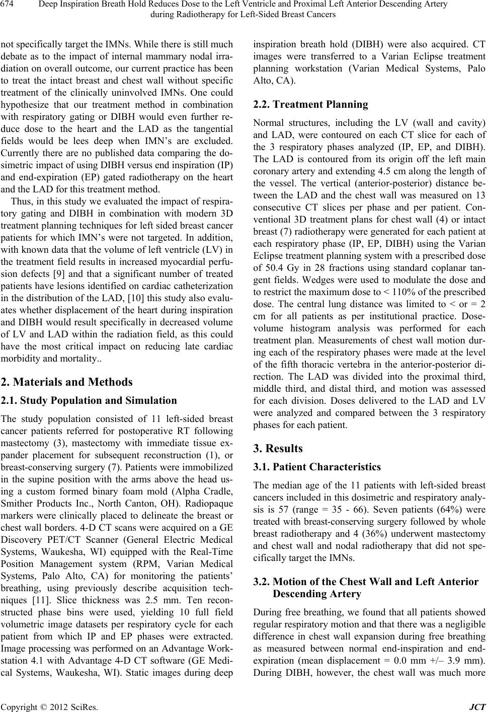
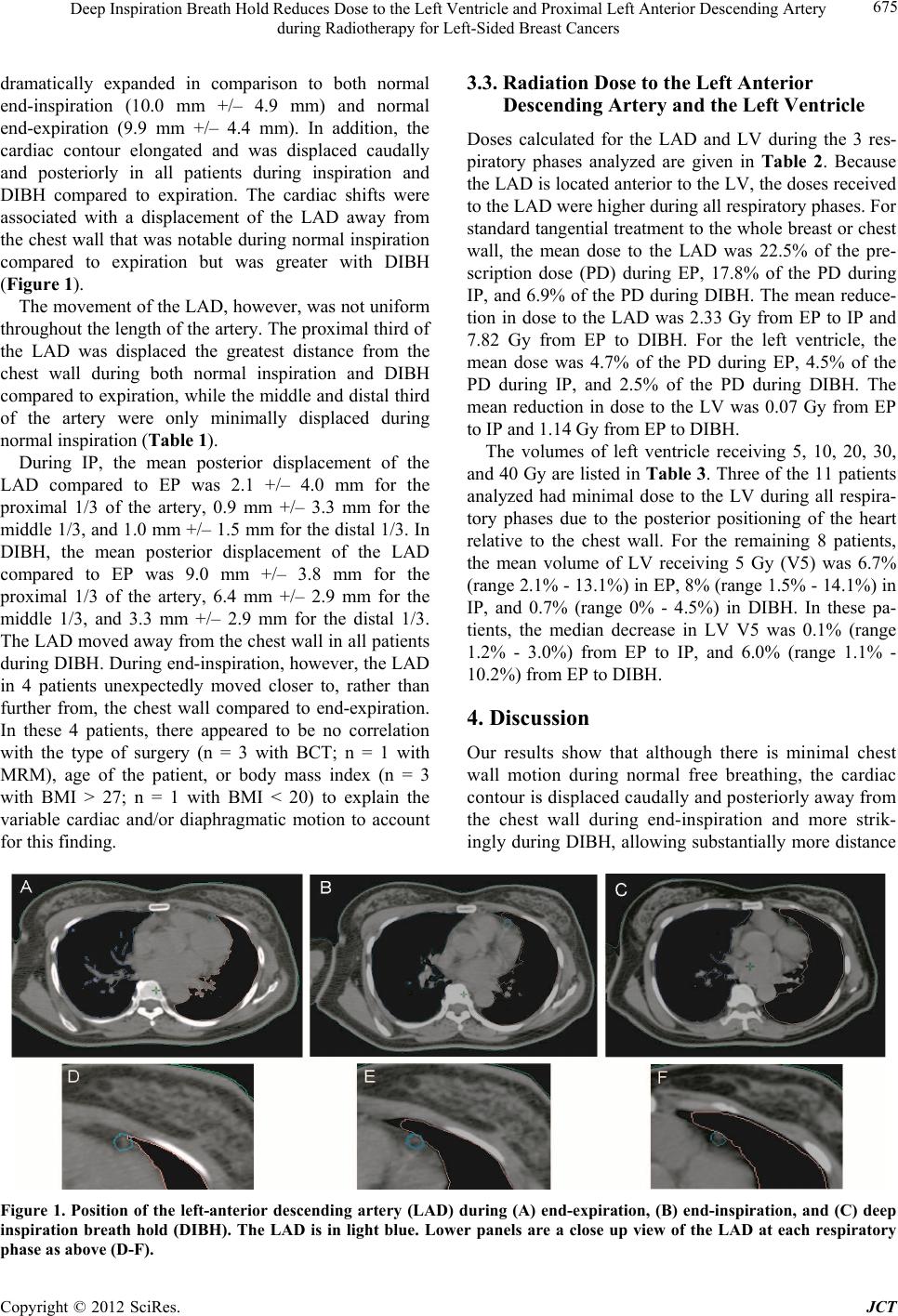
In conclusion, treatment of left-sided breast cancers
during end-inspiration or DIBH can substantially reduce
the radiation dose to the left ventricle and left-anterior
descending artery compared to treatment during end-
expiration. In particular, inspiration and especially DIBH
cause a displacement of the origin and proximal 2/3 of
the LAD away from the chest wall, resulting in the po-
tential to decrease radiation dose to the most critical
segment of the artery during tangent field radiotherapy.
In order to minimize a patient’s long-term risk of coro-
nary artery disease and cardiac morbidity, we recom-
mend tailoring the delivery of left-sided breast radio-
therapy to the patient’s anatomy, using a respiratory-
gated CT for evaluation of heart position at baseline and
the amount of displacement with inspiration or DIBH. In
addition to improved 3D treatment planning techniques,
even patients receiving radiotherapy for left-sided breast
cancers without specific targeting of the IMNs may
benefit from the use of respiratory-gated treatments.
5. Conflict of Interest
We certify that regarding this paper, no actual or poten-
tial conflicts of interests exist; the work is original, has
not been accepted for publication nor is concurrently
under consideration elsewhere, and will not be published
elsewhere without the permission of the Editor. All the
authors have contributed directly to the planning, execu-
tion or analysis of the work reported or to the writing of
the paper.
REFERENCES
[1] Early Breast Cancer Trialists’ Collaborative Group, “Ef-
fect s o f Ra di ot he r apy and Sur gery i n Ea r ly Bre ast Ca nc er .
An Overview of the Randomized Trials,” The New Eng-
land Journal of Medicine, Vol. 333, No. 22, 1995, pp.
1444-1455. doi:10.1056/NEJM199511303332202
[2] M. Clarke, R. Collins, S. Darby, C. Davies, P. Elphin-
stone, et al., “Effects of Radiotherapy and of Differences
in the Extent of Surgery for Early Breast Cancer on Local
Recurrence and 15-Year Survival: An Overview of the
Randomised Trials,” Lancet, Vol. 366, No. 9503, 2005,
pp. 2087-2106.
[3] C. R. Correa, H. I. Litt, W. T. Hwang, V. A. Ferrari, L. J.
Solin, et al., “Coronary Artery Findings after Left-Sided
Compared with Right-Sided Radiation Treatment for Early-
Stage Breast Cancer,” Journal of Clinical Oncology, Vol.
25, No. 21, 2007, pp. 3031-3037.
doi:10.1200/JCO.2006.08.6595
[4] E. E. Harris, C. Correa, W. T. Hwang, J. Liao, H. I. Litt,
et al., “Late Cardiac Mortality and Morbidity in Early-
Stage Breast Cancer Patients after Breast-Conservation
Treatment,” Journal of Clinical Oncology, Vol. 24, No.
25, 2006, pp. 4100-4106.
doi:10.1200/JCO.2005.05.1037
[5] E. A. Krueger, M. J. Schipper, T. Koelling, R. B. Marsh,
J. B. Butler, et al., “Cardiac Chamber and Coronary Ar-
tery Doses Associated with Postmastectomy Radiother-
apy Techniques to the Chest Wall and Regional Nodes,”
International Journal of Radiation Oncology, Biology
and Physics, Vol. 60, No. 4, 2004, pp. 1195-1203.
doi:10.1016/j.ijrobp.2004.04.026
[6] V. M. Remouchamps, F. A. Vicini, M. B. Sharpe, L. L.
Kestin, A. A. Martinez, et al., “Significant Reductions in
Heart and Lung Doses Using Deep Inspiration Breath
Hold with Active Breathing Control and Intensity-
Modulated Radiation Therapy for Patients Treated with
Locoregional Breast Irradiation,” International Journal of
Radiation Oncology, Biology and Physics, Vol. 55, No. 2,
2003, pp. 392-406. doi:10.1016/S0360-3016(02)04143-3
[7] S. S. Korreman, A. N. Pedersen, L. R. Aarup, T. J.
Nottrup, L. Specht, et al., “Reduction of Cardiac and
Pulmonary Complication Probabilities after Breathing
Adapted Radiotherapy for Breast Cancer,” International
Journal of Radiation Oncology, Biology and Physics, Vol.
65, No. 5, 2006, pp. 1375-1380.
doi:10.1016/j.ijrobp.2006.03.046
[8] S. S. Korreman, A. N. Pedersen, T. J. Nottrup, L. Specht,
and H. Nystrom, “Breathing Adapted Radiotherapy for
Breast Cancer: Comparison of Free Breathing Gating
with the Breath-Hold Technique,” Radiotherapy & On-
cology, Vol. 76, No. 3, 2005, pp. 311-318.
doi:10.1016/j.radonc.2005.07.009
[9] L. B. Marks, X. Yu, R. G. Prosnitz, S. M. Zhou, P. H.
Hardenbergh, et al., “The Incidence and Functional Con-
sequences of RT-Associated Cardiac Perfusion Defects,”
International Journal of Radiation Oncology, Biology
and Physics, Vol. 63, No. 1, 2005, pp. 214-223.
doi:10.1016/j.ijrobp.2005.01.029
[10] E. S. Evans, R. G. Prosnitz, X. Yu, S. M. Zhou, D. R.
Hollis, et al., “Impact of Patient-Specific Factors, Irradi-
ated Left Ventricular Volume, and Treatment Set-Up Er-
rors on the Development of Myocardial Perfusion Defects
after Radiation Therapy for Left-Sided Breast Cancer,”
International Journal of Radiation Oncology, Biology
and Physics, Vol. 66, No. 4, 2006, pp. 1125-1134.
doi:10.1016/j.ijrobp.2006.06.025
[11] T. Pan, T. Y. Lee, E. Rietzel and G. T. Chen, “4D-CT
Imaging of a Volume Influenced by Respiratory Motion
on Multi-Slice CT,” Medical Physics, Vol. 31, No. 2,
2004, pp. 333-340. doi:10.1118/1.1639993
[12] S. H. Giordano, Y. F. Kuo, J. L. Freeman, T. A. Buchholz,
G. N. Hortobagyi, et al., “Risk of Cardiac Death after
Adjuvant Radiotherapy for Breast Cancer,” Journal of the
National Cancer Institute, Vol. 97, No. 6, 2005, pp. 419-
424. doi:10.1093/jnci/dji067
[13] L. F. Paszat, W. J. Mackillop, P. A. Groome, K. Schulze
and E. Holowaty, “Mortality from Myocardial Infarction
following Postlumpectomy Radiotherapy for Breast Can-
cer: A Population-Based Study in Ontario, Canada,” In-
ternational Journal of Radiation Oncology, Biology and
Physics, Vol. 43, No. 4, 1999, pp. 755-762.
Copyright © 2012 SciRes. JCT
Deep Inspiration Breath Hold Reduces Dose to the Left Ventricle and Proximal Left Anterior Descending Artery
during Radiotherapy for Left-Sided Breast Cancers
Copyright © 2012 SciRes. JCT
679
doi:10.1016/S0360-3016(98)00412-X
[14] P. H. Hardenbergh, M. T. Munley, G. C. Bentel, R.
Kedem, S. Borges-Neto, et al., “Cardiac Perfusion Changes
in Patients Treated for Breast Cancer with Radiation
Therapy and Doxorubicin: Preliminary Results,” Interna-
tional Journal of Radiation Oncology, Biology and Phys-
ics, Vol. 49, No. 4, 2001, pp. 1023-1028.
doi:10.1016/S0360-3016(00)01531-5
[15] X. Yu, R. R. Prosnitz, S. Zhou, P. H. Hardenberg, A.
Tisch, et al., “Symptomatic Cardiac Events following
Radiation Therapy for Left-Sided Breast Cancer: Possible
Association with Radiation Therapy-Induced Changes in
Regional Perfusion,” Clinical Breast Cancer, Vol. 4, No.
3, 2003, pp. 193-197.
[16] P. A. Lind, R. Pagnanelli, L. B. Marks, S. Borges-Neto, C.
Hu, et al., “Myocardial Perfusion Changes in Patients Ir-
radiated for Left-Sided Breast Cancer and Correlation
with Coronary Artery Distribution,” International Jour-
nal of Radiation Oncology, Biology and Physics, Vol. 55,
No. 4, 2003, pp. 914-920.
doi:10.1016/S0360-3016(02)04156-1
[17] K. S. I. J. Campbell, J. S. Douglas Jr. and J. M. Bradford,
“Prevalence and Distribution of Disease in Patients Ca-
theterized for Suspected Coronary Disease,” McGraw-
Hill, New York, 1985.
[18] V. M. Remouchamps, N. Letts, F. A. Vicini, M. B.
Sharpe, L. L. Kestin, et al., “Initial Clinical Experience
with Moderate Deep-Inspiration Breath Hold Using an
Active Breathing Control Device in the Treatment of Pa-
tients with Left-Sided Breast Cancer Using External
Beam Radiation Therapy,” International Journal of Ra-
diation Oncology, Biology and Physics, Vol. 56, No. 3,
2003, pp. 704-715. doi:10.1016/S0360-3016(03)00010-5
Abbreviations
DIBH: deep-inspiration breath hold; LAD: left anterior
descending artery; LV: left ventricle; EP: end-expiration;
IP: end-inspiration ; IMN: intern al mammary n odes.