 Advances in Bioscience and Biotechnology, 2012, 3, 782-787 ABB http://dx.doi.org/10.4236/abb.2012.326098 Published Online October 2012 (http://www.SciRP.org/journal/abb/) Prevention of beta cell death in chronic pancreatitis Huansheng Dong, Katherine A. Morgan, David B. Adams, Hongjun Wang* Department of Surgery, Medical University of South Carolina, Charleston, USA Email: *wangho@musc.edu Received 29 August 2012; revised 30 September 2012; accepted 6 October 2012 ABSTRACT Chronic pancreatitis is best described as a relentless, continuous inflammatory destruction of the pancreas parenchyma, characterized by irreversible destruc- tion of the exocrine tissues, fibrosis, and at the late stage, the destruction of endocrine cells. Current therapies for chronic pancreatitis patients focus on pain relief by medical and minimally invasive endo- scopic treatment as well as surgical management with resection of diseased parenchyma and drainage of obstructed ducts. Radical treatment of chronic pan- creatitis has been successful with total pancreatic- tomy and islet autotransplantation (TP-IAT) that may prevent maladaptive intractable pain pathways and also avoid pancreatogenic diabetes in the well- selected patient. Distinct loss of pancreatic islet cells occurs in about 30% - 50% of patients during the progression of chronic pancreatitis when severe fi- brosis develops at the late stage of the disease. Pro- found β cell apoptosis induced by stresses encoun- tered during islet isolation and transplantation fur- ther compromises β cell survival and function after TP-IAT. The molecular mechanisms that lead to β cell dysfunction in chronic pancreatitis remain largely undelineated. In this review, we summarize factors that may contribute to β cell apoptosis during the disease progress and after TP-IAT and discuss poten- tial interventional approaches that may prevent islet cell death during these processes. Such information is critical to the development of therapeutic protocols that can preserve the viability and function of β cell in patients with chronic pancreatitis. Keywords: Chronic Pancreatitis; Islet Autotransplantation; Beta Cell Apoptosis; Protective Gene 1. INTRODUCTION Chronic pancreatitis (CP) is characterized by long- standing inflammation of the pancreas that is notable for the development of progressive pain, fibrosis and loss of exocrine and endocrine function [1]. Approximately 15,000 Americans are diagnosed with chronic pancreatic- tis each year. The pathophysiology of chronic pancreatic- tis is dominated by acinar cell death and loss of exocrine function of the pancreas. Endocrine insufficiency that occurs much later in the disease can lead to type 3c dia- betes [2,3]. Current therapies for chronic pancreatitis patients focus on pain relief by medications and a variety of minimally invasive endoscopic procedures as well as surgical treatment with resection of diseased parenchyma and drainage of obstructed ducts [3,4]. Radical treatment of chronic pancreatitis has been successful with total pancreatectomy and islet autotransplantation (TP-IAT) that may prevent maladaptive intractable pain pathways and also avoid pancreatogenic diabetes in the well-se- lected patient [5]. Although the pathogenesis and biological behavior of chronic pancreatitis has been widely studied, mecha- nisms of apoptosis and proliferation of endocrine (e.g., acinar) and endocrine (e.g., insulin-secreting ) cells un- der the conditions of chronic pancreatitis remain poorly defined. Profound apoptosis of acinar cells are observed in chronic pancreatitis patients, while cells have been found less vulnerable than acinar cells at the early stage of chronic pancreatitis [6,7]. Nevertheless, cell apop- tosis over a prolonged period of inflammation that influ- ences insulin secretion develops late in the disease proc- ess. Diabetes occurs in around 30% - 50% of patients diagnosed with long-term chronic pancreatitis [8]. In advanced stages o f chron ic pancreatitis, reductio n of islet cells corresponds with severe cell dysfunction due to prolonged exposur e to inflammator y cytokines, oxid ative stresses and gene dysfunction [7,9]. Moreover, non-im- mune related stresses encountered during islet isolation and transplantatio n also resu lts in a significant nu mber of islets undergoing apoptosis after TP-IAT, which further comprise the function of cells [10]. In this review, we summarize molecular mechanisms that lead to cell dysfunction during the progress of chronic pancreatitis and after TP-IAT and discuss inter- ventional therapies that might improve the viability and function of cells. Understanding such mechanisms will *Corresponding author. OPEN ACCESS  H. S. Dong et al. / Advances in Bioscience and Biotechnology 3 (2012) 782-787 783 not only lead us to a better understanding of the patho- genetic mechanisms leading to cell death in chronic pancreatitis, but also help develop efficient therapeutic treatment protocols for this disease. 2. MECHANISMS OF CELL DEATH DURING THE PROGRESS OF CHRONIC PANCREATITIS Long-term inflammation in patients with chronic pan- creatitis impairs cell function and as many as 30% - 50% of patients develop a type of diabetes that differs from type 1 and type 2 diabetes [8]. The diabetes associ- ated with chronic pancreatitis has been labeled as “type 3c” diabetes [11]. Type 3c diabetes is caused by the apoptosis of β cells and is generally not diag nosed in the early stages of chronic pancreatitis, but manifests itself in a later stages of the disease irrespective of the etiology of chronic pancreatitis [12 ]. At the early stage of chronic pancreatitis, the damage to the pancreas is highly specific for the exocrine com- partment and affects the endocrine islets to a less extent [13]. For example, Fas (CD95)/Fas ligand (CD95L)- mediated islet cell destruction by CD95L expressing cy- totoxic T cells is an important mechanism in the devel- opment of type 1 diabetes, but does not occur at the same extent in chronic pancreatitis patient. Exocrine cells ex- press CD95, and shed their death by binding to the death ligands (CD95L) on infiltration T lymphocytes in the presence of IFN-γ, while islet cells secured themselves by expressing CD95L instead of CD95. Thus, T cells infiltrates are prevalent within acinar cells but rare with in islets in pancreas with chronic pancreatitis [13-15]. Moreover, a strong induction of TNF-related apoptosis- inducing ligand (TRAIL) receptor 1 and 2 (TRAIL-R1 and R2) is observed in exocrine cells, while islet cells only express TRAIL-R4 [15]. TRAIL released locally by activated pancreatic stellate cells binds to TRAIL-R1 and R2 specifically, but not TRAIL-4R, and selectively lead to exocrine apoptosis [16]. In addition, islet cells have been shown to retain their “immune-privileged” status by activated anti-apoptotic programs through NF-B [17- 19]. As the disease progresses, chronic inflammation can lead to β cell apoptosis and dysfunction. Chronic in- flammation causes increasing stress and cytokine secre- tion by both macrophages and T lymphocytes with marked fibrosis that eventually leads to β cell apoptosis and clinical diabetes [20]. The changes in the internal milieu of pancreatic tissue in chronic pancreatitis arising from the chronic inflammation and cytokine release (e.g., IFN-γ, TNF-α) that leads to deranged cellular crosstalk and signaling mechanisms and altered function of islet cells. Qualitative and quantitative changes in cytokine signaling pathways determine the fate of β cell to live and proliferate or to undergo apoptosis [21]. The apop- tosis of β cell are likely caused by the cytokine expres- sion, oxidative stress, and reduced expression of certain genes (e.g., pancreatic duodenal homeobox 1) in the pancreas [22-24]. Apoptosis of β cells, when it occurs, has been ascribed to immune processes initiated by CD8+ T cells or CD4+ T cells dependent on Fas/FasL as well as cytokines (e.g., IFN-γ and TNF-α). Apparently, cellular and molecular events leading to β cell apoptosis represent the adaptive response of normal islets towards the noxious environment caused by proinflammatory cytokines. 3. DEATH OF CELLS AFTER TP-IAT TP-IAT is being increasingly investigated and used as a treatment option for pain relief, and it is also used to prevent pancreatogenic diabetes in chronic pancreatitis [25]. Among all the treatment options, TP-IAT is a safe and effective option for chronic pancreatitis patients and has the potential to eliminate pancreatic pain without total sacrifice of the endocrine function of the pancreas [4,26,27]. Islets transplanted into the liver via the portal vein were found to have improved functions with time [28]. However, even though TP-IAT can improve quality of life and decreased narcotic requirement, more than 60% of patients require long-term insulin treatment [4, 27,29-31]. Non-immune related stresses encountered during islet isolation and transp lantation results in a large number of islets undergoing apoptosis immediately after transplantation, and as many as 50% - 60% of islet cell apoptosis happens at 2 - 3 days post transplantation even under optimal conditions [32,33]. The death of pancre- atic islets are likely due to transplantation-associated stress which include hypoxia, nutrient deprivation, reac- tive oxygen species, pro-inflamma tory cytokines ind uced during harvesting, isolation , and implantation of the islet cell mass [10,34]. 4. APPROACHES TO PROTECT β CELL FROM APOPTOSIS IN CHRONIC PANCREATITIS PATIENT In order to prevent the chronic inflammation in chronic pancreatitis patient, altering the environment where islets reside may help relieve the stress and improve islet sur- vival and function. For example, strategies such as die- tary modification, oral hypoglycemics, and exogenous insulin can help alleviate the stress of β cell, and thus preserve islet fun ction (Figure 1) [35,36]. It has been postulated that patients with chronic pan- creatitis should be considered for TP-IAT before severe islet destruction occurs. Patient islet quality and quantity determines β cell function after transplantation. Insulin Copyright © 2012 SciRes. OPEN ACCESS  H. S. Dong et al. / Advances in Bioscience and Biotechnology 3 (2012) 782-787 784 Figure 1. Approach to improve islet survival and function dur- ing chronic pancreatitis progress and during TP-IAT. requirement is well-known to be associated with lower islet cell yield after transplantation [25,37]. Low islet yields are common in patients with long-term chronic pancreatitis, as inflammation and fibrosis lead to pancre- atic endocrine failure over time [38,39]. In the early stages of chronic pancreatitis, islet/β-cell remains intact morphologically and functionally and pa tients usually do not have diabetes [17]. In contrast, the number of islet cells were shown to be significantly reduced corre- sponding to the stage of the disease before the onset of diabetes [7,9]. Both the exocrine and endocrine tissues have greater destruction in chronic pancreatitis patients compared to patients with minimal duct disease or non- dilated chronic pancreatitis [40]. Thus, islet transplanta- tion early in the progression of chronic pancreatitis can maximally preserve the endocrine function of the islets [37]. 5. APPROACHES THAT CAN IMPROVE THE FUNCTION OF ISLETS DURING TP-IAT Strategies that enable β cell resistance to stresses during TP-IAT would prevent β cell apoptosis, thereby improv- ing clinical application of islet transplantation [4,27,29]. Several approaches are currently being explored to im- prove islet survival after transp lantation including induc- tion of protective genes expression ex vivo in islets dur- ing harvest, physically isolating islets from insults using encapsulation techniques, transplanting islets to a better site that promote survival, and so on (Figure 1). Induction expression of protective gene has been s h o wn able to protect islet cells from stress-induced apoptosis [41]. A protective gene is a gene that is upregulated in response to stress through specific signaling cascades and transcription factor regulation that when induced participate in promoting cell survival [41]. Many protec- tive genes including heme oxygenase (HO-1), A20, B-cell lymphoma 2 (Bcl-2), Bcl-x, heat shock proteins, biliverdin reductase (BVR), and antioxidant enzymes have been found to be expressed in pancreatic islets, and their induction leads to protection against apoptosis and other injuries while their absence leads to a heightened response to stress [41-45]. Most protective genes exert their protection via their anti-inflammatory property and prevention of cell apoptosis. Islet encapsulation with biocompatible materials have been shown function as an immunoisolation approach to facilitate survival of syngeneic, allogeneic and xenoge- neic islets for decades [47-49]. Islet encapsulation can exert both “isolation” and “modulation” effects by physi- cally isolating islets from complement molecules, IgG and host immune cells while delivering cytoprotective molecules locally to the islets to p rotect those islets from stress-induced apoptosis. Islets from chronic pancreatitis patients are extremely fragile due to long-term inflame- mation in the pancreas. Therefore coating islets with nanoparticles loaded with protective molecules may well preserve their function after islet autotransplantatio n. Significant progress has been made in developing suitable materials with reliable biocompatibility, me- chanical and chemical stability, and required permselec- tivity for cell encapsulation [50-53]. Islet encapsulation with microcapsules prevents them from apoptosis [52,54]. Modifying islet surfaces with bioreactive chemicals pre- vents blood-mediated inflammatory responses [55] and prolonged survival of islet allograft [56]. Coating islets with FDA-approved poly(lactide-co-glycolide) (PLGA) nanoparticles increase islet function [57,58]. PLGA has been developed for many years and approved by the FDA for drug delivery based on its biodegradability, drug biocompatibility, suitable biodegradation kinetics and mechanical properties and ease of processing [59-61]. Loading drugs into nanoparticles where drugs are only active in the target area of the body, such as locally to islets, can avoid toxicity and side effects when adminis- tered systemically [62]. In addition, transplanting islets into other sites less stressful may promote their survival. Currently islets are transplanted into the liver of patient via portal vein infu- sion. However, the liver is not the ideal site for islet sur- vival after transplantation due to the instant blood-medi- ated inflammatory reaction (IBMIR), hypoxia and in- flammatory cytokine release by surrounding tissue in- duced by capillary bed occlusion in hepatic microvascu- lature [63,64]. In addition islets implanted in the liver are exposed to a non-native mechanical stress and exposure to toxins filtered through the liver that further impedes islet survival and function [65,66]. Other promising al- ternative sites currently being explored include subcuta- neous, intramuscular, omental, and bone marrow sites [46,67-71]. In conclusion, current work has demonstrated apop- tosis of β cell in patients with chronic pancreatitis d ue to inflammation and inflammatory cytokines. These find- Copyright © 2012 SciRes. OPEN ACCESS  H. S. Dong et al. / Advances in Bioscience and Biotechnology 3 (2012) 782-787 785 ings serve to explain the late onset of diabetes in patients with long-standing chronic pancreatitis. When islets in patients with chronic pancreatitis are autologously trans- planted, additional islet stressors result in β cell loss through apoptosis. Strategies that can prevent β cell death during these processes can benefit patients with chronic pancreatitis and perhaps more importantly lead to new understanding of diabetes of all causes. 6. ACKNOWLEDGEMENTS This study was supported in part by the South Carolina Clinical & Translational Research (SCTR) Institute, with an academic home at the Medical University of South Carolina CTSA, NIH/NC RR Grant Num- ber UL1RR029882 and the JDRF grant 5-2012-149 (to H.W. and D.A.). REFERENCES [1] Talukdar, R., Saikia, N., Singal, D.K. and Tandon, R. (2006) Chronic pancreatitis: Evolving paradigms. Pan- creatology, 6, 440-449. doi:10.1159/000094561 [2] Dixon, E., Graham, J.S., Sutherland, F. and Mitchell, P.C. (2004) Splenic injury following endoscopic retrograde cholangiopancreatography: A case report and review of the literature. Journal of the Society of Laparoendoscopic Surgeons, 8, 275-277. [3] Manciu, N., Beebe, D.S., T ran, P., Gruessner, R., Suther- land, D.E. and Belani, K.G. (1999) Total pancreatectomy with islet cell autotransplantation: Anesthetic implica- tions. Journal of Clinical Anesthesia, 11, 576-582. doi:10.1016/S0952-8180(99)00100-2 [4] Dixon, J., DeLegge, M., Morgan, K.A. and Adams, D.B. (2008) Impact of total pancreatectomy with islet cell transplant on chronic pancreatitis management at a dis- ease-based center. The American Surgeon, 74, 735-738. [5] Wahoff, D.C., Papalois, B. E., Najarian, J.S., et al. (1995) Autologous islet transplantation to prevent diabetes after pancreatic resection. Annals of Surgery, 222, 562-575. [6] Bhanot, U.K. and Moller, P. (2009) Mechanisms of par- enchymal injury and signaling pathways in ectatic ducts of chronic pancreatitis: Implications for pancreatic car- cinogenesis. Laboratory Investigation, 89, 489-497. doi:10.1038/labinvest.2009.19 [7] Schrader, H., et al. (2009) Reduced pancreatic volume and beta-cell area in patients with chronic pancreatitis. Gastroenterology, 136, 513-522. doi:10.1053/j.gastro.2008.10.083 [8] Mitchell, R.M., Byrne, M.F. and Baillie, J. (2003) Pan- creatitis. Lancet, 361, 1447-1455. doi:10.1016/S0140-6736(03)13139-X [9] Mitnala, S., Pondugala, P.K., et al. (2010) Reduced ex- pression of PDX-1 is associated with decreased beta cell function in chronic pancreatitis. Pancreas, 39, 856-862. doi:10.1097/MPA.0b013e3181d6bc69 [10] Davalli, A.M., Scaglia, L., Zangen, D.H., et al. (1996) Vulnerability of islets in the immediate posttransplanta- tion period. Dynamic changes in structure and function, Diabetes, 45, 1161-1167. doi:10.2337/diabetes.45.9.1161 [11] Cui, Y. and Andersen, D.K. (2011) Pancreatogenic dia- betes: Special considerations for management. Pancrea- tology: Official Journal of the International Association of Pancreatology, 11, 279-294. doi:10.1159/000329188 [12] Lankisch, P.G. (2001) Natural course of chronic pan- creatitis. Pancreatology: Official Journal of the Interna- tional Association of Pancreatology, 1, 3-14. doi:10.1159/000055786 [13] Bateman, A.C., Turner, S.M., et al. (2002) Apoptosis and proliferation of acinar and islet cells in chronic pancreati- tis: Evidence for differential cell loss mediating preserva- tion of islet function. Gut, 50, 542-548. doi:10.1136/gut.50.4.542 [14] Hasel, C., Rau, B., et al. (2001) Differential and mutually exclusive expression of CD95 and CD95 ligand in epithe- lia of normal pancreas and chronic pancreatitis. Journal of Technical Methods and Pathology, 81, 317-326. [15] Hasel, C., Durr, S., et al. (2003) In chronic pancreatitis, widespread emergence of TRAIL receptors in epithelia coincides with neoexpression of TRAIL by pancreatic stellate cells of early fibrotic areas. Journal of Technical Methods and Pathology, 83, 825-836. [16] Lamhamedi-Cherradi, S.E., Zheng, S., Tisch, R.M. and Chen, Y.H. (2003) Critical roles of tumor necrosis fac- tor-related apoptosis-inducing ligand in type 1 diabetes. Diabetes, 52, 2274-2278. doi:10.2337/diabetes.52.9.2274 [17] Bhanot, U.K. and Moller, P. (2009) Mechanisms of par- enchymal injury and signaling pathways in ectatic ducts of chronic pancreatitis: Implications for pancreatic car- cinogenesis. Journal of Technical Methods and Patho- logy, 89, 489-497. [18] Degli-Esposti, M.A., Dougall, W.C., et al. (1997) The novel receptor TRAIL-R4 induces NF-kappaB and pro- tects against TRAIL-mediated apoptosis, yet retains an incomplete death domain. Immunity, 7, 813-820. doi:10.1016/S1074-7613(00)80399-4 [19] Chen, X., Ji, B., Han, B., Ernst, S.A., Simeone, D. and Logsdon, C.D. (2002) NF-kappaB activation in pancreas induces pancreatic and systemic inflammatory response. Gastroenterology, 122, 448-457. doi:10.1053/gast.2002.31060 [20] Butler, A.E., Janson, J. , et al. (2003) Beta-cell de ficit and increased beta-cell apoptosis in humans with type 2 dia- betes. Diabetes, 52, 102-110. doi:10.2337/diabetes.52.1.102 [21] Donath, M.Y., Storling, J., et al. (2008) Cytokines and beta-cell biology: From concept to clinical translation. Endocrine Reviews, 29, 334-350. doi:10.1210/er.2007-0033 [22] Tandon, R.K. and Garg, P.K. (2011) Oxidative stress in chronic pancreatitis: Pathophysiological relevance and management. Antioxidants & Redox Signaling, 15, 2757- 2766. doi:10.1089/ars.2011.4115 [23] Pavan Kumar, P., Radhika, G., et al. (2012) Interferon Copyright © 2012 SciRes. OPEN ACCESS  H. S. Dong et al. / Advances in Bioscience and Biotechnology 3 (2012) 782-787 786 gamma and glycemic status in diabetes associated with chronic pancreatitis. Pancreatology, 12, 65-70. [24] Mitnala, S., Pondugala, P.K., et al. (2010) Reduced ex- pression of PDX-1 is associated with decreased beta cell function in chronic pancreatitis. Pancreas, 39, 856-862. doi:10.1097/MPA.0b013e3181d6bc69 [25] Blondet, J.J., Carlson, A.M., et al. (2007) The role of total pancreatectomy and islet autotransplantation for chronic pancreatitis. The Surgical clinics of North Ame- rica, 87, 1477-1501. doi:10.1016/j.suc.2007.08.014 [26] Argo, J.L., Contreras, J.L., Wesley, M.M. and Christein, J.D. (2008) Pancreatic resection with islet cell autotrans- plant for the treatment of severe chronic pancreatitis. The American Surgeon, 74, 530-536. [27] Nath, D.S., Kellogg, T.A. and Sutherland, D.E. (2004) Total pancreatectomy with intraportal auto-islet trans- plantation using a temporarily exteriorized omental vein. Journal of the American College of Surgeons, 199, 994- 995. doi:10.1016/j.jamcollsurg.2004.07.033 [28] Bellin, M.D. and Sutherland, D.E. (2010) Pediatric islet autotransplantation: Indication, technique, and outcome. Current Diabetes Reports, 10, 326-331. doi:10.1007/s11892-010-0140-4 [29] Kobayashi, T., Manivel, J.C., et al. (2011) Correlation of histopathology, islet yield, and islet graft function after islet autotransplantation in chronic pancreatitis. Pancreas, 40, 193-199. doi:10.1097/MPA.0b013e3181fa4916 [30] Farney, A.C., Najarian, J.S., et al. (1991) Autotransplan- tation of dispersed pancreatic islet tissue combined with total or near-total pancreatectomy for treatment of chro- nic pancreatitis. Surgery, 110, 427-437. [31] Robertson, R.P., Lanz, K.J., et al. (2001) Prevention of diabetes for up to 13 years by autoislet transplantation after pancreatectomy for chronic pancreatitis. Diabetes, 50, 47-50. doi:10.2337/diabetes.50.1.47 [32] Biarnes, M., Montolio, M., et al. (2002) Beta-cell death and mass in syngeneically transplanted islets exposed to short- and long-term hyperglycemia. Diabetes, 51, 66-72. doi:10.2337/diabetes.51.1.66 [33] Ahn, Y.B., Xu, G., et al. (2007) Changes in gene expres- sion in beta cells after islet isolation and transplantation using laser-capture microdissection. Diabetologia, 50, 334-342. doi:10.1007/s00125-006-0536-5 [34] Davalli, A.M., Scaglia, L., et al. (1995) Early changes in syngeneic islet grafts: effect of recipient’s metabolic con- trol on graft outcome. Transplantation Proceedings, 27, 3238-3239. [35] Bhardwaj, P., Thareja, S., et al. (2004) Micronutrient antioxidant intake in patients with chronic pancreatitis, Tropical gastroenterology. Journal of the Digestive Dis- eases Foundation, 25, 69-72. [36] Hsu, J.T., Yeh, C.N., et al. (2005) Outcome of pancreati- coduodenectomy for chronic pancreatitis. Journal of the Formosan Medical Association, 104, 811-815. [37] Ong, S.L., Gravante, G., et al. (2009) Total pancreatec- tomy with islet autotransplantation: an overview. Journal of the International Hepato Pancreato Biliary Associa- tion, 11, 613-621. doi:10.1111/j.1477-2574.2009.00113.x [38] Howes, N., Lerch, M.M., et al. (2004) Clinical and ge- netic characteristics of hereditary pancreatitis in Europe. Clinical Gastroenterology and Hepatology, 2, 252-261. doi:10.1016/S1542-3565(04)00013-8 [39] Ammann, R.W. (2006) Diagnosis and management of chronic pancreatitis: Current knowledge. Swiss Medical Weekly, 136, 166-174. [40] De Cock, H.E., Forman, M.A., et al. (2007) Prevalence and histopathologic characteristics of pancreatitis in cats. Veterinary Pathology, 44, 39-49. doi:10.1354/vp.44-1-39 [41] Wang, H., Ferran, C., et al. (2011) Induction of protective genes leads to islet survival and function. Journal of Transplantation, 2011, 141898. doi:10.1155/2011/141898 [42] Wang, H., Wu, H., et al. (2012) Activation of peroxisome proliferator-activated receptor gamma prolongs islet al- lograft survival, cell transplantation. Cell Transplant, 21, 2111-2118. [43] Emamaullee, J.A. and Shapiro, A.M. (2006) Interven- tional strategies to prevent beta-cell apoptosis in islet transplantation. Diabetes, 55, 1907-1914. doi:10.2337/db05-1254 [44] Grey, S.T., Arvelo, M.B., et al. (1999) A20 inhibits cyto- kine-induced apoptosis and nuclear factor kappaB-de- pendent gene activation in islets. The Journal of Experi- mental Medicine, 190, 1135-1146. doi:10.1084/jem.190.8.1135 [45] Riachy, R., Vandewalle, B., et al. (2002) 1,2,5-dihy- droxyvitamin D3 protects RINm5F and human islet cells against cytokine-induced apoptosis: Implication of the antiapoptotic protein A20. Endocrinology, 143, 4809- 4819. doi:10.1210/en.2002-220449 [46] Christoffersson, G., Carlsson, P.O. and Phillipson, M. (2011) Intramuscular islet transplantation promotes re- stored islet vascularity. Islets, 3, 69-71. doi:10.4161/isl.3.2.14997 [47] Basta, G. and Calafiore, R. (2011) Immunoisolation of pancreatic islet grafts with no recipient’s immunosup- pression: Actual and future perspectives. Current Diabe- tes Reports, 11, 384-391. doi:10.1007/s11892-011-0219-6 [48] Fort, A., Fort, N., et al. (2008) Biohybrid devices and encapsulation technologies for engineering a bioartificial pancreas. Cell Transplantation, 17, 997-1003. doi:10.3727/096368908786991498 [49] Giraldo, J.A., Weaver, J.D. and Stabler, C.L. (2010) Tis- sue engineering approaches to enhancing clinical islet transplantation through tissue engineering strategies. Journal of Diabetes Science and Technology, 4, 1238- 1247. [50] Trivedi, N., Keegan, M., et al. (2001) Islets in alginate macrobeads reverse diabetes despite minimal acute insu- lin secretory responses. Transplantation, 71, 203-211. doi:10.1097/00007890-200101270-00006 [51] van Schilfgaarde, R. and de Vos, P. (1999) Factors influ- Copyright © 2012 SciRes. OPEN ACCESS  H. S. Dong et al. / Advances in Bioscience and Biotechnology 3 (2012) 782-787 Copyright © 2012 SciRes. 787 OPEN ACCESS encing the properties and performance of microcapsules for immunoprotection of pancreatic islets. Journal of Molecular Medicine, 77, 199-205. doi:10.1007/s001090050336 [52] Sandberg, J.O., Olsson, N., et al. (1995) Immunosuppres- sion, macroencapsulation and ultraviolet-B irradiation as immunoprotection in porcine pancreatic islet xenotrans- plantation. Pharmacology & Toxicology, 76, 400-405. doi:10.1111/j.1600-0773.1995.tb00169.x [53] Pollok, J.M., Ibarra, C., et al. (1998) Immuno-isolation of xenogenic islands of Langerhans in a tissue engineered autologous cartilage capsule. Zentralblatt fur Chirurgie, 123, 830-833. [54] Qi, M., Gu, Y., et al. (2004) PVA hydrogel sheet macro- encapsulation for the bioartificial pancreas. Biomaterials, 25, 5885-5892. doi:10.1016/j.biomaterials.2004.01.050 [55] Teramura, Y. and Iwata, H. (2008) Islets surface modifi- cation prevents blood-mediated inflammatory responses. Bioconjugate Chemistry, 19, 1389-1395. doi:10.1021/bc800064t [56] Lee, D.Y., Park, S.J., et al. (2007) Highly poly(ethylene) glycolylated islets improve long-term islet allograft sur- vival without immunosuppressive medication. Tissue En- gineering, 13, 2133-2141. doi:10.1089/ten.2006.0009 [57] Basarkar, A. and Singh, J. Poly (2009) (lactide-co-gly- colide)-polymethacrylate nanoparticles for intramuscular delivery of plasmid encoding interleukin-10 to prevent autoimmune diabetes in mice. Pharmaceutical Research, 26, 72-81. [58] Mao, G.H., Chen, G.A., et al. (2009) The reversal of hyperglycaemia in diabetic mice using PLGA scaffolds seeded with islet-like cells derived from human embry- onic stem cells. Biomaterials, 30, 1706-1714. doi:10.1016/j.biomaterials.2008.12.030 [59] Jain, R., Shah, N.H., et al. (1998) Controlled drug deli- very by biodegradable poly(ester) devices: Different pre- parative approaches. Drug Development and Industrial Pharmacy, 24, 703-727. doi:10.3109/03639049809082719 [60] Jain, R.A. (2000) The manufacturing techniques of vari- ous drug loaded biodegradable poly(lactide-co-glycolide) (PLGA) devices. Biomaterials, 21, 2475-2490. doi:10.1016/S0142-9612(00)00115-0 [61] Lu, J.M., Wang, X., et al. (2009) Current advances in research and clinical applications of PLGA-based nano- technology. Expert Review of Molecular Diagnostics, 9, 325-341. doi:10.1586/erm.09.15 [62] Athanasiou, K.A., Niederauer, G.G. and Agrawal, C.M. (1996) Sterilization, toxicity, biocompatibility and clini- cal applications of polylactic acid/polyglycolic acid co- polymers. Biomaterials, 17, 93-102. doi:10.1016/0142-9612(96)85754-1 [63] Yin, D., Ding, J.W., et al. (2006) Liver ischemia contri- butes to early islet failure following intraportal transplant- tation: Benefits of liver ischemic-preconditioning. Ameri- can Journal of Transplantation, 6, 60-68. doi:10.1111/j.1600-6143.2005.01157.x [64] Wilson, J.T. and Chaikof, E.L. (2008) Thrombosis and inflammation in intraportal islet transplantation: A review of pathophysiology and emerging therapeutics. Journal of Diabetes Science and Technology, 2, 746-759. doi:10.1097/01.tp.0000290388.70019.6e [65] Cardani, R., Pileggi, A., et al. (2007) Allosensitization of islet allograft recipients. Transplantation, 84, 1413-1427. [66] Rickels, M.R., Kamoun, M., et al. (2007) Evidence for allograft rejection in an islet transplant recipient and ef- fect on beta-cell secretory capacity. The Journal of Cli- nical Endocrinology and Metabolism, 92, 2410-2414. doi:10.1210/jc.2007-0172 [67] Lund, T., Korsgren, O., et al. (2010) Sustained reversal of diabetes following islet transplantation to striated muscu- lature in the rat. The Journal of Surgical Research, 160, 145-154. doi:10.1016/j.jss.2008.11.009 [68] Salazar-Banuelos, A., Wright, J.R., et al. (2008) Pancre- atic islet transplantation into the bone marrow of the rat. American Journal of Surgery, 195, 674-678. doi:10.1016/j.amjsurg.2007.12.040 [69] Berman, D.M., O’Neil, J.J., et al. (2009) Long-term sur- vival of nonhuman primate islets implanted in an omental pouch on a biodegradable scaffold. American Journal of Transplantation, 9, 91-104. doi:10.1111/j.1600-6143.2008.02489.x [70] van der Windt, D.J., Echeverri, G.J., et al. (2008) The choice of anatomical site for islet transplantation. Cell Transplantation, 17, 1005-1014. doi:10.3727/096368908786991515 [71] Pileggi, A., Molano, R.D., et al. (2006) Reversal of dia- betes by pancreatic islet transplantation into a subcuta- neous, neovascularized device. Transplantation, 81, 1318- 1324. doi:10.1097/01.tp.0000203858.41105.88
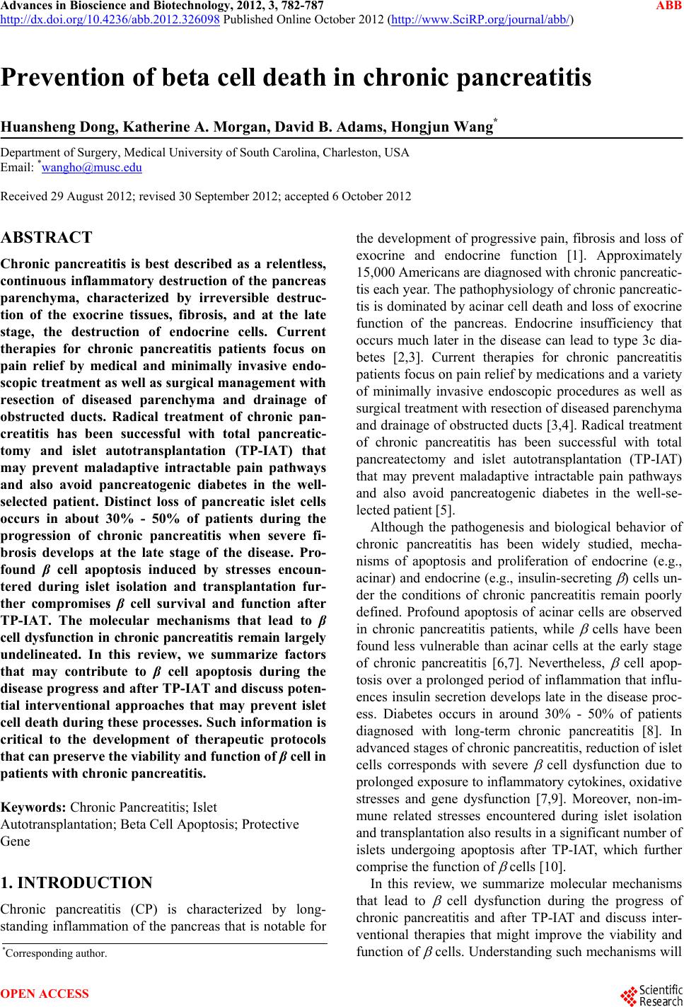
|