H. SAĞLAM ET AL.
Copyright © 2012 SciRes. OJU
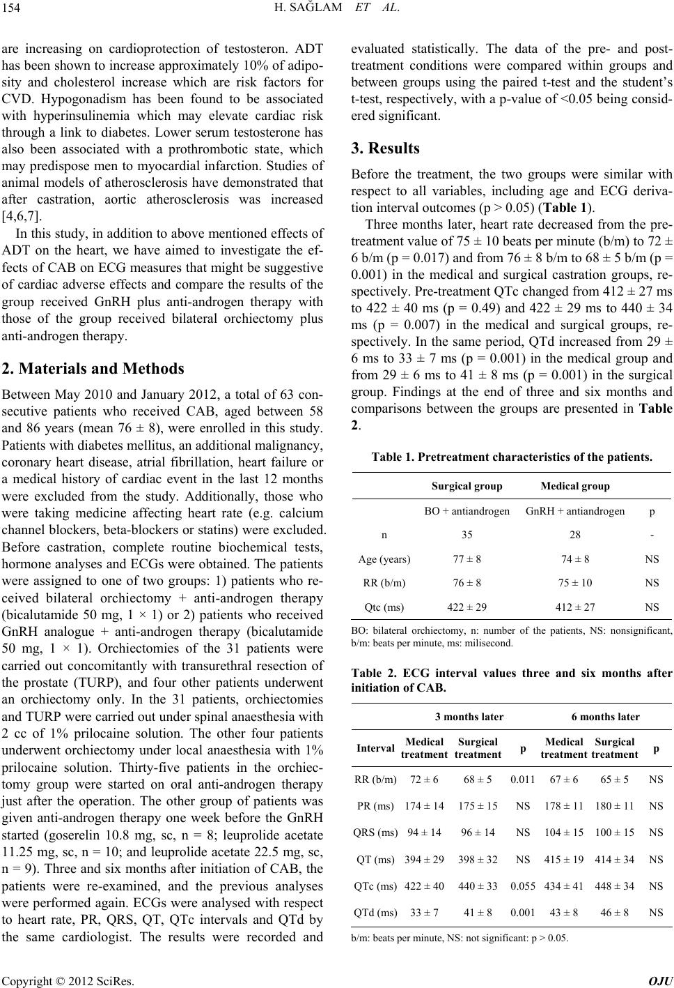
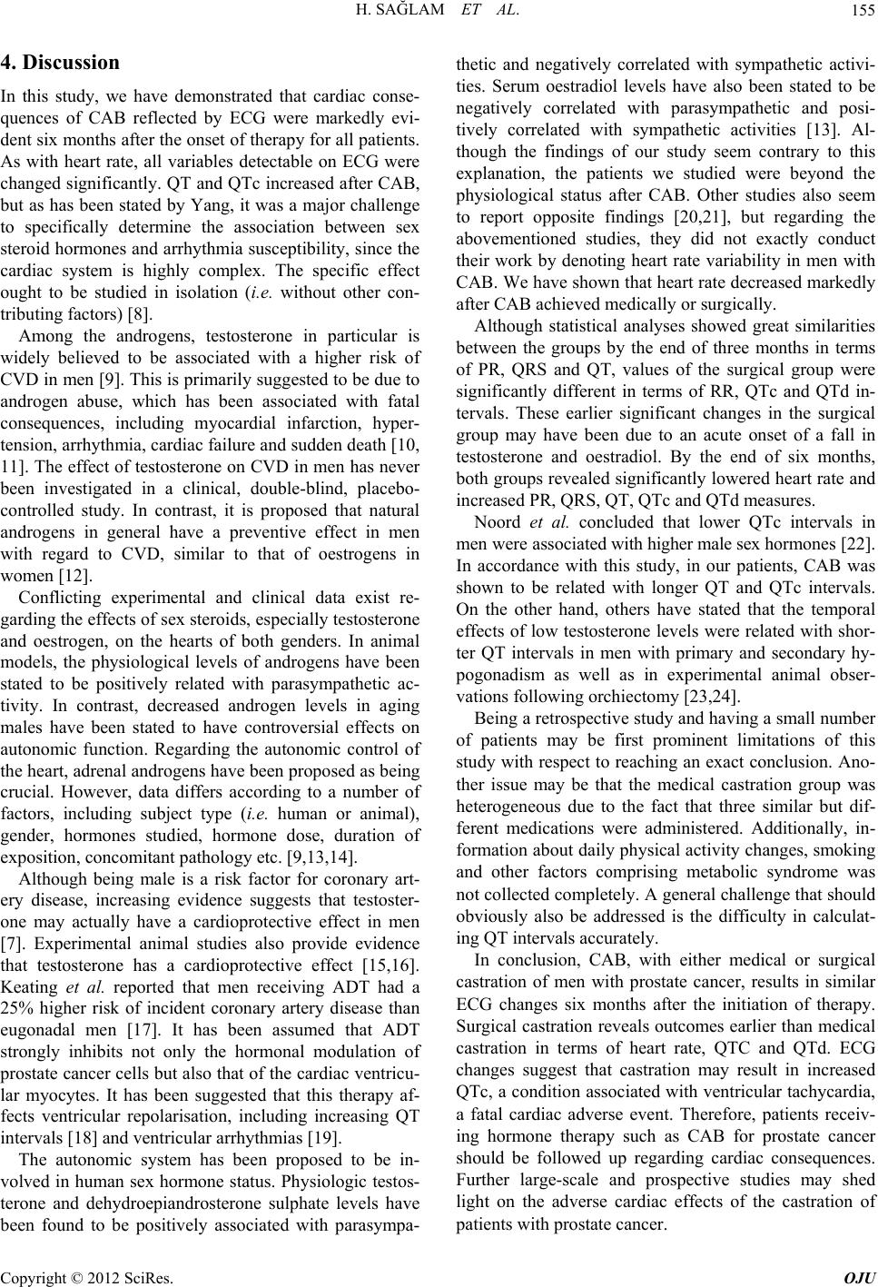
156
REFERENCES
[1] A. Jemal, R. Siegel, E. Ward, T. Murray, J. Xu and M. J.
Thun, “Cancer Statistics,” CA Cancer Journal of Clini-
cians, Vol. 57, No. 1, 2007, pp. 43-66.
doi:10.3322/canjclin.57.1.43
[2] A. Jemal, F. Bray, M. Center, J. Ferlay, E. Ward and D.
Forman, “Global Cancer Statistics,” CA Cancer Journal
of Clinicians, Vol. 61, No. 2, 2011, pp. 69-90.
doi:10.3322/caac.20107
[3] A. Karl and B. Konety, “Androgen Deprivation Therapy
for Prostate Cancer: Indications, Contraindications and
Possible Consequences,” F1000 Medicine Reports, 2009.
http://www.ncbi.nlm.nih.gov/pmc/articles/PMC2920692
[4] G. W. Chodak, T. Keane and L. Klotz, “Critical Evalua-
tion of Hormonal Therapy for Carcinoma of the Prostate,”
Urology, Vol. 60, No. 2, 2002, pp. 201-208.
doi:10.1016/S0090-4295(02)01677-1
[5] V. B. Shahinian, Y. F. Kuo, J. L. Freeman and J. S.
Goodwin, “Risk of Fracture after Androgen Deprivation
for Prostate Cancer,” The New England Journal of Medi-
cine, Vol. 352, No. 2, 2005, pp. 154-164.
doi:10.1056/NEJMoa041943
[6] F. A. S. Chutz and W. K. Oh, “Neoadjuvant and Adjuvant
Therapies in Prostate Cancer,” Urologic Clinics of North
America, Vol. 37, No. 1, 2010, pp. 97-104.
doi:10.1016/j.ucl.2009.11.012
[7] C. S. Saigal, J. L. Gore, T. L. Krupski, J. Hanley, M. Schon-
lau and M. S. Litwin, “Androgen Deprivation Therapy
Increases Cardiovascular Morbidity in Men with Prostate
Cancer,” Cancer, Vol. 110, No. 7, 2007, pp. 1493-1500.
doi:10.1002/cncr.22933
[8] P.-C. Yang, J. Kurokawa, T. Furukawa and C. E. Clancy,
“Acute Effects of Sex Steroid Hormones on Susceptibility
to Cardiac Arrhythmias: A Simulation Study,” PLOS
Computational Biology, Vol. 6, No. 1, 2010, Article ID:
e1000658.
http://www.ncbi.nlm.nih.gov/pmc/articles/PMC2813260
[9] M. F. Kalin and B. Zumoff, “Sex Hormones and Cor-
onary Disease: A Review of the Clinical Studies,” Ster-
oids, Vol. 55, No. 8, 1990, pp. 30-52.
doi:10.1016/0039-128X(90)90058-J
[10] W. Weidemann and H. Hanke, “Cardiovascular Effects of
Androgens,” Cardiovascular Drug Reviews, Vol. 20, No.
3, 2002, pp. 175-198.
doi:10.1111/j.1527-3466.2002.tb00086.x
[11] R. W. Rockhold, “Cardiovascular Toxicity of Anabolic
Steroids,” Annual Review of Pharmacology and Toxi-
cology, Vol. 33, 1993, pp. 497-520.
doi:10.1146/annurev.pa.33.040193.002433
[12] P. Alexandersen, J. Haarbo, I. Byrjalsen, H. Lawaetz and
C. Christiansen, “Natural Androgens Inhibit Male Ather-
osclerosis: A Study in Castrated, Cholesterol-Fed Rab-
bits,” Circulation Research, Vol. 87, No. 7, 1999, pp.
813-819. doi:10.1161/01.RES.84.7.813
[13] M. T. Doğru, M. M. Başar, E. Yuvanç, V. Simşek and O.
Sahin, “The Relationship between Serum Sex Steroid Le-
vels and Heart Rate Variability Parameters in Males and
the Effect of Age,” Turk Kardiyol Dern Ars, Vol. 38, No.
7, 2010, pp. 459-465.
[14] T. M. Saleh, A. E. Cribb and B. J. Connell, “Role of Es-
trogen in Central Nuclei Mediating Stroke-Induced
Changes in Autonomic Tone,” Journal of Stroke and
Cerebrovascular Diseases, Vol. 12, No. 9, 2003, pp. 182-
195. doi:10.1016/S1052-3057(03)00080-6
[15] S. Tsang, S. Wu, J. Liu and T. M. Wong, “Testosterone
Protects Rat Hearts against Ischaemic Insults by Enhanc-
ing the Effects of Alpha(1)-Adrenoceptor Stimulation,”
British Journal of Pharmacology, Vol. 153, No. 4, 2008,
pp. 693-709. doi:10.1038/sj.bjp.0707624
[16] P. Y. Liu, A. K. Death and D. J. Handelsman, “Andro-
gens and Cardiovascular Disease,” Endocrine Reviews,
Vol. 24, No. 3, 2003, pp. 313-340.
doi:10.1210/er.2003-0005
[17] N. L. Keating, A. J. O’Malley and M. R. Smith, “Diabe-
tes and Cardiovascular Disease during Androgen Depri-
vation Therapy for Prostate Cancer,” Journal of Clinical
Oncology, Vol. 24, No. 27, 2006, pp. 4448-4456.
doi:10.1200/JCO.2006.06.2497
[18] K. Ezaki, M. Nakagawa, Y. Taniguchi, Y. Nagano, Y.
Teshima, K. Yufu, et al., “Gender Differences in the ST
Segment: Effect of Androgen-Deprivation Therapy and
Possible Role of Testosterone,” Circulation Journal, Vol.
74, No. 11, 2010, pp. 2448-2454.
doi:10.1253/circj.CJ-10-0221
[19] S. M. Al-Khatib, N. M. LaPointe, J. M. Kramer and R. M.
Califf, “What Clinicians Should Know About the QT
Interval,” The Journal of the American Medical Asso-
ciation, Vol. 289, No. 16, 2003, pp. 2120-2127.
doi:10.1001/jama.289.16.2120
[20] M. Zitzmann and E. Nieschlag, “Androgen Receptor Gene
CAG Repeat Length and Body Mass Index Modulate the
Safety of Long-Term Intramuscular Testosterone Unde-
canoate Therapy in Hypogonadal Men,” The Journal of
Clinical Endocrinology & Metabolism, Vol. 92, No. 10,
2007, pp. 3844-3853. doi:10.1210/jc.2007-0620
[21] S. N. Culos-Reed, J. L. Robinson, H. Lau, K. O’Connor
and M. R. Keats, “Benefits of a Physical Activity Inter-
vention for Men with Prostate Cancer,” Journal of Sport &
Exercise Psychology, Vol. 25, No. 1, 2007, pp. 118-127.
[22] C. van Noord, M. Dörr, M. C. Sturkenboom, S. M. Straus,
T. Reffelmann, S. B. Felix, et al., “The Association of
Serum Testosterone Levels and Ventricular Repolariza-
tion,” European Journal of Epidemiology, Vol. 25, No. 1,
2010, pp. 21-28. doi:10.1007/s10654-009-9406-z
[23] G. F. Pecori, P. M. Toja, B. Filippini, J. Michailidis, M.
Scacchi, M. S. Badiale, et al., “Increased Prevalence of
Prolonged QT Interval in Males with Primary or Second-
ary Hypogonadism: A Pilot Study,” International Journal
of Andrology, Vol. 33, No. 1, 2010, pp. e132-e138.
doi:10.1111/j.1365-2605.2009.00985.x
[24] A. Kirilmaz, E. Bolu, F. Kilicaslan, K. Erinc, M. Uzun, E.
Isik, et al., “Comparison of Electrocardiographic Repo-
larization Patterns between Hypogonad Males and Nor-
mal Subjects,” Annals of Noninvasive Electrocardiology,
Vol. 8, No. 4, 2003, pp. 284-288.
doi:10.1046/j.1542-474X.2003.08404.x