J. D. FISHER ET AL.
138
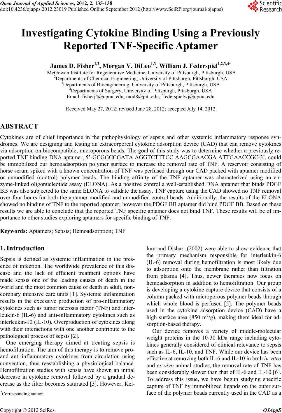
4. Discussion
The ability of CADs packed with aptamer-immobilized
or unmodified beads to capture TNF from horse serum
was tested. The results in Figure 1 show that TNF cap-
ture with unmodified and aptamer-immobilized beads
was negligible. The aptamer-immobilized on the surface
of the PSDVB beads was reported to specifically bind
TNF, therefore we expected that the aptamer-immobi-
lized beads would display a significantly higher ability to
capture TNF than the control beads. Based on the surface
density of carboxyl groups on the beads, we calculated
that if successfully coupled there would be at least a 10
molar excess of aptamer to TNF. Therefore, one possible
explanation is that the TNF aptamer was not successfully
coupled to the surface of the PSDVB beads. This is
unlikely however, as we have successfully coupled anti-
bodies to the PSDVB beads using the same chemistry.
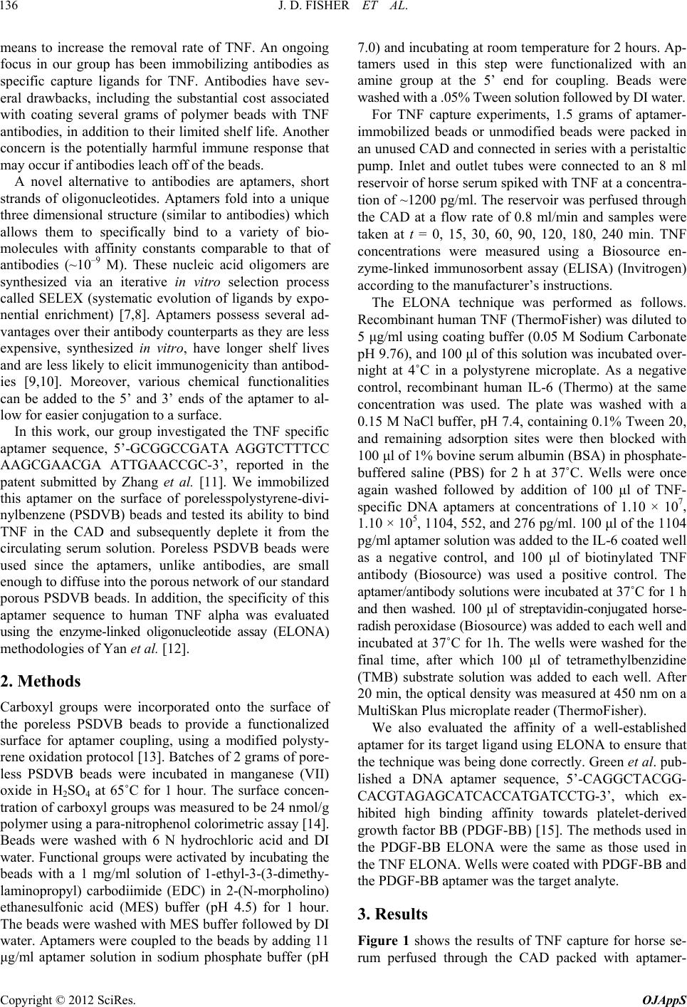
Another possible explanation was that the reported ap-
tamer did not bind to TNF. To characterize the affinity of
the published aptamer for TNF, we utilized a previously
reported enzyme-linked oligonucleotide assay (ELONA)
[12].
From the ELONA data we are able to conclude that
the TNF aptamer sequence does not specifically bind
TNF. There are several possible explanations for this
finding. The discrepancies in data could be a result of
differences in the protein at which the aptamer was tar-
geted. Our group used commercially available recombi-
nant human TNF from ThermoFisher Scientific, but the
recombinant protein used by Zhang et al. was produced
in their laboratory. The target proteins were synthesized
in different environments, which may suggest that the
three dimensional structure of the proteins may have dif-
fered enough to impact the aptamer’s affinity toward
TNF. The TNF used in our work was in its correct three-
dimensional shape, as evidenced by our positive control,
a TNF antibody, being able to bind TNF in the ELONA.
A TNF antibody was not used as a positive control in the
group’s patent or published description of the RNA ap-
tamer [11,12].
The aptamer sequence, 5’-GCGGCCGATA AGGTC-
TTTCC AAGCGAACGA ATTGAACCGC-3’, reported
by Zhang and coworkers does not appear to bind com-
mercially available recombinant TNF. While this result is
a negative finding, we believe that this correspondence
provides important data to other investigators who may
be studying specific ligands for TNF.
REFERENCES
[1] M. Schoenberg, M. Weiss and P. Radermacher, “Out-
come of Patients with Sepsis and Septic Shock after ICU
Treatment,” Langenbeck’s Archives Surgery, Vol. 383,
No. 1, 1998, pp. 44-48. doi:10.1007/s004230050090
[2] J. A. Kellum, L. Kong, M. P. Fink, L. A. Weissfield, D.
M. Yealy, M. R. Pinsky, J. Fine, A. Krichevsky, R. L.
Delude and D. C. Angus, “Understanding the Inflamma-
tory Cytokine Response in Pneumonia and Sepsis,” Ar-
chives of International Medicine, Vol. 167, No. 15, 2007,
pp. 1655-1663. doi:10.1001/archinte.167.15.1655
[3] J. Kellum, “Immunomodulation in Sepsis: The Role of
Hemofiltration,” Minerva Anestesiologica, Vol. 65, No. 6,
1999, pp. 410-418.
[4] J. A. Kellum and M. K. Dishart, “Effect of Hemofiltration
Filter Adsorption on Circulating IL-6 Levels in Septic
Rats,” Critical Care, Vol. 6, No. 5, 2002, pp. 429-433.
doi:10.1186/cc1528
[5] M. Song, J. Winchester, R. L. Albright, V. J. Capponi, M.
D. Choquette and J. A. Kellum, “Cytokine Removal with
a Novel Adsorbent Polymer,” Blood Purification, Vol. 22,
No. 5, 2004, pp. 428-434. doi:10.1159/000080235
[6] M. V. DiLeo, J. Kellum and W. J. Federspiel, “A Simple
Mathematical Model of Cytokine Capture Using a He-
moadsorption Device,” Annals of Biomedical Engineer-
ing, Vol. 37, No. 1, 2009, pp. 222-229.
doi:10.1007/s10439-008-9587-8
[7] R. Stoltenburg, C. Reinemann and B. Strehlitz, “SELEX—
A (r)Evolutionary Method to Generate High-Affinity Nu-
cleic Acid Ligands,” Biomolecular Engineering, Vol. 24,
No. 4, 2007, pp. 381-403. doi:10.1016/j.bioeng.2007.06.001
[8] S. J. Klug and M. Famulok, “All You Wanted to Know
about SELEX,” Molecular Biology Reports, Vol. 20, No.
2, 1994, pp. 97-107. doi:10.1007/BF00996358
[9] X. Yang and D. G. Gorenstein, “Progress in Thioaptamer
Development,” Current Drug Targets, Vol. 5, No. 8, 2004,
pp. 705-715. doi:10.2174/1389450043345074
[10] J. W. Guthrie, C. L. A. Hamula, H. Zhang and X. C. Le,
“Assays for Cytokines Using Aptamers,” Methods, Vol. 38,
No. 4, 2006, pp. 324-330. doi:10.1016/j.ymeth.2006.01.001
[11] Z. Zhang, X. Yan and C. Xu, “Oligonuecleotide Antago-
nist for Human Tumor Necrosis Factor A (TNF-a),” In-
stitute for Viral Disease Control and Prevention Chinese
Center for Disease Control and Prevention, United States,
2007.
[12] X. Yan, X. Gao and Z. Zhang, “Isolation and Characteri-
zation of 2’-Amino-Modified RNA Aptamers for Human
TNFa,” Genomics Proteomics & Bioinformatics, Vol. 2,
No. 1, 2004, pp. 32-42.
[13] N. Zammatteo, C. Girardeaux, D. Delforge, J.-J. Pireaux
and J. Remacle, “Amination of Polystyrene Microwells:
Application to the Covalent Grafting of DNA Probes for
Hybridization Assays,” Analytical Biochemistry, Vol. 236,
No. 1, 1996, pp. 85-94. doi:10.1006/abio.1996.0135
[14] M. Matteucci and M. Caruthers, “Synthesis of Deoxyoli-
gonucleotides on a Polymer Support,” Journal of the Ameri-
can Chemical Society, Vol. 103, No. 11, 1981, pp. 3185-
3191. doi:10.1021/ja00401a041
[15] L. S. Green, D. Jellinek, R. Jenison, A. Ostman, C.-H.
Heldin and N. Janjic, “Inhibitory DNA Ligands to Plate-
let-Derived Growth Factor B-Chain,” Biochemistry, Vol.
35, No. 45, 1996, pp. 14413-14424.
doi:10.1021/bi961544+
Copyright © 2012 SciRes. OJAppS