T. CORDOVA-FRAGA ET AL. 133
the lungs in expanding work as a reservoir (increase the
volume of pulmonary blood), so that, left ventricular filling
does not increase during inspiration. During aspiration,
however, is produced the opposite effect: the thoracic
cavity volume decreases, because the chest wall is re-
tracts and the diaphragm up. This produces a pressure
increase intrapulmonary, causing a decrease in lung
volume, the heart and the thoracic vena cava. Therefore,
the accumulated blood in the pulmonary reservoir during
the inspiration is forced to pass into the atrium and left
ventricle, which increases left ventricular filling and
stroke volume of ejection into the aorta artery, in other
words, when inhaled, there is an increase in venous
pressure and when exhaling, there is a decrease in this,
these small variations are also detected by the PPC.
Gravity, temperature and metal objects also affect the
m
se that suggest the possibility that
ha
ca
5. Conclusions
s shown an excellent signal, w
cu
th BMI indicating a
de
sc
ore measurements com-
pa
6. Acknowledgements
artial support to the DAIP
REFERENCES
[1] S. I. Fox, “Fisaw-Hill, New York,
otti, M. Lanti, M. Angeletti, G. Botta, M. Cirilio
a1.html
html
5
. E.
. Paez, C. Galarza and G. Waisman,
0229
easurements. Gravity has a significant effect on blood
reservoirs and changes the values of pressure for a person
in a supine position, semi-fowler or perfectly straight.
The temperature changes the levels distensible of the
vessel and metal objects are detected by the PPC due to
their magnetic nature.
There are no databa
s been measured the pressure central with a noninvasive
method. The PPC has an advantage at this point, because
when positioned a magnet properly according to the
anatomy over a vein, can be record the movement of this
in a voltage-time signal, considering the magnet as an
extension of that place where it is placed, for healthy
subjects the vein is best seen in semi-fowler position.
For graduation of the units between PPC and the 2003.
[2] A. Men
rdiac catheterization in the arteries, can be associate a
“size” of the curve of blood recorded according to the
protocol specified by the pressure units. The size of the
curve for both methods is qualitatively and quantitatively
similar. It is noteworthy that what is related and it makes
sense is talk about the “size” of the curve in the PPC, since
it is very sensitive to movement between measurements
and also the curve is shifted with respect to a reference
level and for another subject does not match the voltage
obtained with other measurements of the same subject,
however the magnitude or the “size” of the curves can be
related to high accuracy.
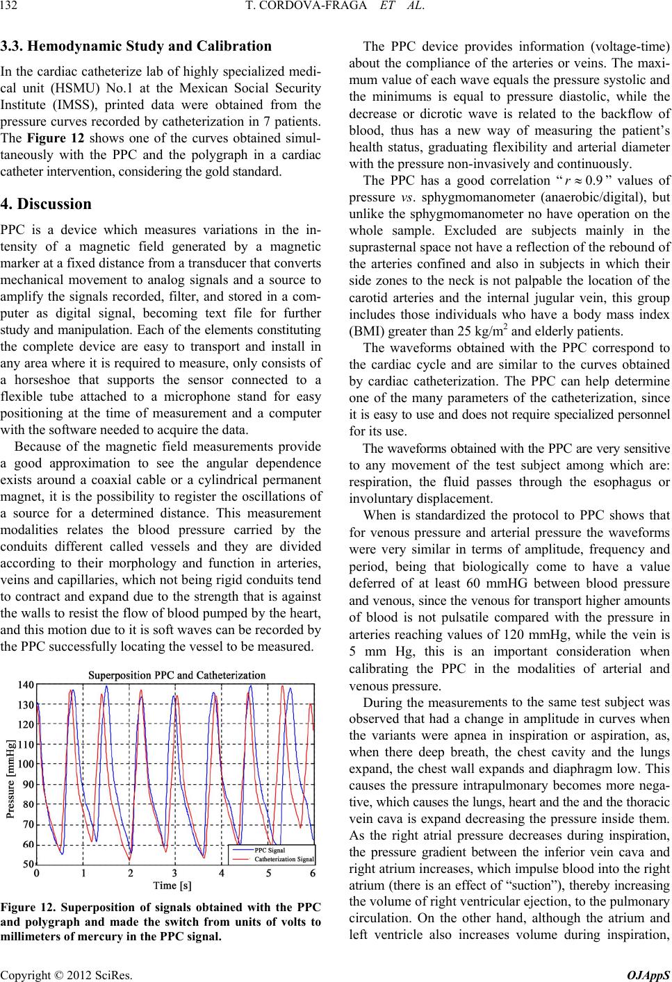
The PPC device hahich “Hemodynamic Significance of High Brachial Pulse Pres-
sure in Young Men,” Clinical Hypertension, Vol. 26, No.
3, 2004, pp. 199-207.
describes qualitatively and unequivo cally form elevations
and depressions corresponding to waves of arterial and
venous pressure by noninvasive methods, however is af-
fected by characteristics of the medium and particularly
by respiration, although it has proven highly correlated
with pressur e points o f a sphygmomano meter digital [13]
while the gold standard in measuring blood pressure [14].
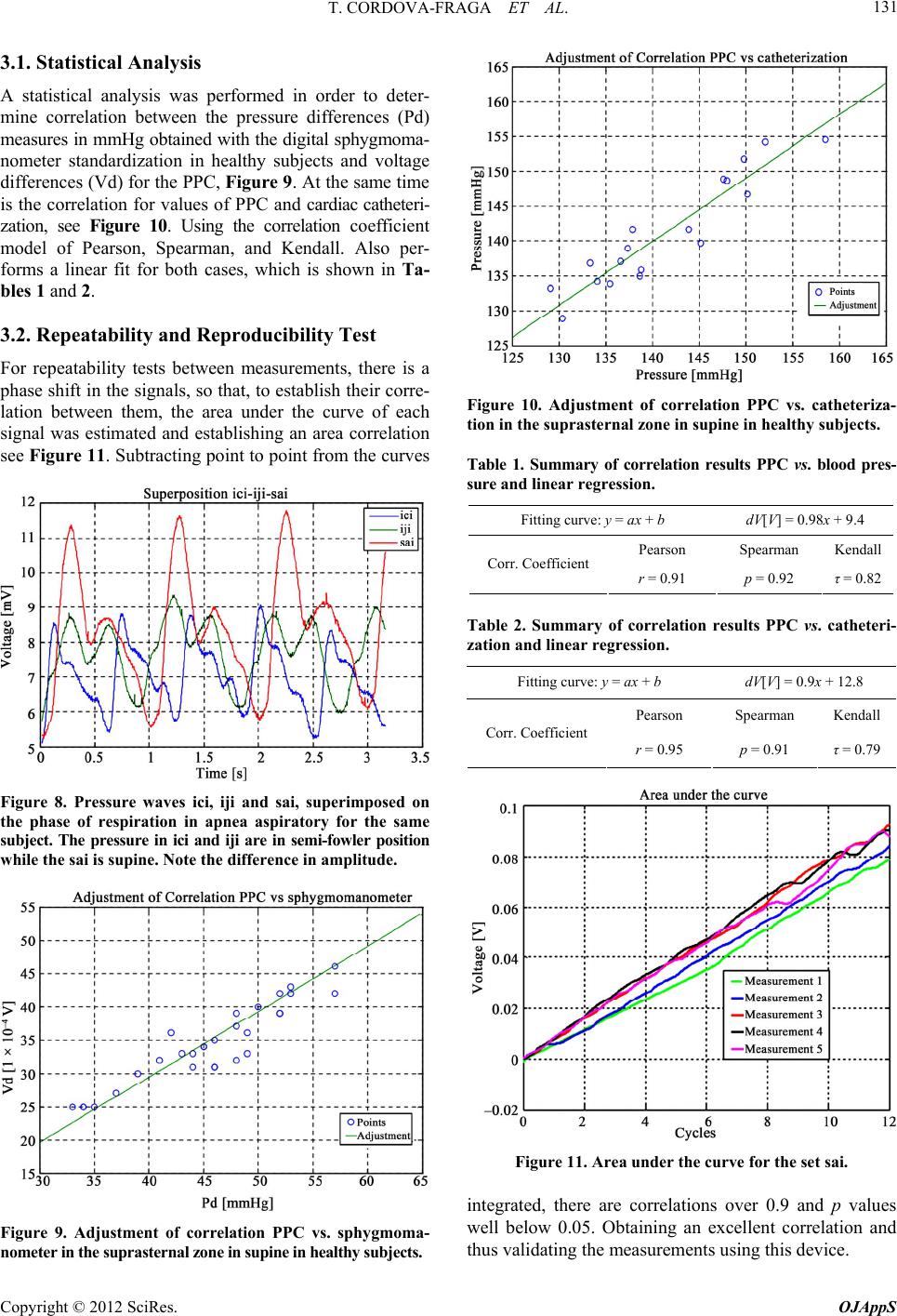
It has more meaning find the magnitude under the
rve, see Figure 11, to relate maximum and minimum
to a coordinate of voltage as the device shows a wide
variability in their measurements if used at different
times with a variation period of 24 h, These results in a
descriptive curve outdated even for the same subject,
with the same values of pressure.
It cannot be used in individuals wi
gree of obesity, because both the suprasternal area as
the lateral neck show no oscillatory motion of venous
and arteries by the large amount of fatty material which
causes i t to lo se t he sp rea d of t he m ovem ent of vas odilat io n.
If it want to be used for pressure measurement, two
ales are needed depending on the type of vessel to
measure, because biologically both vessel s sho w pr essure
values and curved very characteristic, for th e PPC measur -
ing in (voltage-time) and which is already associated
with a curve (mmHg-time).
It is proposed to carry out m
ring the signal acquired by the PPC vs. cardiac cathe-
terization, Doppler echocardiography and oscillometric
methods. For the venous study a larger sample is needed.
The authors acknowledg e the p
No. 017/2010 and Universidad De La Salle Bajío P/2011.
iología Humana,” McGr
and M. Laurenzi, “Twenty-Year Cardiovascular and All-
Cause Mortality Trends and Changes in Intravascular
Risk Factors,” Vol. 27, 2009, pp. 266-274.
[3] www.udec.cl/~ofem/revista/revista02/revist
[4] www.juntadeandalucia.es/averroes/2970/salud/servet.
[5] www.es.wikipedia.org/wiki/William_Harvey
[6] www.annualreviews.org/doi/pdf/10.1146/annurev
[7] www.udec.cl/~ofem/revista/revista02/revista1.html
[8] J. C. R. Pascual, M. R. Romero, R. C. Cerda and J
Morales, “Test de Reac tividad Vascular Pulmonary Blan-
hir,” Neumología y Cirugía de Tórax, Vol. 65, No. S4,
2006, pp. S43-S50.
[9] J. Alfie, C. Majul, O
doi:10.1081/CEH-12003
Scandurra, G. J. Meschino
. Devera and F. C. Y R.
[10] F.M. Clara, A. Casarini, A. G.
and A. R. Introzzi, “Evaluación De Hipertensos en Base a
Registros de Variación de diÁmetro Arterial Radial,”
Medicina, Vol. 66, No. 6, 2006.
[11] D. Bia, I. Aguirre, Y. Zocalo, L
Armentano, “Diferencias Regionales en Viscosidad, Ela-
Copyright © 2012 SciRes. OJAppS