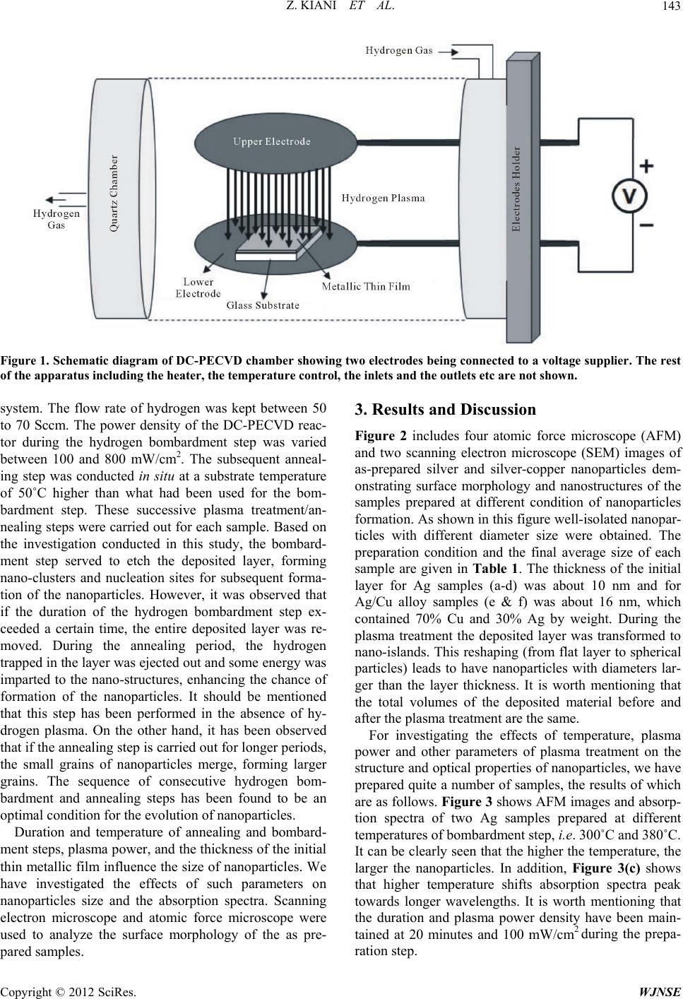
World Journal of Nano Science and Engineering, 2012, 2, 142-147
http://dx.doi.org/10.4236/wjnse.2012.23018 Published Online September 2012 (http://www.SciRP.org/journal/wjnse)
Low Temperature Formation of Silver and Silver-Copper
Alloy Nano-Particles Using Plasma Enhanced
Hydrogenation and Their Optical Properties
Zeinab Kiani, Yaser Abdi, Ezatollah Arzi*
Nanophysics Laboratory, Department of Physics, University of Tehran, Tehran, Iran
Email: *arzi@khayam.ut.ac.ir
Received November 22, 2011; revised January 12, 2012; accepted May 23, 2012
ABSTRACT
In this paper, a novel method of producing nanoparticles at low temperatures using hydrogen bombardment of thin
films, deposited on glass substrates, is introduced. Silver nanoparticles were obtained by this method in our Plasma En-
hanced Chemical Vapor Deposition system. Optical and morphological characteristics of these nanoparticles were ex-
tensively studied for various conditions of plasma treatment, such as plasma power density, temperature, duration of
hydrogen bombardment, thickness of the initial thin metallic film etc. In addition, Ag-Cu alloy nanoparticles on glass
substrates were also achieved. The process of nanoparticle formation in this method shows that several kinds of metals
and semiconductors nanoparticles can be obtained using this approach. Scanning Electron Microscopy, Atomic Force
Microscopy and Transmission Electron Microscopy were used to analyze the nanostructures.
Keywords: Nanoparticles; Plasma; Hydrogen; PECVD; Silver; Alloy
1. Introduction
Because of their linear and nonlinear optical properties,
metal nanoparticles have been widely studied in recent
years. They are suitable candidates for various devices
because of their unique optical [1], electronic [2], cataly-
sis [3], chemical [4] properties. Many different methods
were applied for producing nanoparticles such as chemi-
cal synthesis [5], exploding wire [6], and laser ablation
[7]. We have used a novel different method that produces
nanoparticles at a temperature much lower than the
melting point of their respective bulk using hydrogen
plasma in Enhanced Chemical Vapor Deposition system.
In this work, silver nanoparticles and bimetallic alloy of
silver-copper nanoparticles were produced by this plasma
treatment method. This method can also be used for the
formation of other metallic or semiconductors nanoparti-
cles by PVD method on glass or silicon or other sub-
strates, out of which two examples of nickel and silicon
nanoparticles are presented.
Optical properties of metal nanoparticles are related to
the excitation of surface plasmon. The parameters such
as size and shape [8,9], surrounding medium [10], metal-
lic species [11] of nanoparticles change the excitation
energy of surface plasmons. We have investigated the
effects of size on the absorption spectra of the nanoparti-
cles.
2. Experimental
The work started by cleaning the glass substrate in the
standard RCA1 solution, which contained NH3/
H2O2/DI-H2O with relative volume proportions of 1:1:5,
and followed by the deposition of a thin layer of silver
with the thickness of about 10 nm in a thermal evapora-
tion system at the base pressure of 5 × 10⁶ torr [Forma-
tion of bimetallic alloy nanoparticles needs to evaporate
two metallic sources simultaneously in the physical va-
por deposition (PVD) system]. The silver-coated glass
was then put in the direct current plasma enhanced
chemical vapor deposition (DC-PECVD) apparatus, as
shown schematically in Figure 1. This DC-PECVD
setup has two flat electrodes, made of iron, which hy-
drogen plasma forms between them by the application of
a voltage between the electrodes. The sample was placed
on the cathode (lower electrode) and during the plasma
treatment was bombarded by hydrogen radicals. We
made an electrical connection between the metallic layer
and the cathode to prevent the build up of positive
charges on glass substrate that might repel the plasma.
The substrate temperature was varied between 200˚C and
400˚C and the hydrogen pressure was maintained at 0.4 -
2 torr during the Plasma treatment in the DC-PECVD
*Corresponding author.
C
opyright © 2012 SciRes. WJNSE
Z. KIANI ET AL. 143
Figure 1. Schematic diagram of DC-PECVD chamber showing tw o electrodes being conne cted to a voltage supplier. The rest
of the apparatus including the heater, the temperature control, the inlets and the outlets etc are not shown.
system. The flow rate of hydrogen was kept between 50
to 70 Sccm. The power density of the DC-PECVD reac-
tor during the hydrogen bombardment step was varied
between 100 and 800 mW/cm2. The subsequent anneal-
ing step was conducted in situ at a substrate temperature
of 50˚C higher than what had been used for the bom-
bardment step. These successive plasma treatment/an-
nealing steps were carried out for each sample. Based on
the investigation conducted in this study, the bombard-
ment step served to etch the deposited layer, forming
nano-clusters and nucleation sites for subsequent forma-
tion of the nanoparticles. However, it was observed that
if the duration of the hydrogen bombardment step ex-
ceeded a certain time, the entire deposited layer was re-
moved. During the annealing period, the hydrogen
trapped in the layer was ejected out and some energy was
imparted to the nano-structures, enhancing the chance of
formation of the nanoparticles. It should be mentioned
that this step has been performed in the absence of hy-
drogen plasma. On the other hand, it has been observed
that if the annealing step is carried out for longer periods,
the small grains of nanoparticles merge, forming larger
grains. The sequence of consecutive hydrogen bom-
bardment and annealing steps has been found to be an
optimal condition for the evolution of nanoparticles.
Duration and temperature of annealing and bombard-
ment steps, plasma power, and the thickness of the initial
thin metallic film influence the size of nanoparticles. We
have investigated the effects of such parameters on
nanoparticles size and the absorption spectra. Scanning
electron microscope and atomic force microscope were
used to analyze the surface morphology of the as pre-
pared samples.
3. Results and Discussion
Figure 2 includes four atomic force microscope (AFM)
and two scanning electron microscope (SEM) images of
as-prepared silver and silver-copper nanoparticles dem-
onstrating surface morphology and nanostructures of the
samples prepared at different condition of nanoparticles
formation. As shown in this figure well-isolated nanopar-
ticles with different diameter size were obtained. The
preparation condition and the final average size of each
sample are given in Table 1. The thickness of the initial
layer for Ag samples (a-d) was about 10 nm and for
Ag/Cu alloy samples (e & f) was about 16 nm, which
contained 70% Cu and 30% Ag by weight. During the
plasma treatment the deposited layer was transformed to
nano-islands. This reshaping (from flat layer to spherical
particles) leads to have nanoparticles with diameters lar-
ger than the layer thickness. It is worth mentioning that
the total volumes of the deposited material before and
after the plasma treatment are the same.
For investigating the effects of temperature, plasma
power and other parameters of plasma treatment on the
structure and optical properties of nanoparticles, we have
prepared quite a number of samples, the results of which
are as follows. Figure 3 shows AFM images and absorp-
tion spectra of two Ag samples prepared at different
temperatures of bombardment step, i.e. 300˚C and 380˚C.
It can be clearly seen that the higher the temperature, the
larger the nanoparticles. In addition, Figure 3(c) shows
that higher temperature shifts absorption spectra peak
towards longer wavelengths. It is worth mentioning that
the duration and plasma power density have been main-
tained at 20 minutes and 100 mW/cm2 during the prepa-
ration step.
Copyright © 2012 SciRes. WJNSE
Z. KIANI ET AL.
144
Figure 4 is a comparison between the samples prepared
at two different plasma power densities of 100 and 600
mW/cm2. As shown in this figure, at lower plasma power
density during the bombardment, smaller nanoparticles
Figure 2. AFM and SEM images of silver nanoparticles
(a)-(d) and Ag/Cu alloy samples ((e) & (f)) formed on glass
substrates using hydrogen plasma treatment, showing that
different conditions of sample preparation lead to different
sizes of nanoparticles.
are achieved and correspondingly, the absorption peak
appears at a wavelength shorter than that of the sample
prepared at higher plasma power density. Thus, we see
that changes in morphology and sizing of nanoparticles
caused by temperature and plasma power density lead to
variations in their optical properties. It seems that smaller
particles have their absorption peak at shorter wave-
lengths range. We have also investigated the effects of
other ranges of plasma power density on our samples. It
is observed that raising the power density to values above
800 mW/cm2 will lead to total etching of the deposited
layer. On the other hand, no grain formation was ob-
served at power densities below 100 mW/cm2, as con-
firmed by SEM and AFM results.
Comparison between two samples, one of them bom-
barded for 20 minutes and the other one for 45 minutes
but both at the same temperature of 300˚C with the same
plasma power density of 150 mW/cm2, is represented in
Figure 5. This comparison demonstrates that longer du-
ration of bombardment results in smaller nanoparticles
and hence the absorption spectrum gets blue shifted.
Further experiments revealed that the thickness of the
initial deposited layer is very important. It is found that
increasing the layer thickness above 50 nm leads to ob-
serve no grain formation. Figure 6 shows SEM images
of three samples having silver layer thickness of 70, 40
and 10 nm prepared at the same condition of plasma
treatment and annealing step, confirming the above men-
tioned conclusion. In addition, Figure 6(d) shows ab-
sorption spectra of these samples showing no absorption
peak in the spectrum of the sample with 70 nm initial
layer thickness.
We have used this approach not only to produce silver
nanoparticles, but also for other materials such as nickel
and silicon nanoparticles, their SEM images are shown in
Figure 7. Thus we believe that this method is suitable for
producing nanoparticles of various metallic and semi-
conductor materials.
Table 1. Sample preparation conditions for Figure 2.
Bombardment condition
Average particle size (nm) Plasma power density (mW/cm2)
Temperature (˚C) Duration (minutes)
Sample
75 200 300 15 a
50 180 300 20 b
65 230 300 20
*c
70 300 380 20 d
145 120 220 20 e
55 130 300 15 f
*
It was then annealed at 400˚C for 20 minutes.
Copyright © 2012 SciRes. WJNSE
Z. KIANI ET AL. 145
(a) (b)
(c)
Figure 3. AFM images of two Ag samples prepared at dif-
ferent temperature of the bombardment step, (a) 300˚C and
(b) 380˚C; (c) Absorption spectra of the two above-mention-
ed samples.
(a)
(b)
Figure 4. (a) SEM images of the above-mentioned samples,
formed at plasma power density of 100 mW/cm2 (left) and
600 mW/cm2 (right). These SEM images and the corre-
sponding absorption spectra show that lower power density
of plasma during bombardment leads to the formation of
smaller nanoparticles; (b) Absorption spectra of the Ag
samples that have been formed at the plasma power density
of 100 and 650 mW/cm2.
Figure 5. Absorption spectra of samples having bombarded
for different periods: 20 and 45 minutes. Comparison be-
tween them shows that longer time of bombardment leads
to form smaller nanoparticles.
(a) (b) (c)
(d)
Figure 6. SEM images of silver samples prepared on silicon
substrates at the same condition of bombardment treat-
ment, except that the initial thickness of deposited layer was
(a) 10 nm; (b) 40 nm and (c) 70 nm; (d) Absorption spectra
of the above mentioned samples.
(a) (b)
Figure 7. SEM images of (a) semiconductor silicon nano-
particles; (b) metallic nickel nanoparticles, produced by the
proposed hydrogen tre atment method.
Copyright © 2012 SciRes. WJNSE
Z. KIANI ET AL.
146
To form alloy nanoparticles, silver and copper were
simultaneously evaporated by PVD system as two com-
ponents of bimetallic alloy on glass substrate. AFM im-
ages of plasma treated silver/copper nanoparticles are
represented in Figure 8. Figure 9 shows TEM images of
silver/copper alloy nanoparticles that were produced by
this novel hydrogen bombardment method, which clearly
shows the formation of alloy nanoparticles. By looking
carefully at this image, one can see that some parts of
some grains in the TEM image are darker, which means
that these grains contain the two component materials
and thus the alloy of silver/copper was formed in this
sample. Therefore, the method is capable of producing
alloys. Another method for depicting the structure is the
optical analysis [12]. Optical results of the as-pre- pared
samples depict two different structures for these nanos-
tructures. Figure 10 shows the absorption spectra of two
Ag/Cu nanoparticles prepared at two different tempera-
tures of 200˚C and 300˚C during the bombardment step.
As shown in this figure, the sample prepared at the lower
temperature has two isolated peaks corresponding to the
absorption peaks of pure silver and pure copper nanopar-
ticles whilst the other one (dashed curve) has a single
(a) (b)
Figure 8. AFM images of Ag/Cu alloy nanoparticles with
initial layer thickness of about 15 nm that have been pre-
pared in different condition, (a) contains 30% Ag and 70%
Cu by weight, bombarded for 20 minutes at plasma power
density of 100 mW/cm2 at 300˚C and (b) contains 50% Ag
and 50% Cu by weight, bombarded for 20 minutes at
plasma power density of 140 mW/cm2 at 370˚C.
Figure 9. TEM images of silver/copper alloy nanoparticles
showing that the grains were formed by this method. The
darker color of some parts of the grains shows that the
grains contain more than one material, and thus the sil-
ver/copper alloy nanoparticles were formed.
peak at a wavelength between the two above-mentioned
peaks. It depicts that the sample with two peaks is just a
mixture of Ag nanoparticles and Cu nanoparticles but the
other one is an alloy of Ag/Cu nanoparticles. Figure 11
shows the absorption spectra of the alloys with different
mixing ratio. As shown in this figure, shifting the ab-
sorption peak towards the blue or red regions depends on
the mass percentage of silver or copper components in
the alloy.
Figure 10. Absorption spectra of samples having different
temperature in their bombardment steps. The sample that
exhibits two peaks in its absorption spectrum (solid line),
bombarded at 200˚C, shows the formation of separated Ag
and Cu nanoparticles, not alloy. The other sample , which is
bombarded at 300˚C, shows one peak in its spectrum
(dashed line) that can be attributed to alloy for mation.
Figure 11. Comparison between the absorption spectra of
Ag/Cu alloy nanoparticles having different weight ratio of
Cu; all prepared at the same condition. It shows that by
increasing the Cu ratio in the nanoparticles, the absorption
spectrum gets a red shift.
Copyright © 2012 SciRes. WJNSE
Z. KIANI ET AL.
Copyright © 2012 SciRes. WJNSE
147
The results of the visible light spectroscopy presented
in this paper show the wavelength dependent absorption
in the fabricated nanostructures. It can be explained by
the Langmuir theory describing the collective oscillations
of surface charges. In metal particles with subwavelength
dimensions there are in fact dipole electron oscillations
bounded by the nanoscopic particle. From quantum me-
chanical point of view, the charge oscillations are easily
excited by the incident photons with energy of ћωp,
where ωp is the resonance frequency of the charge oscil-
lations depends on the size, material and shape of the
nanoparticle. The source of absorption peaks in the pre-
sented spectra is the light induced quantum excitation of
such oscillations.
4. Conclusion
In summary, we have successfully produced the silver
and silver-copper alloy nanoparticles using a low tem-
perature plasma bombardment method, well below the
melting point of the sample constituents. The method can
be used for any other materials to produce nanoparticles
of them on any arbitrary substrates (even flexible sub-
strates). It is easy to make a patterned structure of
nanoparticles by this method. We believe that the pro-
posed method can produce clusters of nanoparticles and
may be the produced films can be used in device fabrica-
tions such as transistors, single electron transistors,
nanoparticles-based gas sensors etc. Optical measure-
ments of as-prepared nanoparticles confirm the quantum
behavior of the samples arising from decreasing the size
and confinement.
5. Acknowledgements
We would like to thank the research council of the Uni-
versity of Tehran for partial financial support.
REFERENCES
[1] W. H. Weber and G. W. Ford, “Propagation of Optical
Excitations by Dipolar Interactions in Metal Nanoparticle
Chains,” Physical Review B, Vol. 70, No. 12, 2004, pp.
125429.1-125429.8. doi:10.1103/PhysRevB.70.125429
[2] S. Link and M. A. El-Sayed, “Spectral Properties and
Relaxation Dynamics of Surface Plasmon Electronic Os-
cillations in Gold and Silver Nanodots and Nanorods,”
The Journal of Physical Chemistry B, Vol. 103, No. 40,
1999, pp. 8410-8426. doi:10.1021/jp9917648
[3] W. P. Zhou, A. Lewera, R. Larsen, R. I. Masel, P. S. Ba-
gus and A. Wieckowski, “Size Effects in Electronic and
Catalytic Properties of Unsupported Palladium Nanopar-
ticles in Electrooxidation of Formic Acid,” The Journal of
Physical Chemistry B, Vol. 110, No. 27, 2006, pp.
13393-13398. doi:10.1021/jp061690h
[4] P. Waszczuk, T. M. Barnard, C. Rice, R. I. Masel and A.
Wieckowski, “A Nanoparticle Catalyst with Superior Ac-
tivity for Electrooxidation of Formic Acid,” Electro-
chemistry Communications, Vol. 4, No. 7, 2003, pp. 599-
603. doi:10.1016/S1388-2481(02)00386-7
[5] B. Choi and H.-H. Lee, “Characterization of the Optical
Properties of Silver Nanoparticle Films,” Nanotechnology,
Vol. 18, No. 7, 2007, Article ID: 075706.
[6] T. K. Sindhu, R. Sarathi and S. R. Chakravarthy, “Under-
standing Nanoparticle Formation by a Wire Explosion
Process through Experimental and Modeling Studies,”
Nanotechnology, Vol. 19, No. 2, 2008, Article ID:.
025703.
[7] S. H. Ko and Y. Choi, “Nanosecond Laser Ablation of
Gold Nanoparticle Films,” Applied Physics Letters, Vol.
89, No. 14, 2006, p. 141126. doi:10.1063/1.2360241
[8] M. Valden, X. Lai and D. W. Goodman, “Onset of Cata-
lytic Activity of Gold Clusters on Titania with the Ap-
pearance of Nonmetallic Properties,” Science, Vol. 281,
No. 5383, 1998, pp. 1647-1650.
doi:10.1126/science.281.5383.1647
[9] X. Y. Xu, K. K. Caswell, E. Tucker, S. Kabisatpathy, K.
L. Brodhacker and W. A. Scrivens, “Size and Shape
Separation of Gold Nanoparticles with Preparative Gel
Electrophoresis,” Journal of Choromatography A, Vol.
1167, No. 1, 2007, pp. 35-41.
doi:10.1016/j.chroma.2007.07.056
[10] N. Nath and A. Chilkoti, “A Colorimetric Gold Nanopar-
ticle Sensor to Interrogate Biomolecular Interactions in
Real Time on a Surface,” Analytical Chemistry, Vol. 74,
No. 3, 2002, pp. 504-509. doi:10.1021/ac015657x
[11] K. Esumi, T. Matsumoto, Y. Seto and T. Yoshimura,
“Preparation of Gold-, Gold/Silver-Dendrimer Nanocom-
posites in the Presence of Benzoin in Ethanol by UV Irra-
diation,” Journal of Colloid and Interface Science, Vol.
284, No. 1, 2005, pp. 199-203.
doi:10.1016/j.jcis.2004.09.020
[12] H. J. Jiang, K. Moon and C. P. Wong, “Synthesis of
Ag-Cu Alloy Nanoparticles for Lead-Free Interconnect
Materials,” Proceedings of International Symposium on
Advanced Packaging Materials: Processes, Properties
and Interfaces, Irvine, 16-18 March 2005, pp. 173-177.
doi:10.1109/ISAPM.2005.1432072