Case Reports in Clinical Medicine
Vol.2 No.2(2013), Article ID:31788,2 pages DOI:10.4236/crcm.2013.22048
Large number of Fasciola hepatica discovered during endoscopic retrograde cholangiopancreatography (ERCP)
![]()
Kurdistan Center for Gastroenterology & Hepatology, Sulaymaniyah, Iraq; *Corresponding Author: shaikhanimohammad@gmail.com
Copyright © 2013 Mohamed Alshekhani et al. This is an open access article distributed under the Creative Commons Attribution License, which permits unrestricted use, distribution, and reproduction in any medium, provided the original work is properly cited.
Received 30 September 2012; revised 1 April 2013; accepted 16 April 2013
Keywords: Fasiolahepatics; Obstructivejaundice; Triclabendazole
ABSTRACT
Fascioliasis is a zoonotic infection of herbiviores, occasionally affecting humans who ingest fresh water plants. We report a case of a young woman who presented to our center with vague abdominal discomfort & discovered on abdominal ultrasound to have gall stones & dilated common bile duct. She was scheduled for ERCP which revealed a large number of fasciola worms draining from the common bile duct to the duodenum.
1. INTRODUCTION
Fascioliasis is a zoonotic infection of the liver & biliary tract & sometimes affecting ectopic sites distant from the hepatobilairy system during the migration of the larva from the intestine towards its final distination namely the bilaiary tract. The diagnosis of the disease is very difficult during the acute hepatic parachymal phase & ectopic phase [1]. Even during the chronic biliary phase the diagnosis is frequently done during ERCP or even surgery. Making things more difficult that biliary faciolosis can mimic & can cause common causes of biliary obstruction namely cholelithiais & cholangiocarcinoma. Experienced abdominal & endoscopic sonographists can diagnose the infection if they observe moving objects in the gall bladder or common bile duct during their search for hepatobilairy pathologies & frequebtly they are gaining experience in that respect [2,3].
2. CASE REPORT
We report a case of 35 years old woman from rural Iraqi Kurdistan near the Iran boarder, who was referred to our center for upper abdominal discomfort. On examination the patient slightly pale but not jaundiced. Abdominal examination revealed no organomegaly or ascites. Complete blood picture & blood film revealed 8% esosinophilia. Liver function tests revealed normal bilirubin & liver enzymes, but high alkaline phsphataase. Abdominal ultrasound dilated common bile ducts of 12 mms & gall bladder echogenic shadows suggestive of gall stones. During ERCP, following endoscopic sphicterotomy & trail of basket stone extraction, unexpectedly large number of Fasciola hepatica was drained from the common bile duct to the duodenum (endoscopic photos), some of which were retrieved endoscopically to outside the body while the others flew down to the more distal small intestine (Figures 1-3). No stone was extracted. The patient improved clinically after the ERCP. Triclabendazole was prescribed to kill any remaining Fasiolas. The patient admitted that she was advised to increase the intake of green vegetable because of her anemia & she increased the intake of fresh water plants available in her rural area. After ERCP, ELISA serological test was sent for the patient & proved to be positive.
3. DISCUSSION
Although Human Fasciolosis occurs in the developing world, but it was not reported in Iraq until the last few years & still these case reports are from the Iraqi Kurdistan & not the other parts of Iraq [4,5]. These case appeared after the mini epidemic that occurred in Krminshah, Iran & increasingly case reports from Van area, Turkey [6-8], both of which areas are close to Iraqi Kurdistan & we think that the herbivores trafficking across the boarders had participated in transporting the infection to our area. The case under discussion & most of our cases are discovered during ERCP or surgery unexpectedly,

Figure 1. Fasciola hepatica intact worm drained to the duodenum after endoscopic shinterotomy during ERCP.
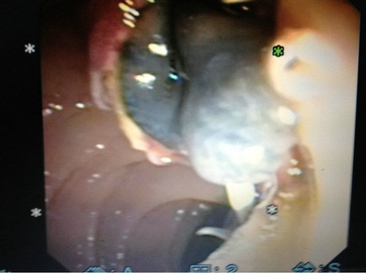
Figure 2. Closer view of Fasciola hepatica intact worm drained to the duodenum after endoscopic shinterotomy during ERCP.
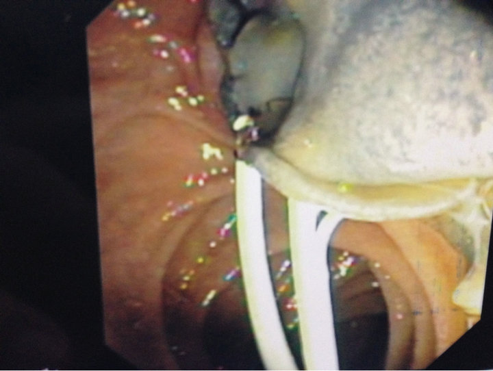
Figure 3. Closer view of Fasciola hepatica intact worm drained to the duodenum after endoscopic shinterotomy during ERCP.
because the infection was not prevalent before & the clinicians, surgeons & radiologists are still they not be familiar with the disease, in addition to the fact that its clinical & imaging features may simulate other hepatobilairy diseases [3,9].
REFERENCES
- Dusak, A., Onur, M.R., Cicek, M., Firat, U., Ren, T. and Dogra, V.S. (2012) Radiological imaging features of fasciola hepatica infection—A pictorial review. Journal of Clinical Imaging Science, 2, 2. doi:10.4103/2156-7514.92372
- Kaya, M., Beştaş, R. and Cetin, S. (2011) Clinical presentation and management of Fasciola hepatica infection: Single-center experience. World Journal of Gastroenterology, 17, 4899-4904. doi:10.3748/wjg.v17.i44.4899
- Mohammad Alizadeh, A.H., Roshani, M., Lahmi, F., Davoodi, N.A., Rostami Nejad, M., Seyyedmajidi, M.R. and Zali, M.R. (2011) Cholangiocarcinoma in magnetic resonance cholangiopancreatography and fascioliasis in endoscopic ultrasonography. Case Reports in Gastroenterology, 5, 569-577. doi:10.1159/000333229
- Karabuli, T.A., Shaikhani, M.A.R., Karadaghi, S.H.S. and Kasnazan, K.H. (2009) Hepatobiliary and pancreatic: Fascioliasis. Journal of Gastroenterology and Hepatology, 24, 1309.
- Hawrami, T.A. (2010) Case report: Fasciola hepatica worm swims in the gallbladder. Zanco Journal of Medical Sciences, 14, 1-4.
- Taheri, M.S., Aminzade, Z., Shokohi, Sh., Birang, Sh. and Aghazade, K. (2007) Case report: Hepatobiliary fascioliasis: Clinical and radiological features. Iranian Journal of Parasitology, 2, 48-55.
- Hatami, H. (2012) The report of 1st outbreak of fascioliasis in Kermanshah. 4th Congress of Zoonotic Disease, Tehran, 1379-1384.
- Karahocagil, M.K., Akdeniz, H., Sunnetcioglu, M., Cicek, M., Mete, R., Akman, N., Ceylan, E., Karsen, H. and Yapici, K. (2011) A familial outbreak of fascioliasis in Eastern Anatolia: A report with review of literature. Acta Tropica, 118, 177-183. doi:10.1016/j.actatropica.2008.08.013
- Deveci, U., Oztürk, T. and Ustün, C. (2011) A case of radiologically diagnosed pediatric Fasciola hepatica. Turkiye Parazitolojii Dergisi, 35, 117-119. doi:10.5152/tpd.2011.29