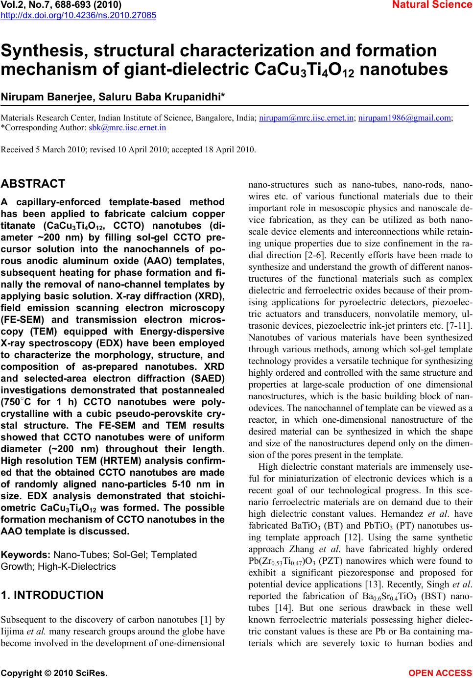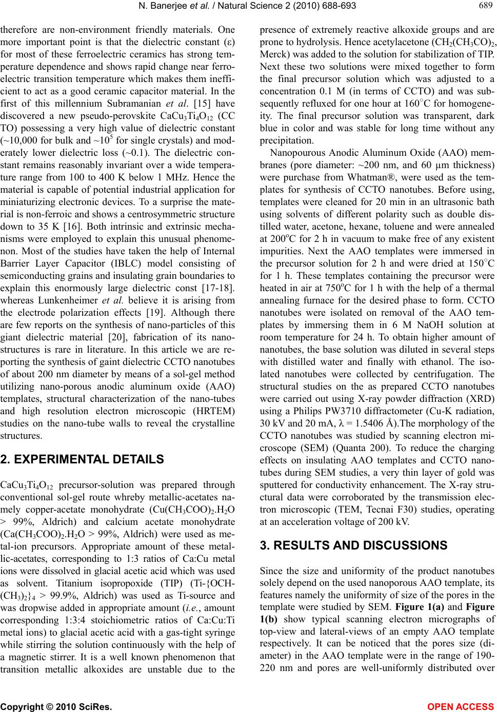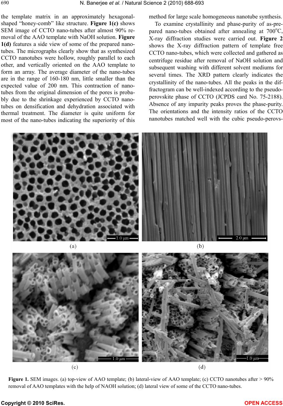
N. Banerjee et al. / Natural Science 2 (2010) 688-693
Copyright © 2010 SciRes. OPEN ACCESS
693
693
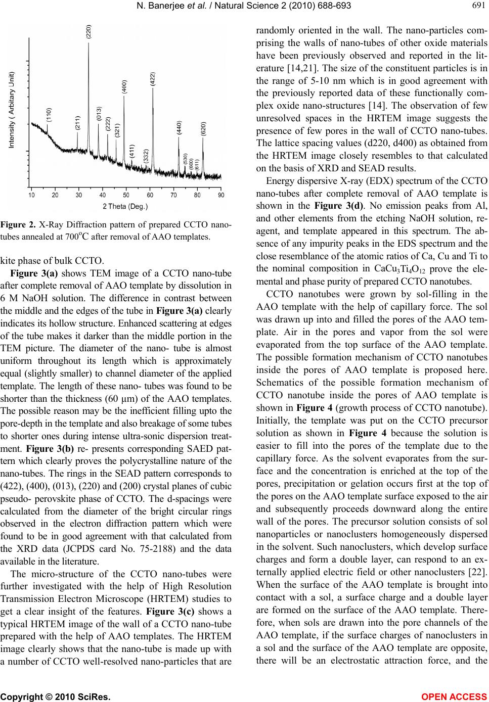
nanoclusters will preferentially deposit on the surface of
the pore channel, resulting in the formation of nano-
tubes.
4. CONCLUSIONS
In summary, CCTO nanotubes have been prepared suc-
cessfully by sol-gel method using the closely packed
porous nanochannel alumina templates. The crystallinity
and phase purity of the CCTO nanotubes were con-
firmed via XRD and SAED analysis. EDX analysis cor-
roborated the stoichiometric CaCu3Ti4O12 formation.
The walls of the nanotubes were found to be made up of
nanoparticles as confirmed by the HRTEM studies. The
nanoparticles embedded in the wall were found in the
range of 5-10 nm. This facile method of preparing the
CaCu3Ti4O12 nanotubes at a large scale might be impor-
tant for many applications in nanodevices.
5. ACKNOWLEDGEMENTS
N. B. thanks to Int. PH.D program, Indian Institute of Science for
financial assistances and Institute Nano-Science Initiative for micro-
scopic facilities.
REFERENCES
[1] Iijima, S. (1991) Helical microtubules of graphitic carbon.
Nature, 354(6348), 56-58.
[2] Hu, J., Odom, T.W. and Lieber, C.M. (1999) Chemistry
and physics in one dimension: Synthesis and properties of
nanowires and nanotubes. Accounts of Chemical Research,
32(5), 435-445.
[3] Atzke, G.R.P., Rumeich, F.K. and Nesper, R. (2002)
Based on oxide nanotubes and nanorods − anisotropic
building blocks for future nanotechnology. Angewandte
Chemie, 114(14), 2554-2571.
[4] Goldberger, J., He, R., Zhang, Y., Lee, S., Yan, H., Choi,
H. and Yang, P. (2003) Single-crystal gallium nitride
nanotubes, Nature, 422(6932), 599-602.
[5] Lee, S.B., Mitchell, D.T., Rofin, L.T., Ne vanen, T.K.,
Sçderlund, H. and Martin, C.R. (2002) Antibody-based
bio-nanotube membranes for enantiomeric drug separa-
tions. Scie nc e , 296(5576), 2198-2200.
[6] Sha, J., Niu, J., Ma, X., Xu, J., Zhang, X., Yang, Q. and
Yang, D. (2002) Silicon nanotubes. Advanced Materials,
14(17), 1219-1224.
[7] Junquera, J. and Ghosez, P. (2003) Critical thickness for
ferroelectricity in perovskite ultrathin films. Nature,
422(6931), 534-539.
[8] Wang, Y. and Santiago-Aviles, J.J. (2004) Synthesis of
lead zirconate titanate nanofibres and the Fourier-tran-
sform infrared characterization of their metallo-organic
decomposition process. Nanotechnology, 15, 32.
[9] Luo, Y., Szafraniak, I., Zakharo, N.D., Nagarajan, V.,
Steinhart, M., Ehrspohn, R.B.W., Endorff, J.H.W.,
Ramesh, R. and Alexe, M. (2003) Nanoshell tubes of
ferroelectric lead zirconate titanate and barium titanate.
Applied Physics Letters, 83(5377), 440-442.
[10] Chu, M.W., Szafraniak, I., Scholz, R., Harnagea, C.,
Hesse, D., Alexe, M. and Gosele, U. (2004) Impact of
misfit dislocations on the polarization instability of
epitaxial nanostructured ferroelectric perovskites. Na-
ture Materials, 3(2), 87-90.
[11] Roelofs, A., Schneller, I., Szot, K. and Waser, R. (2002)
Piezoresponse force microscopy of lead titanate nano-
grains possibly reaching the limit of ferroelectricity. Ap-
plied Physics Letters, 81(27), 5231-5233.
[12] Hernandez, B.A., Chang, K.S., Fisher, E.R. and Dorhout,
P.K. (2002) Sol-Gel template synthesis and characteriza-
tion of batio3 and pbtio3 nanotubes. Chemistry of Mate-
rials, 4, 480.
[13] Zhang, X.Y., Zhao, X., Lai, C.W., Wang, J., Tang, X.G.
and Dai, J.Y. (2004) Synthesis and piezoresponse of
highly ordered Pb(Zr0.53Ti0.47)O3 nanowire arrays. Ap-
plied Physics Letters, 85(18), 4190-4192.
[14] Singh, S. and Krupanidhi, S.B. (2007) Synthesis and
structural characterization of Ba0.6Sr0.4TiO3 nanotubes.
Physics Letter A, 367(4-5), 356-359.
[15] Subramanian, M.A., Li, D., Duan, N., Reisner, B.A. and
Sleight, A.W. (2000) High dielectric constant in ACu3-
Ti 4O12 and ACu3Ti 3FeO12 phase. Journal of Solid State
Chemistry, 151(2), 323-325.
[16] Liu, J. J., Sui, Y.C., Duan, C.-G., Mei, W.-N., Smith,
R.W., and Hardy, J.R. (2006) CaCu3Ti 4O12: Low-tem-
perature synthesis by pyrolysis of an organic solution.
Chemistry Materials, 18(16), 3878-3882.
[17] Sinclair, D.C., Adams, T.B., Morrison, F.D. and West,
A.R. (2002) CaCu3Ti 4O12: One-step internal barrier layer
capacitor. Applied Physics Letters, 80(12), 2153-2155.
[18] Liu, J., Duan, C., Yin, W., Mei, W.N., Smith, R.W. and
Hardy, J.R. (2004) Large dielectric constant and max-
well-wagner relaxation in Bi2⁄3Cu3Ti4O12. Physics Review
B, 70, 144106.
[19] Lunkenheimer, P., Bobnar, V., Pronin, A.V., Ritus, A.I.,
Volkov, A.A. and Loidl, A. (2002) Origin of apparent
colossal dielectric constants. Physics Review B, 66,
052105.
[20] Thomas, P., Dwarakanath, K., Varma, K.B.R. and Kutty,
T.R.N. (2008) Nanoparticles of the giant dielectric mate-
rial, CaCu3Ti4O12 from a precursor route. Journal of
Physics and Chemistry of Solids, 69(10), 2594-2604.
[21] Zhang, X.Y., Lai, C.W., Zhao, X., Wang, D.Y. and Dai,
J.Y. (2005) Synthesis and ferroelectric properties of mul-
tiferroic BiFeO3 nanotube arrays. Applied Physics Letters,
87, 143102.
[22] Wang, Y. and Cao, G. (2007) Synthesis and electro-
chemical properties of InVO4 nanotube arrays. Journal
of Materials Chemistry, 17(2298), 894-899.