Paper Menu >>
Journal Menu >>
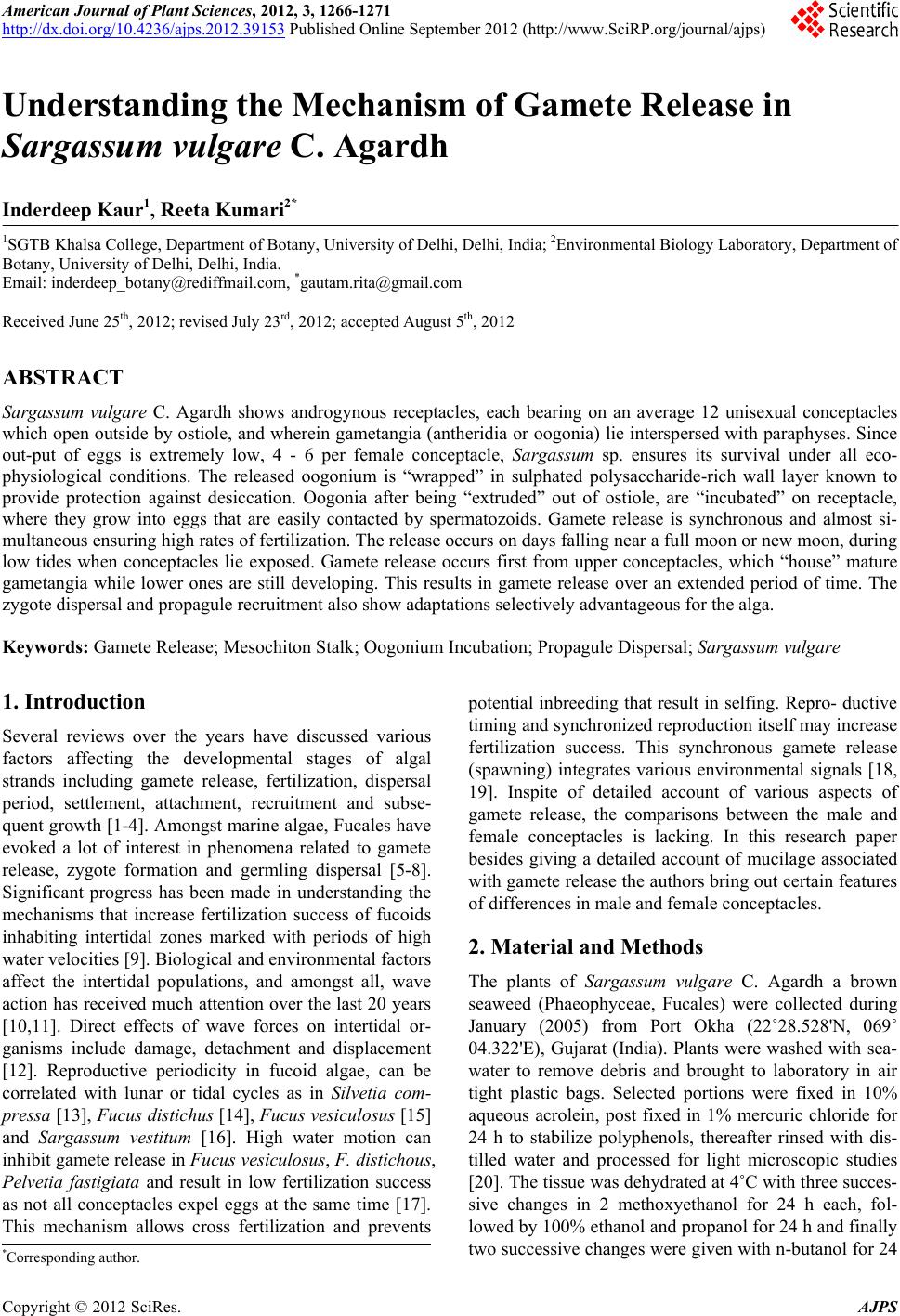 American Journal of Plant Sciences, 2012, 3, 1266-1271 http://dx.doi.org/10.4236/ajps.2012.39153 Published Online September 2012 (http://www.SciRP.org/journal/ajps) Understanding the Mechanism of Gamete Release in Sargassum vulgare C. Agardh Inderdeep Kaur1, Reeta Kumari2* 1SGTB Khalsa College, Department of Botany, University of Delhi, Delhi, India; 2Environmental Biology Laboratory, Department of Botany, University of Delhi, Delhi, India. Email: inderdeep_botany@rediffmail.com, *gautam.rita@gmail.com Received June 25th, 2012; revised July 23rd, 2012; accepted August 5th, 2012 ABSTRACT Sargassum vulgare C. Agardh shows androgynous receptacles, each bearing on an average 12 unisexual conceptacles which open outside by ostiole, and wherein gametangia (antheridia or oogonia) lie interspersed with paraphyses. Since out-put of eggs is extremely low, 4 - 6 per female conceptacle, Sargassum sp. ensures its survival under all eco- physiological conditions. The released oogonium is “wrapped” in sulphated polysaccharide-rich wall layer known to provide protection against desiccation. Oogonia after being “extruded” out of ostiole, are “incubated” on receptacle, where they grow into eggs that are easily contacted by spermatozoids. Gamete release is synchronous and almost si- multaneous ensuring high rates of fertilization. The release occurs on days falling near a full moon or new moon, during low tides when conceptacles lie exposed. Gamete release occurs first from upper conceptacles, which “house” mature gametangia while lower ones are still developing. This results in gamete release over an extended period of time. The zygote dispersal and propagule recruitment also show adaptations selectively advantageous for the alga. Keywords: Gamete Release; Mesochiton Stalk; Oogonium Incubation; Propagule Disp ersal; Sargassum vulgare 1. Introduction Several reviews over the years have discussed various factors affecting the developmental stages of algal strands including gamete release, fertilization, dispersal period, settlement, attachment, recruitment and subse- quent growth [1-4]. Amongst marine algae, Fucales have evoked a lot of interest in phenomena related to gamete release, zygote formation and germling dispersal [5-8]. Significant progress has been made in understanding the mechanisms that increase fertilization success of fucoids inhabiting intertidal zones marked with periods of high water velocities [9]. Biological and environmen tal factors affect the intertidal populations, and amongst all, wave action has received much attention over the last 20 years [10,11]. Direct effects of wave forces on intertidal or- ganisms include damage, detachment and displacement [12]. Reproductive periodicity in fucoid algae, can be correlated with lunar or tidal cycles as in Silvetia com- pressa [13], Fucus distichus [14], Fucus vesiculosus [15] and Sargassum vestitum [16]. High water motion can inhibit gamete release in Fucus vesiculosus, F. distichous, Pelvetia fastigiata and result in low fertilization success as not all conceptacles expel eggs at the same time [17]. This mechanism allows cross fertilization and prevents potential inbreeding that result in selfing. Repro- ductive timing and synchronized reproduction itself may increase fertilization success. This synchronous gamete release (spawning) integrates various environmental signals [18, 19]. Inspite of detailed account of various aspects of gamete release, the comparisons between the male and female conceptacles is lacking. In this research paper besides giving a detailed account of mucilage associated with gamete release the authors bring out certain features of differences in male and female conceptacles. 2. Material and Methods The plants of Sargassum vulgare C. Agardh a brown seaweed (Phaeophyceae, Fucales) were collected during January (2005) from Port Okha (22˚28.528'N, 069˚ 04.322'E), Gujarat (India). Plants were washed with sea- water to remove debris and brought to laboratory in air tight plastic bags. Selected portions were fixed in 10% aqueous acrolein, post fixed in 1% mercuric chloride for 24 h to stabilize polyphenols, thereafter rinsed with dis- tilled water and processed for light microscopic studies [20]. The tissue was dehydrated at 4˚C with three succes- sive changes in 2 methoxyethanol for 24 h each, fol- lowed by 100% ethanol and prop anol for 24 h and fin ally two successive changes were given with n-butanol for 24 *Corresponding a uthor. Copyright © 2012 SciRes. AJPS  Understanding the Mechanism of Gamete Release in Sargassum vulgare C. Agardh 1267 h. The dehydrated material was infiltered and finally embedded in glycol methacrylate. Sections (2 µ thick) were cut with glass knives using a rotary microtome. The sections were stained with Periodic Acid Schiff reagent and 0.5% Toluidine Blue O at pH 4.4 [21]. Total proteins were localized using Coomassie Brilliant Blue [22]. Al- cian blue was used to stain sulphated polysaccharides [23]. For scanning electron microscopy the plants were fixed in 6% Glutaraldehyde, post fixed in OsO4. The por- tions were then dehydrated in ascending acetone series, ranging from 10 through 90% followed by two changes in absolute acetone for 10 min. The material was later passed through dry acetone (absolute acetone in CuSO4) followed by critical point drying at 20˚C and 800 lb/ square inch with pressure for 30 min. This temperature and pressure were raised to 35˚C and 1200 lb/square inch respectively. These were then coated with gold (100 Å thickness) under vacuum on sputter coating unit (Edward coating unit) for 2 min to eliminate charging. The coated material was examined and photographed with LEO 435 VP, variable pressure scanning electron microscope at an accelerating voltage of 15 KV. 3. Results Sargassum vulgare has a perennial existence in intertidal pools where the plants undergo a period of active vegeta- tive growth from July to October and reproductive growth from October to February. When the temperature dips and illumination is low during November, the plants en- ter a period of sexual reproduction which culminates in gamete release and zygote formation in the month of February. The germlings are dispersed around February and March. After an active reproductive phase, the fronds shows senesce and die back leaving only peren- nial holdfast which regenerates in the month of May giving rise to a stipe and leaves. 3.1. Gametogenesis During fertile phase, plants bear abundant androgynous receptacles (Figure 1(A)). This unisexual conceptacles are initiated in the cortical region of receptacles (Figure 1(B)). Amongst several female conceptacles only 3 or 4 male conceptacles are seen in a single receptacle (Figure 1(C)). The young conceptacle is oval to pear- shaped with a narrow neck that opens outside through ostiole (Figure 1(D)). Inside the conceptacle, differentiation of oogonia, (or antheridia) and also the associated paraphyses takes place. With further development, ostiole narrows down while the base widens to accommodate developing game- tangia (Figures 1(E), (F)) and thus the mature concep- tacle assumes a spherical shape. The cells of conceptacle floor wall are the progenitors of gametangia and associ- ated paraphyses. The oogonia arise only from the con- ceptacles cells whereas, antheridia are produced on para- physes from all over the conceptacle lining. Male gametangium or antheridium is unilayered when young but, at maturity acquires a second layer (Figure 1(G)). Later, a space develops between two layers which Figure 1. (A) Scanning electron micrograph showing a re- ceptacle with raised regions that correspond to conceptacles whose ostioles (os) are seen as pores; (B, C) L.S. of recepta- cles showing conceptacles (co) at various stages of develop- ment (arrows). Number of female conceptacles (oo) is greater than male (an) conceptacles in (C). TBO stained; (D) Young female (oo) conceptacle with a narrow ostiole and associated plug (pl) material. TBO stained; (E, F) CBB stained sections showing paraphyses (arrows) in oogonial (oo) conceptacle; In (F) three oogonia (oo) are at different stages of development; (G, H) Male conceptacles showing mature antheridia with two wall layers (arrows) and a sul- phated-rich pad like structure. Ostiole remains closed by compactly arranged cells in (H). TBO stained. Copyright © 2012 SciRes. AJPS  Understanding the Mechanism of Gamete Release in Sargassum vulgare C. Agardh 1268 gets filled with sulphated polysaccharides. The cyto- plasm encloses 64 nuclei that correspond to the number of spermatozoids. The ostiole remains closed due to com- pactly arranged meristoderm cells, until antheridia ma- ture (Figure 1(H)). In S. vulgare approximately, 64 spermatozoids are produced per antheridium and many antheridia per conceptacles, while only one egg per oo- gonium and 4 or 5 oogonia per conceptacle are pr oduced (Figure 1(F)). The young oogonium has an alginate-rich single lay- ered wall known as exochiton (Figure 2(A)). Dur ing fur- ther development two more layers namely mesochiton rich in sulphated polysaccharides and endochiton, a mix of sulphated and carboxylated polysaccharides are formed. Period of mesochiton formation is the most active stage when oogonial nucleus is prominent and nuclear mem- brane is irregular in outlin e (Figure 2(B)). Nuclear blebs are formed which establish a “cross talk” between cyto- plasm and nucleus. The perinuclear region is full of phy- sodes (Figure 2(C)). The conceptacle opening remains occluded with a polysaccharide-rich plug Figure 2(D)). 3.2. Gamete Release and Role of Mucilage Sargassum vulgare releases its gametes profusely in packets on a full moon and new moon day during low tides. The oogonia are released while they are “wrapped” in mucilage (Figure 2(E)). The mesochiton, which acts as a protective covering at this stage shows a granular texture (Figures 2(F), (G)). A maximum of two oogonia may be released through the ostiole at a given time. This is because the released oogonia are held back to the conceptacle floor by a mesochiton stalk and the narrow ostiole which can dilate only a little cannot accommodate more than two stalks (Fi gure 2(H)). On the contrary, the spermatozoids are released “en masse” through ostiole where the meristoderm loosens and the gametes “wrapp ed” in mucilage are released. Th e conceptacle cavity shows scanty mucilage (Figures 3(A), (B)). The antheridial contents take up a tadpole-like shape which facilitates th eir exit through the na rrow osti- ole (Figure 3(C) ). There is copious amount of mucilage in the concept- tacle cavity which houses oogonia in contrast to male conceptacle (Figure 3(B)). The paraphyses which co- exist with oogonia (Figure 3(D)) possess mucilage with staining properties similar to plug material (Figures 3(E), (F)) and are therefore act as a source of polysaccharides. 3.3. Propagule Settlement Mesochiton stalk ho lds the zygote and initial d ivisions in germling formation occur while it is retained on recepta- cle (Figures 3(G), (H)). This “parental care” is extend- edto the germlings that are shed with a well defined rhi- Figure 2. (A) Section through young female conceptacle showing oogonium (oo) with only one layered (exochiton) wall (arrow). TBO stained; (B, C) the nucleus (n) is pro- minent, with undulated (arrow) envelope and a prominent nucleolus. Nuclear blebs (double arrow) are seen in (C); (D) Plug (pl) material from female conceptacles stains well with PAS and Alcian blue (inset) indicating its complex nature; (E) Scanning electron micrograph showing released oogonia with a mesochition stalk (st) and mucilage (mu); (F, G) Oo- gonia at the time of release showing ruptured exochition (arrows), thick granular, mesochition (ms) and physode rich cytoplasm. TBO stained; (H) Two oogonia released from a conceptacle with lightly stained mesochiton stalks (arrows). TBO stained. zoidal end (Figure 3(G)). 4. Discussion Sargassum is one of the most widely investigated alga with various events in reproductive phase having been elucidated [24,25]. Great emphasis has been laid down Copyright © 2012 SciRes. AJPS  Understanding the Mechanism of Gamete Release in Sargassum vulgare C. Agardh 1269 Figure 3. (A)-(C) Sections through male conceptacles where ostiole (os) “gapes” apart in (A); conceptacle has reduced polysaccharides in the cavity (Alcian Blue stained) in (B) and spermatozoids “wrapped” in a mucilage showing their release in (C). (A) and (C) are TBO stained; (D)-(F) Para- physes (pa) from oogonial conceptacles; (D) phase contrast micrograph; (E) stained with Alcian blue; (F) TBO staine d; (G, H) Incubated germlings with a smaller vacuolated rhizoidal (rh) and a larger physode (ph) rich cell. The ar- rows show walls laid down in the germlings. on gamete release and post fertilization recruitment sta- ges [2,26,27]. Reproductive events are governed by a number of environmental cues which evolve as a mecha- nism for populations to increase the probability of fer- tilization by releasing gametes at the same time. Gamete release at low tide period is stated to be a consequence of mechanism selected to permit successful fertilization. Sexual reproduction is enhanced in habitats or at the times where water motion is low and water rela- tively calm [26]. According to Pearson and Brawley [14] ability of Fu- cus distichous to synchronise gamete release during pe- riods of low water motion is an extremely valuable ad- aptation for an organism inhabiting intertidal zone. Low tides provide ideal conditions for making large number of gametes available which increase chances of gamete collision. According to Suto [28], the period of anthero- zoid motility is short and the adaptations are therefore towards quick encounter with eggs. Another feature which ensures a high success rate of fertilization and establishment is the mucilage around th e female gametes and propagules [29 ]. The present study highlights for the first time, that architecture of male and female conceptacles is fine- tuned for sexual act. The male conceptacle in Sargassum heterophyllum is reported to possess an ostiolar plug [29] which plays an important role in determining the time of gamete release. In the present study, male conceptacle in S. vulgare lacks ostiolar plug and remains closed by tightly fitted ostiolar (meristoderm) cells. On the other hand, female conceptacle has a plug. The present study indicates that these plug helps in isolating the developing structures from outer eco-physical pressures and also provides gametangia insulated environment. This regu- lates oogonial release by not allowing female to exit till spermatozoids are made available. Removal of such a plug from female conceptacle might take a little longer because it is not only copious in amount but also a mix- ture of polysaccharides. Thus it needs to be dissolved in order to make way for oogonia being released. Since plug is absent from antheridial conceptacle, the release of spermatozoids is easier and it may have a bearing on temporal difference in gamete release. In such a situation, it is quite likely that gamete release in this androgynous species is protandrous. One feature reported for the first time is the difference in number of paraphyses in male and female concepta- cles. Paraphyses are known to be associated with mu- cilage secretion that fills the cavity, bathes oogonia and occludes ostiole. The role of paraphysial secretions in early oogonial development is to check desiccation. At maturity, the secretions help in making ostiole slippery hence facilitating gamete release [8]. Whether the ostio- lar closure in Sargassum conceptacles has any evolution- ary bearing on the callose plugs during micro and mega sporogenesis of higher plants, needs further investiga- tion. 5. Acknowledgements Inderdeep Kaur gratefully acknowledges the financial support provided by the Department of Science and Technology under their Women Scientist Scheme (WOS- Copyright © 2012 SciRes. AJPS  Understanding the Mechanism of Gamete Release in Sargassum vulgare C. Agardh 1270 A). Reeta Kumari is thankful to the University Grants Commission, New Delhi (India) for award of Rajiv Gandhi National Fellowship for the period 2006-07. REFERENCES [1] A. R. O. Chapman, “Demography,” In: M. Littler, D. S. Littler, Eds., Handbook of Phycological Methods, Eco- logical Field Methods: Macroalgae, Cambridge Univer- sity Press, Cambridge, 1985, pp. 253-268. [2] B. Santelices, “Patterns of Reproduction, Dispersal and Recruitment in Seaweeds,” Oceanography Marine Biol- ogy Annual Review, Vol. 28, 1990, pp. 177-276. [3] M. N. Clayton, “Propagules of Marine Macro Algae: Structure and Development,” British Journal of Phycol- ogy, Vol. 27, No. 3, 1992, pp. 219-232. [4] R. L. Vadas, S. Johnson and T. A. Norton, “Recruitment and Mortality of Early Post-Settlement Stages of Benthic Algae,” British Journal of Phycology, Vol. 27, No. 3, 1992, pp. 331-351. [5] T. A. Norton, “Gamete Expulsion and Release in Sar- gassum muticum,” Botanica Marina, Vol. 24, No. 8, 1981, pp. 465-470. doi:10.1515/botm.1981.24.8.465 [6] P. A. Mooney and V. Staden, “Lunar Periodicity of the Levels of Endogenous Cytokinins in Sargassum hetero- phyllum (Phaeophyceae),” Botanica Marina, Vol. 27, No. 10, 1984, pp. 467-472. doi:10.1515/botm.1984.27.10.467 [7] I. Kaur and M. R. Vijayaraghavan, “Oogonial Develop- ment, Maturation and Release in Sargassum vulgare C. Agardh and S. johnstonii Setchell & Gardner,” Aquatic Botany, Vol. 42, No. 2, 1992, pp. 173-185. doi:10.1016/0304-3770(92)90006-5 [8] I. Kaur and M. R. Vijayaraghavan, “Histochemical Stud- ies on the Mesochiton-Stalk, Egg and Zygote of Sargas- sum vulgare C. Agardh (Phaeophyceae, Fucales),” Japa- nese Journal of Phycology, Vol. 40, 1994, pp. 431-436. [9] S. H. Brawley and L. E. Johnson, “Gametogenesis, Gam- etes and Zygotes: An Ecological Perspective on Sexual Reproduction in the Algae,” British Journal of Phycology, Vol. 27, No. 3, 1992, pp. 233-252. [10] C. L. Hurd, “Water Motion, Marine Macroalgal Physiol- ogy and Production,” Journal of Phycology, Vol. 36, No. 3, 2000, pp. 453-472. doi:10.1046/j.1529-8817.2000.99139.x [11] D. I. Taylor and D. R. Schiel, “Wave-Related Mortality in Zygotes of Habitat-Forming Algae from Different Expo- sures in Southern New Zealand: The Importance of ‘Stickability’,” Journal of Experimental Marine Biology and Ecology, Vol. 290, No, 2, 2003, pp. 229-245. doi:10.1016/S0022-0981(03)00094-7 [12] M. W. Denny, “Predicting Physical Disturbance: Mecha- nistic Approaches to the Study of Survivorship on Wave- Swept Shores,” Ecological Monographs, Vol. 65, No. 4, 1995, pp. 371-418. doi:10.2307/2963496 [13] G. A. Pearson, E. A. Serrao, M. J. Dring and R. Schmid, “Blue and Green Light Signals for Gamete Release in the Brown Alga, Silvetia compressa,” Oceanologia, Vol. 138, No. 2, 2004, pp. 193-201. [14] G. A. Pearson and S. H. Brawley, “Reproductive Ecology of Fucus distichus (Phaeophyceae): An Intertidal Alga with Successful External Fertilization,” Marine Ecology Programme Series, Vol. 143, 1996, pp. 211-223. doi:10.3354/meps143211 [15] G. A. Pearson, E. A. Serrao and S. H. Brawley, “Control of Gamete Release in Fucoid Algae: Sensing Hydrody- namic Conditions via Carbon Acquisition,” Ecology, Vol. 79, No. 5, 1998, pp. 1725-1739. doi:10.1890/0012-9658(1998)079[1725:COGRIF]2.0.CO ;2 [16] D. I. May and M. N. Clayt on, “Oogenesis, the Formation of Oogonial Stalks and Fertilization in Sargassum vesti- tum (Fucales, Phaeophyta) from Southern Australia,” Phycologia, Vol. 30, No. 3, 1991, pp. 243-256. doi:10.2216/i0031-8884-30-3-243.1 [17] S. H. Brawley, L. E. Johnson, G. A. Pearson, V. Sper- ansky, R. Li and E. Serrao, “Gamete Release at Low Tide in Fucoid Algae: Maladaptive or Advantageous?” Ameri- can Zoologist, Vol. 39, 1999, pp. 218-229. [18] G. A. Pearson and E. A. Serrao, “Revisiting Synchronous Gamete Release by Fucoid Algae in the Intertidal Zone: Fertilization Success and Beyond?” Integrative and Com- parative Biology, Vol. 46, No. 5, 2006, pp. 587-597. doi:10.1093/icb/icl030 [19] E. A. Serrao, G. A. Pearson, L. Kautsky and S. H. Braw- ley, “Successful External Fertilization in Turbulent Envi- ronments,” Proceedings of National Academy of Science, USA, Vol. 93, No. 11, 1996, pp. 5286-5290. doi:10.1073/pnas.93.11.5286 [20] N. Feder a nd T . P. O’ Br ie n, “Pl ant Mi cr ote c hni que : Some Principles and New Methods,” American Journal of Bot- any, Vol. 55, No. 1, 1968, pp. 123-142. doi:10.2307/2440500 [21] M. E. McCully, “Histological Studies on the Genus Fucus I. Light Microscopy of the Mature Vegetative Plant,” Protoplasma, Vol. 62, No. 4, 1968, pp. 287-305. doi:10.1007/BF01248267 [22] K. Weber and M. Osborn, “Proteins and Sodium Dodecyl Sulphate: Molecular Weight Determination on Poly- acrylamide Gels and Related Procedures,” In: H. Neurath and R. L. Hill, Eds., The Protein Vol. 1, 3rd Edition, Aca- demic Press, New York, 1975, pp. 179-223. [23] B. C. Parker and A. G. Diboll, “Alcian Stains for Histo- chemical Localization of Acid and Sulphated Polysaccha- rides in Algae,” Phycologia, Vol. 6, No. 1, 1966, pp. 37- 46. doi:10.2216/i0031-8884-6-1-37.1 [24] N. Chikako, M. Taizo and T. Terunober, “Degeneration and Extrusion of Nuclei during Oogenesis in Silvetia babingtonii, Cystoseira hakodatensis and Sargassum con- fusum (Fucales, Phaeophyceae),” Phycologia, Vol. 40, No. 5, 2001, pp. 411-420. doi:10.2216/i0031-8884-40-5-411.1 [25] D. Zou, G. Kunshan and C. Weizhou, “Photosynthetic Carbon Acquisition in Sargassum henslowianum (Fucales, Phaeophyta), with Special Reference to the Comparison between the Vegetative and Reproductive Tissues,” Pho- tosynthetic Research, Vol. 107, No. 2, 2011, pp. 159-168. Copyright © 2012 SciRes. AJPS  Understanding the Mechanism of Gamete Release in Sargassum vulgare C. Agardh Copyright © 2012 SciRes. AJPS 1271 doi:10.1007/s11120-010-9612-2 [26] C. Monteiro, H. E. Aschwin, A. S. Ester and S. Rui, “Habitat Differences in the Timing of Reproduction of the Invasive Alga Sargassum muticum (Phaeophyta, Sargas- saceae) over Tidal and Lunar Cycles,” Journal of Phy- cology, Vol. 45, No. 1, 2009, pp. 1-7. doi:10.1111/j.1529-8817.2008.00619.x [27] B. Santelices, “Recent Advances in Fertilization Ecology of Macro Algae,” Journal of Phycology, Vol. 38, No. 1, 2002, pp. 4-10. doi:10.1046/j.1529-8817.2002.00193.x [28] S. Suto, “Studies on the Shedding, Swimming and Fixing of Spores of Seaweeds,” Bulletin of the Japanese Society of Scientific Fisheries, Vol. 16, No. 1, 1950, pp. 1-9. doi:10.2331/suisan.16.1 [29] A. T. Critchley, V. M. Peddemors and R. N. Pienaar, “Reproduction and Establishment of Sargassum hetero- phyllum (Turner) C. Ag. (Phaeophyceae, Fucales),” Euro- pean Journal of Phycology, Vol. 26, 1991, pp. 303-314. doi:10.1080/00071619100650271 |