 Advances in Bioscience and Biotechnology, 2012, 3, 585-591 ABB http://dx.doi.org/10.4236/abb.2012.35076 Published Online September 2012 (http://www.SciRP.org/journal/abb/) Effects of Baicalin and Ligustrazine on airway inflammation and remodeling and underlying mechanism in asthmatic rats Shi-Man Wu1,2*, Hai-Yan Wu2, Yong-Jie Wu2, Li Liu2, Ren-Ping Cai2, Yong-Jian Xu1* 1Respiratory Department, Tongji Hospital, Tongji Medical College, Huazhong University of Science and Technology, Wuhan, China 2Respiratory Department, The First Hospital of Shanxi Medical University, Taiyuan, China Email: *docwushiman@yahoo.com, *yjxu@tjh.tjmu.edu.cn Received 25 June 2012; revised 27 July 2012; accepted 10 August 2012 ABSTRACT Aim: To explore the effects of Baicalin and Ligus- trazine on airway inflammation and construction and underlying mechanisms through the expressions of GATA-3, IL-5, MMP-9 and TIMP-1 in asthmatic rats. Methods: 30 Wistar rats were randomly divided equally into five groups. Lung tissues were sliced. WBC and Eos in lung tissue were estimated by HE stain and the expressions of IL-5, GATA-3, MMP-9, TIMP-1 and collagen type IV in lung tissue were ob- served by immunohistochemistry. The airway wall and airway smooth muscle thicknesses were meas- ured by computed image analysis system. Results: Compared with asthma group, EOS counts and the expression of IL-5 and GATA-3 in the lung tissue were significantly lower in normal controlled groups (P < 0.05 or P < 0.01). Additionally, the thickness of airway wall and airway smooth muscle did signifi- cantly increase, and the expressions of collagen type IV, MMP-9 and TIMP-1 were significantly higher in asthma group (P < 0.05). With the intervention of Baicalin or Ligustrazine, EOS decreased, and the thicknesses of airway wall and airway smooth muscle became thinner compared with asthma group. Mean- while, the expression of collagen type IV, IL-5, GATA- 3, MMP-9 and TIMP-1 significantly decreased (P < 0.05). Airway wall thickness and collagen type IV were associated with Eos, IL-5, TAGA-3, MMP-9, TIMP-1 and MMP-9/TIMP-1. Conclusion: Two herbs could diminish infiltration of EOS with inhibiting the expressions of IL-5, and GATA-3, meanwhile, de- crease the deposition of collagen type IV and the thickness of the airway smooth muscle through regu- lating MMP-9, TIMP-1 level and the balance between MMP-9 and TIMP-1, additionally, had synergetic effects. Keywords: Asthma; Airway Remodeling; Airway Inflammation; Baicalin; Ligustrazine 1. INTRODUCTION Asthma is a chronic airway inflammatory disease char- acterized by airway remodeling and reversible airflow obstruction and involved the infiltrations of eosinophiles (EOS), mast cells, T lymphocytes and cytokines. Airway inflammation and remodeling are two important patho- physiologic processes of asthma. The airway tissue in- jury and repair from acute and chronic airway inflamma- tion resulted in airway remodeling, which was a main cause of irreversible airflow obstruction and a factor that made it difficulty to treat the patients with asthma. In addition, airway remodeling aggravated airway inflam- mation in asthma, vice versa, in this way, those two pathological processes were impacted each other [1]. In asthmatic patients, EOS is a crucial effect cell for the particular inflammation in airway mucous membrane, even so, IL-5 plays an essential role in the inflammation, for IL-5 could regulate EOS function through GATA-3 known as a transcription factor in TH2 lymphocytes, and IL-5 makes EOS recruiting, activating, chemotaxis in the inflammation site [2]. Many components involved in air- way remodeling, including disorder of Extra cellular matrix (ECM) degradation and deposition, hyperplasia of smooth muscle in asthma. Matrix metalloproteinases were main limited enzymes to regulate ECM metabolism and influenced ECM deposition and hyperplasia of smooth muscle [1]. So far, there has been no efficient approach to treat or cure airway remodeling in asthma. Herb is the Chinese traditional medicine, and plays the peculiar parts in preventing and treating the patients with asthma. Though herb had little side effect in treating dis- eases, refining herb was so difficult. This experiment chose two herbs (Baicalin and Ligustrazine) refined, and observed the infiltration of EOS, the expressions of IL-5 *Corresponding authors. OPEN ACCESS 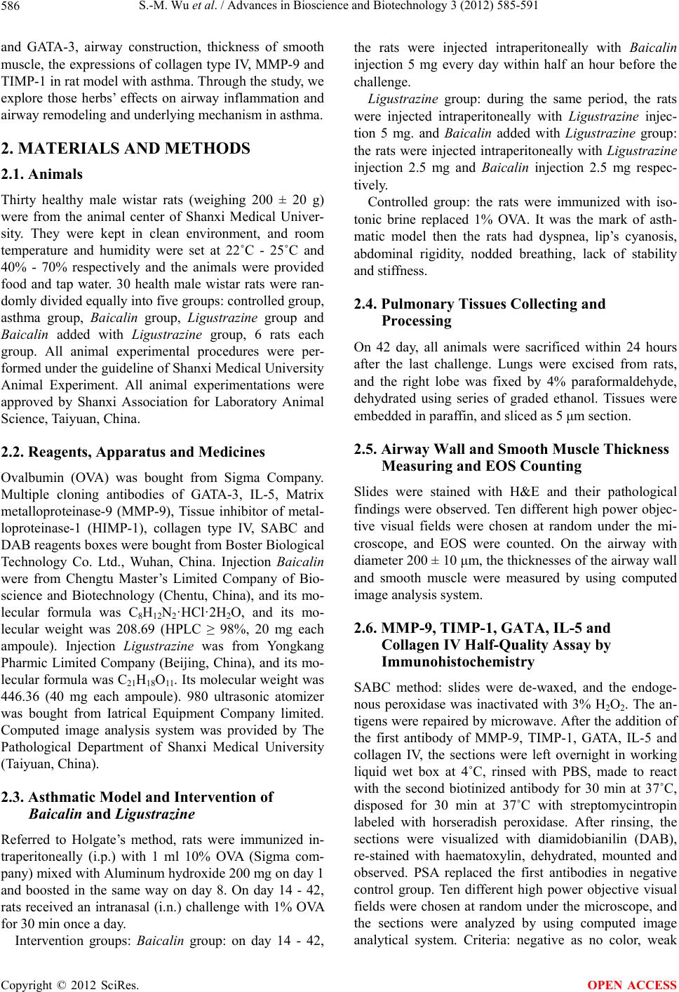 S.-M. Wu et al. / Advances in Bioscience and Biotechnology 3 (2012) 585-591 586 and GATA-3, airway construction, thickness of smooth muscle, the expressions of collagen type IV, MMP-9 and TIMP-1 in rat model with asthma. Through the study, we explore those herbs’ effects on airway inflammation and airway remodeling and underlying mechanism in asthma. 2. MATERIALS AND METHODS 2.1. Animals Thirty healthy male wistar rats (weighing 200 ± 20 g) were from the animal center of Shanxi Medical Univer- sity. They were kept in clean environment, and room temperature and humidity were set at 22˚C - 25˚C and 40% - 70% respectively and the animals were provided food and tap water. 30 health male wistar rats were ran- domly divided equally into five groups: controlled group, asthma group, Baicalin group, Ligustrazine group and Baicalin added with Ligustrazine group, 6 rats each group. All animal experimental procedures were per- formed under the guideline of Shanxi Medical University Animal Experiment. All animal experimentations were approved by Shanxi Association for Laboratory Animal Science, Taiyuan, China. 2.2. Reagents, Apparatus and Medicines Ovalbumin (OVA) was bought from Sigma Company. Multiple cloning antibodies of GATA-3, IL-5, Matrix metalloproteinase-9 (MMP-9), Tissue inhibitor of metal- loproteinase-1 (HIMP-1), collagen type IV, SABC and DAB reagents boxes were bought from Boster Biological Technology Co. Ltd., Wuhan, China. Injection Baicalin were from Chengtu Master’s Limited Company of Bio- science and Biotechnology (Chentu, China), and its mo- lecular formula was C8H12N2·HCl·2H2O, and its mo- lecular weight was 208.69 (HPLC ≥ 98%, 20 mg each ampoule). Injection Ligustrazine was from Yongkang Pharmic Limited Company (Beijing, China), and its mo- lecular formula was C21H18O11 . Its molecular weight was 446.36 (40 mg each ampoule). 980 ultrasonic atomizer was bought from Iatrical Equipment Company limited. Computed image analysis system was provided by The Pathological Department of Shanxi Medical University (Taiyuan, China). 2.3. Asthmatic Model and Intervention of Baicalin and Ligustrazine Referred to Holgate’s method, rats were immunized in- traperitoneally (i.p.) with 1 ml 10% OVA (Sigma com- pany) mixed with Aluminum hydroxide 200 mg on day 1 and boosted in the same way on day 8. On day 14 - 42, rats received an intranasal (i.n.) challenge with 1% OVA for 30 min once a day. Intervention groups: Baicalin group: on day 14 - 42, the rats were injected intraperitoneally with Baicalin injection 5 mg every day within half an hour before the challenge. Ligustrazine group: during the same period, the rats were injected intraperitoneally with Ligustrazine injec- tion 5 mg. and Baicalin added with Ligustrazine group: the rats were injected intraperitoneally with Ligustrazine injection 2.5 mg and Baicalin injection 2.5 mg respec- tively. Controlled group: the rats were immunized with iso- tonic brine replaced 1% OVA. It was the mark of asth- matic model then the rats had dyspnea, lip’s cyanosis, abdominal rigidity, nodded breathing, lack of stability and stiffness. 2.4. Pulmonary Tissues Collecting and Processing On 42 day, all animals were sacrificed within 24 hours after the last challenge. Lungs were excised from rats, and the right lobe was fixed by 4% paraformaldehyde, dehydrated using series of graded ethanol. Tissues were embedded in paraffin, and sliced as 5 μm section. 2.5. Airway Wall and Smooth Muscle Thickness Measuring and EOS Counting Slides were stained with H&E and their pathological findings were observed. Ten different high power objec- tive visual fields were chosen at random under the mi- croscope, and EOS were counted. On the airway with diameter 200 ± 10 μm, the thicknesses of the airway wall and smooth muscle were measured by using computed image analysis system. 2.6. MMP-9, TIMP-1, GATA, IL-5 and Collagen IV Half-Quality Assay by Immunohistochemistry SABC method: slides were de-waxed, and the endoge- nous peroxidase was inactivated with 3% H2O2. The an- tigens were repaired by microwave. After the addition of the first antibody of MMP-9, TIMP-1, GATA, IL-5 and collagen IV, the sections were left overnight in working liquid wet box at 4˚C, rinsed with PBS, made to react with the second biotinized antibody for 30 min at 37˚C, disposed for 30 min at 37˚C with streptomycintropin labeled with horseradish peroxidase. After rinsing, the sections were visualized with diamidobianilin (DAB), re-stained with haematoxylin, dehydrated, mounted and observed. PSA replaced the first antibodies in negative control group. Ten different high power objective visual fields were chosen at random under the microscope, and the sections were analyzed by using computed image analytical system. Criteria: negative as no color, weak Copyright © 2012 SciRes. OPEN ACCESS  S.-M. Wu et al. / Advances in Bioscience and Biotechnology 3 (2012) 585-591 587 positive as wheat color, positive as yellow, strong posi- tive as brown. 2.7. Statistical Analysis All data were presented as mean ± standard deviation (SD). The results were compared by one way analysis of variance (AVONA). The differences between groups were analyzed with LSD method. Multiple Liner regres- sion analysis was employed to analyze the relationship among the parameters (stepwise). SPSS 11.0 software package was used for the evaluation of the statistical significance of the data. Differences were considered significant at P < 0.05. 3. RESULTS 3.1. Pathomorphological Changes There were no inflammation changes in controlled group, however, there were smooth muscle hypertrophy and hyperplasia, thickened basement membrane and airway wall, infiltration of bronchial and parabronchial tissue with lymphocytes and eosinophils, increased plica mu- cosa, desquamation of epithelium and the narrow of air- way lumen in asthma group. In addition, there were less inflammation in Ba icalin, Ligustrazine group and Bai- calin added with Ligustrazine group than those in asthma group (Figures 1-5). 3.2. The Ratio of Airway Wall Luminal to Outer Diameters, the Thicknesses of Smooth Muscle and Collagen Type IV (Table 1) The thicknesses of airway wall and smooth muscle were Figure 1. There was no significant inflammation in controlled group, including no infiltration of bronchial and parabronchial tissues with inflammation cell such as eosinophiles, monocytes and lymphocyte, no smooth muscle hypertrophy and hyperpla- sia, thickened basement membrane, mucus plug, and desqua- mation of epithelia in airway. Figure 2. Airway inflammation was found in asth- matic group, mainly included smooth muscle hyper- trophy and hyperplasia, thickened basement member, oedematous submucosa with infiltration of granulo- cytes, hyperplasia of mucus glands, desquamation of epithelia, and mucus plug. Figure 3. Mild airway inflammation was found in Bai- calin group, included a few of inflammation cells, no or a little of smooth muscle hypertrophy and hyperplasia, and thickened basement member and airway. Figure 4. Light airway inflammation was found in Ligustrazine group, a few of granulocytes infiltrated submucosa, there was no thickened basement mem- ber and airway. Copyright © 2012 SciRes. OPEN ACCESS  S.-M. Wu et al. / Advances in Bioscience and Biotechnology 3 (2012) 585-591 Copyright © 2012 SciRes. 588 than those in Baicalin added with Ligustrazine group; furthermore, collagen IV was higher in asthma group than those in the other groups, as illustrated in Table 1. 3.3. EOS Counts, the Expression of GATA-3 and IL-5 in Lung Tissues (Table 2) EOS count was higher in asthma group than those in the other groups, and EOS count was lower in controlled group than those in the other groups, as also, GATA-3 and IL-5 expressions in lung tissues were higher in asthma group than in the other groups (Table 2). 3.4. The Expressions of MMP-9 and TIMP-1 and MMP-9/TIMP-1 in Lung Tissues (Table 3) Figure 5. No significant inflammation was found in Baicalin added with Ligustrazine group; there was no significant difference in pathological changes, com- pared with the controlled group. The expressions of MMP-9 and TIMP-1 in lung tissues were higher in asthma group than those in the other groups. MMP-9/TIMP-1 was normal in controlled group and Ba icalin added with Ligustrazine group, but it was ot normal in the other groups (Table 3). thicker in asthma group than those in the other groups, and were thicker in Baicalin and Ligustrazine group n Table 1. The ratio of airway wall luminal to outer diameters, the thicknesses of smooth muscle and collagen type IV. Groups n Ratio of luminal to outer airway diameter Thickness of smooth muscle (μm) Collagen IV Controlled 6 0.81 ± 0.04 10.39 ± 0.21 51.76 ± 0.22 Asthma 6 0.63 ± 0.04▲ 19.83 ± 0.57▲ 54.32 ± 1.04▲ Baicalin 6 0.75 ± 0.03▲△● 11.52 ± 0.27▲△○ 53.31 ± 0.58△ Ligustrazine 6 0.77 ± 0.03▲△ 11.33 ± 0.36▲△○ 52.67 ± 0.18△ Baicalin added with Ligustrazine 6 0.74 ± 0.05▲△● 10.69 ± 0.28△ 51.92 ± 0.18△ Compared with controlled group, ▲P < 0.05, compared with asthma group, △P < 0.05, compared with Ligustrazine group, ●P < 0.05, compared with Baicalin added with Ligustrazine group, ○P < 0.05. Table 2. EOS counts, the expression of GATA-3 and IL-5 in lung tissues. Groups N EOS (number/mm2) GATA-3 IL-5 Controlled 6 2.70 ± 0.43 0.083 ± 0.016 0.068 ± 0.019 Asthma 6 31.17 ± 0.83▲ 0.347 ± 0.015▲ 0.330 ± 0.019▲ Baicalin 6 16.58 ± 3.18▲△ 0.294 ± 0.033▲△ 0.261 ± 0.014 Ligustrazine 6 14.07 ± 0.84▲△# 0.192 ± 0.017▲△# 0.187 ± 0.020 Baicalin added with Ligustrazine 6 6.83 ± 0.98▲△#* 0.165 ± 0.016▲△#* 0.137 ± 0.021△ Compared with controlled group, ▲P < 0.000, compared with asthma group, △P < 0.000, compared with Baicalin group, #P < 0.01, compared with Ligustrazine group, *P < 0.000. Table 3. The expressions of MMP-9 and TIMP-1 and MMP-9/TIMP-1 in lung tissues. Groups N MMP-9 TIMP-1 MMP-9/TIMP-1 Controlled 6 53.51 ± 0.25 52.84 ± 0.25 1.01 ± 0.01 Asthma 6 54.99 ± 0.83▲ 60.24 ± 3.02▲ 0.92 ± 0.11▲ Baicalin 6 53.22 ± 0.27△ 54.03 ± 0.69△ 0.99 ± 0.02 Ligustrazine 6 52.70 ± 0.49△ 54.18 ± 1.03△ 0.97 ± 0.03 Baicalin added with Ligustrazine 6 53.31 ± 0.18△ 52.41 ± 0.41△ 1.02 ± 0.02△ Compared with controlled group, ▲P < 0.02, compared with asthma group, △P < 0.01. OPEN ACCESS  S.-M. Wu et al. / Advances in Bioscience and Biotechnology 3 (2012) 585-591 589 3.5. Multiple Liner Regression Analysis for EOS, Thickness of Smooth Muscle and Collagen IV (Multiple Liner Regression Equation) EOS = 96.625IL-5 + 0.982MMP-9 – 57 .2 38 Regression coefficients is 0.957 P < 0.000 Thickness of smooth muscle = –2.639MMP-9 + 2.985TIMP-1 + 185.661MMP-9/TIPM-1 – 0.4collagen IV + 0.39E OS – 9.468GATA-3 – 174.207 Regression coefficients is 0.966 P < 0.000 CollagenIV = –1.113MMP-9 + 1.150TIMP-1 + 67.097MMP-9/TIPM-1 – 2.9EOS – 15.08GATA-3 + 26.432IL-5 – 17.8 8 3 Regression coefficients is 0.771 P < 0.001 4. DISCUSSION Asthama is a serious global disease that cannot be cured effectively so far. Both inhaled glucocorticosteroids and β2-agonists are considered as two effective medicines to treat asthma. However, the shortcomings brought by them cannot be ignored. For example, both two medi- cines have side effects and it is still not clear that they can improve the airway remodeling. Herb, as the tradi- tional Chinese medicine, has significant advantages such as little side effects and more focuses on the balance be- tween location and the whole body. But the deficiency of hard to extract is the main obstruction of putting herb into practice. However, Baicalin and Lingustrazine have the feature of extracting easily which are not shared by most of the herbs. The following is going to discuss how the two medicines control the airway inflammation and improve airway remodeling. Bronchial asthma is an allergic disease characterized with airway inflammation, airway remodeling and airway hyperresponsiveness. Chronic airway inflammation was considered as the essential of asthma, and EOS played an important role in chronic airway inflammation which was associated with asthma severeness [3]. In addition, IL-5 played a key part in activating EOS and regulating EOS function, and could specifically induce EOS and make EOS growing, differentiating and having activities [4]. Airway remodeling of bronchial asthma involved the subepithelial collagen deposition, thickened basement membrane and smooth muscle hypertrophy or hyperplasia in the biopsy of bronchial mucosa in patients with asthma. Airway structure change and airflow obstruction could not be improved through anti-inflammation medication and bronchodilator so it resulted in airway remodeling which aggravated airway narrow, airflow resistance, and airway hyperresponsiveness [5]. The extracellular matrix (ECM) in the bronchial wall was recently reported to be an important component in the maintenance of histological homeostasis of airway tract by balancing synthesis and degradation of the struc- tural component of the composition. Balance between MMPs and TIMPs is a determinant to maintain the bal- ance between synthesis and degradation of ECM [6]. MMP-9 and TIMP-1 are main members in MMPs and TIMPs families respectively. MMP-9 could degrade ECM, and TIMP-1 is the physiological inhibitor of MMP-9. Normal cells secret MMP-9 and TIMP-1 in the ratio of 1 to 1, and they combined dissoluble non-covalence with each other, which is irreversible and stable. In addition, the research showed that the balance between MMP-9 and TIMP-1 was associated with airway remodeling in asthma [6]. Baicalin is one kind of herbs, and acts as anti-in- flammation and against allergy. The reports found that Baicalin could decrease the EOS count in BALF, sputum and blood and WBC count in BALF in sensitized Guinea pig [7]. Ligustrazine is a herb too, it can improve circula- tion of blood and anticoagulation, and treated the patients with COPD, cardiac-encephalic vascular diseases, throm- boembolism disease [8]. A few reports represented the two herbs could treat airway inflammation and remodel- ing in patients with asthma. This experiment found that the ratio of luminal to outer airway diameter was fewer in asthma group than in the other groups; meanwhile, the thicknesses of smooth muscle and the expression of collagen IV were higher in asthma group than in the other groups. The intervention with Ba icalin and Ligustrazine made the thicknesses of smooth muscle and the expression of collagen IV de- creased, and ratio of luminal to outer airway diameter increased. Furthermore, the thickness of smooth muscle was thinner in Baicalin added with Ligustrazine group than in Baicalin group and Ligustrazine group, which proved that Baicalin and Ligustrazine had a synergetic effect on ameliorating the thickness of smooth muscle. This study proved that two herbs reduced airway in- flammation (decreased Eosinophil infiltration of airway) through inhibiting expressiones of GATA-3 and IL-5. The experiment results found that EOS, GATA-3 and IL-5 were higher in asthma group than in the other groups. GATA-3 and EOS were higher in Baicalin and Ligustrazine groups than in Baicalin added with Ligus- trazine group, and IL-5 was higher in Baicalin group than in Baicalin added with Ligustrazine group. The re- cent research showed that GATA-3 was a specific tran- scription factor of TH2 lymphocyte, and induced TH2 lymphocyte to secrete cytokine. Inhibiting the expression of GATA-3 can decrease the inflammation of airway led by TH2 lymphocyte [9,10]. These results did also con- firm that Baicalin and Ligustrazine could decrease the Eosinophil infiltration of airway by inhibiting the ex- pressiones of GATA-3 and IL-5. In addition, Baicalin added with Ligustrazine group excelled Baicalin and Copyright © 2012 SciRes. OPEN ACCESS  S.-M. Wu et al. / Advances in Bioscience and Biotechnology 3 (2012) 585-591 590 Ligustrazine groups at decreasing EOS infiltration and the expressions of GATA-3 and IL-5. Two herbs had synergetic effect. This experiment showed that the expressions of MMP-9 and TIMP-1 in the airway wall of rats were higher in asthma group than in the other groups, and the ratio of MMP-9/TIMP-1 was abnormal in asthma group, and it was significantly lower than in both controlled group and Baicalin added with Ligustrazine groups. The ratio of MMP-9/TIMP-1 was normalized in Baicalin added with Ligustrazine group. However, the ratio of MMP-9/ TIMP-1 in Baicalin group and Ligustrazine group was not significant compared with asthma group, so the in- tervention of Baicalin added with Ligustrazine could have a synergetic effect in regulating the ratio of MMP- 9/TIMP-1. Studies showed that MMP-9, TIMP-1 and MMP-9/ TIMP-1 played crucial roles in airway remodeling [8,9]. To understand underlying mechanism of airway in- flammation and remodeling, multiple liner regression analysis was made. ESO infiltration of airway was pre- sented as the characteristic of airway inflammation, and the thickness of smooth muscle and the content of colla- gen IV were presented as the marks of airway remodel- ing. Furthermore, their metabolism was influenced with airway inflammation and remodeling cytokines. Multiple liner regression analysis found the positive correlation between EOS and IL-5 and MMP-9, which showed that airway inflammation cytokine (IL-5) was associated with EOS infiltration of airway, in addition, EOS increasing with higher expression of MMP-9 proved airway in- flammation associated with ECM degraded. There were the positive correlations between the thickness of smooth muscle and TIMP-1, MMP-9/TIMP-1 and EOS, however, and the negative correlation between the thicknesses of smooth muscle and MMP-9, collagen IV, and GATA-3, meanwhile, the positive correlation between the expres- sions of collagen IV and TIMP-1, MMP-9/TIMP-1, IL-5, nevertheless, the negative correlation between the ex- pressions of collagen IV and MMP-9. Those results showed that the higher expression of TIMP-1 and the abnormal ratios of MMP-9/TIMP-1 increased the thick- ness of smooth muscle and the expression of collagen IV, however, the higher expression of MMP-9 decreased them. The result proved that MMP-9 degraded ECM, and TIMP-1 inhibited activity of MMP-9. In addition, these results also proved that airway inflammation (EOS infil- tration) induced higher expression of MMP-9, then the expression of TIMP-1 did compensative increased, so that MMP-9/TIMP-1 kept from unbalance to balance. In same way, repeated airway inflammation and asthma exacerbation made MMP-9/TIMP-1 unbalance and bal- ance in turn. Finally, these pathological changes resulted in the decompensation of collagen (deposition of colla- gen IV), the hypertrophy of smooth muscle and the changes of airway structure, teamed airway remodeling. Those results were similar to Xu’s [11] and Hoshino’s [12] ones. The results manifested that EOS infiltration of airway, the thickness of smooth muscle and the content of collagen IV played an important part in airway in- flammation, maintaining airway structure and airway remodeling respectively. With the intervention of Bai- calin and Ligustrazine, the expressions of MMP-9 and TIMP-1 were decreased, and MMP-9/TIMP-1 tended to be normal. In addition, MMP-9/TIMP-1 in Baicalin added with Ligustrazine group was not significant com- pared with controlled group, but significant compared with asthma group. However, MMP-9/TIMP-1 in both Baicalin group and Ligustrazine group was not signifi- cant compared with asthma group, so the result proved that Baicalin and Ligustrazine had synergetic effect on regulating MMP-9/TIMP-1. In summary, the results found that EOS infiltration in airway in asthma group was more than in the other groups; smooth muscle thickness in asthma group was thicker than in the other groups; the expression of colla- gen IV in asthma group was higher than in the other groups. Baicalin and Ligustrazine could inhibite airway inflammation, decrease smooth muscle thickness, dimin- ish the deposition of collagen IV and increase the ratio of luminal to outer airway diameter through regulating air- way inflammation cytokine (IL-5, GATA-3), infiltration of EOS and airway remodeling cytokine (MMP-9, TIMP- 1 and MMP-9/TIMP-1). Furthermore, Baicalin and Ligustrazine had synergetic effect on treating asthma, especially airway remodeling of asthma, and both herbs will be the effective medicines for treating asthma in future. 5. CONCLUSION Airway inflammation and airway wall remodeling were two important aspects in asthma, and they were influ- enced by each other. Eos counts and the expression of IL-5 and GATA-3 in the lung tissues in asthma group were significantly higher than the other groups; mean- while, the expressions of MMP-9 and TIMP-1 signifi- cantly were higher too. Additionally, the thicknesses of airway wall and airway smooth muscle in asthma group significantly increased. After intervention with Baica lin or Ligustrazine, the thicknesses of airway wall and air- way smooth muscle became thinner than those in asthma group, at the same time, the expression of MMP-9 and TIMP-1 significantly decreased. Smooth muscle thick- ness and collagen type IV were associated with EOS, IL-5, TAGA-3, MMP-9, TIMP-1 and MMP-9/TIMP-1. Two herbs could inhibit the expressions of IL-5 and GATA-3 and the infiltration of EOS and decrease the Copyright © 2012 SciRes. OPEN ACCESS  S.-M. Wu et al. / Advances in Bioscience and Biotechnology 3 (2012) 585-591 Copyright © 2012 SciRes. 591 deposition of collagen type IV and reduce the airway smooth muscle thickness through regulating MMP-9 and TIMP-1 level and influencing the balance between MMP- 9 and TIMP-1, and had synergetic effect. This will pro- vide new therapeutic way to asthmatic patients. OPEN ACCESS 6. ACKNOWLEDGEMENTS We wish to thank the Shanxi Science and Technology Department for the funding of the study. We are also grateful to professor Jun-mai Zhao for help in the computed image analysis. Foundation: International Cooperative Project of Shanxi Province, China, 30370608. REFERENCES [1] Holgate, S.T., Peters-Goiden, M., Jpanettieri, R.A., et al. (2003) Roles of cysteinyl leukotrienes in airway inflam- mation, smooth muscle function, and remodeling. Journal of Allergy and Clinical Immunology, 111, S18-S34. doi:10.1067/mai.2003.25 [2] Menzies-Gow, A. and Robinson, D.S. (2002) Eosinophils eosinophilis cytokines (interleukin-5), and antieosinophlllc therapy in asthma. Current Opinion in Pulmonary Medi- cine, 8, 33-38. doi:10.1097/00063198-200201000-00006 [3] Duncan, C.J.A., Lawrie, A., Blaylock, H.G., et al. (2003) Reduced eosinophil apoptosis in induced sputum corre- lates with asthma severity. European Respiratory Journal, 22, 484-490. doi:10.1183/09031936.03.00109803a [4] Humbles, A.A., Lloyd, C.M., Mc Millan, S.J., et al. (2004) A critical role of eosinophile in allergic airways remod- eling. Science, 305, 1776-1779. doi:10.1126/science.1100283 [5] Vignola, A.M., Riccobono, L., Mirabella, A., et al. (1998) Sputum metalloproteinase-9/tissue inhibitor of metallo- proteinase-1 ratio correlates with airflow obstruction in asthma and chronic bronchitis. American Journal of Res- piratory and Critical Care Medicine, 158, 1945-1950. [6] Cataldo, D.D., Tournoy, K.G., Vermaelen, K., et al. (2002) Matrix metalloproteinase-9 deficiency impairs cel- lular infilatration and bronchial hyperresponsiveness dur- ing allergen induced airway inflammation. American Journal of Pathology, 161, 491-498. doi:10.1016/S0002-9440(10)64205-8 [7] Jiang, B., Zhang, S.M., Li, Q., et al. (2001) Protection of Baicalin in asthmatic model of guinea-pigs. Pharmaceu- tical Care and Research, 1, 36-39. [8] Ni, Q. and Dong, J.C. (2004) Experimental study of the effect of components of three traditional Chinese medi- cines on airway allegic inflammation in guinea pigs with bronchial asthma. Chinese Journal of Experimental Tra- ditional Medical Formulae, 10, 49-51. [9] Finotto, S., De Sanctis, C.T., Lehr, H.A., et al. (2001) Treatment of allergic airway inflammation and hyperre- sponsiveness by antisense-induced local blockade of GATA-3 expression. The Journal of Experimental Medi- cine, 193, 1247-1260. doi:10.1084/jem.193.11.1247 [10] Palmans, E., Kips, J.C. and Paumwels, R.A. (2000) Pro- longed allergen exposure induces structural airway changes in sensitized rats. American Journal of Respiratory and Critical Care Medicine, 161, 627-635. [11] Xu, S.Y., Xu, Y.J., Zhang, Z.X., et al. (2006) Pathologi- cal features and mechanism of airway remodeling in asthmatic rats. Journal of Huazhong University of Sci- ence and Technology (Health Sciences), 35, 465-468. [12] Hoshino, M., Takehashi, M., Takai, Y., et al. (1999) In- haled corticosteroids decrease subepithelial collagen depo- sition by modulation of the balance between matrix met- alloproteinase-9 and inhibitor of metalloproteinase-1 ex- pression in asthma. Journal of Allergy and Clinical Im- munology, 104, 356-363. doi:10.1016/S0091-6749(99)70379-9
|