M. Irshad et al. / Advances in Bioscience and Biotechnology 3 (2012) 580-584 581
peel waste was obtained from the local fruit market, Gu-
jrat, Pakistan and used as a growth supported solid sup-
port. Before to use substrate was first crushed into pieces,
oven dried and finally ground to fine particle size.
2.2. Fungal Culture and Inoculum Development
Fungal strain T. viride was available in the Molecular
Biotechnology Laboratory, Department of Biochemistry
and Molecular Biology, University of Gujrat, Pakistan. A
spore suspension inoculum of T. viride was developed in
an Erlenmeyer flask containing 30 mL of Potato Dex-
trose broth at 30˚C ± 1˚C for 5 days after sterilizing at 15
lbs/in2 pressure and 121˚C.
2.3. Pretreatment of Orange Peel Waste
10 g of moisture free orange peel was pretreated with 2%
HCl by adopting thermal treatment methodology as de-
scribed previously [1]. After pretreatment the slurry of
the substrate was filtered using Whatman No. 1 filter pa-
per. Residues were washed 3 times with distilled water to
remove extra acidity and used for production of Exo 1,
4-β glucanase under optimum fermentation conditions.
2.4. Solid-State Fermentation
For the production of Exo 1, 4-β glucanase 10 g pre-
treated orange peel was moist with Basel salt media in an
Erlenmeyer flask (250 mL) capacity. The major con-
stituents of the Basel media were: (NH4)2SO4, 10 g·l–1;
KH2PO4, 4 g·l–1; MgSO4·7H2O, 0.5 g·l–1 and CaCl2, 0.5
g·l–1. Orange peel based sterilized Solis-State medium
was inoculated with 5 mL of freshly prepared fungal spore
suspension and incubated at 30˚C ± 1˚C for stipulated
fermentation time period under still culture conditions.
2.5. Extraction of Exo 1, 4-β Glucanase
At the end of selected incubation period, Exo 1, 4-β glu-
canase was extracted from the fermented biomass by
adding 100 mL of 0.1 M succinate buffer of pH 5 and the
flasks were shaken at 120 rpm for 30 min. The contents
were filtered and filtrates were centrifuged at 10,000 × g
(4˚C) for 10 min. A carefully collected supernatants were
and used to determine enzyme activity and for purifica-
tion purposes.
2.6. Determination of Exo 1, 4-β Glucanase
Activity and Protein Contents
Exo 1, 4-β glucanase was assayed according to the method
of Deshpande et al. [11], using 1% salicin as reaction
substrate with DNS as coupling reagent. The reaction
mixture contained 0.1 mL of enzyme extract with 1 mL
of 1% salicin and 1 mL of 0.1 M succinate buffer of pH 5.
The mixture was incubated for 30 min at 50˚C and the
reaction was then terminated by adding DNS reagent (2
mL). The reaction mixtures were heated for 15 min in a
boiling water bath followed by cooling in ice. The ab-
sorbance was measured at 540 nm against reagent blank.
One unit of enzyme activity was defined as the amount
of glucose (μmol) released by 1 mL of enzyme solution
per min. To determine the protein contents of the crude
and purified enzyme extracts bovine serum albumin was
used as standard.
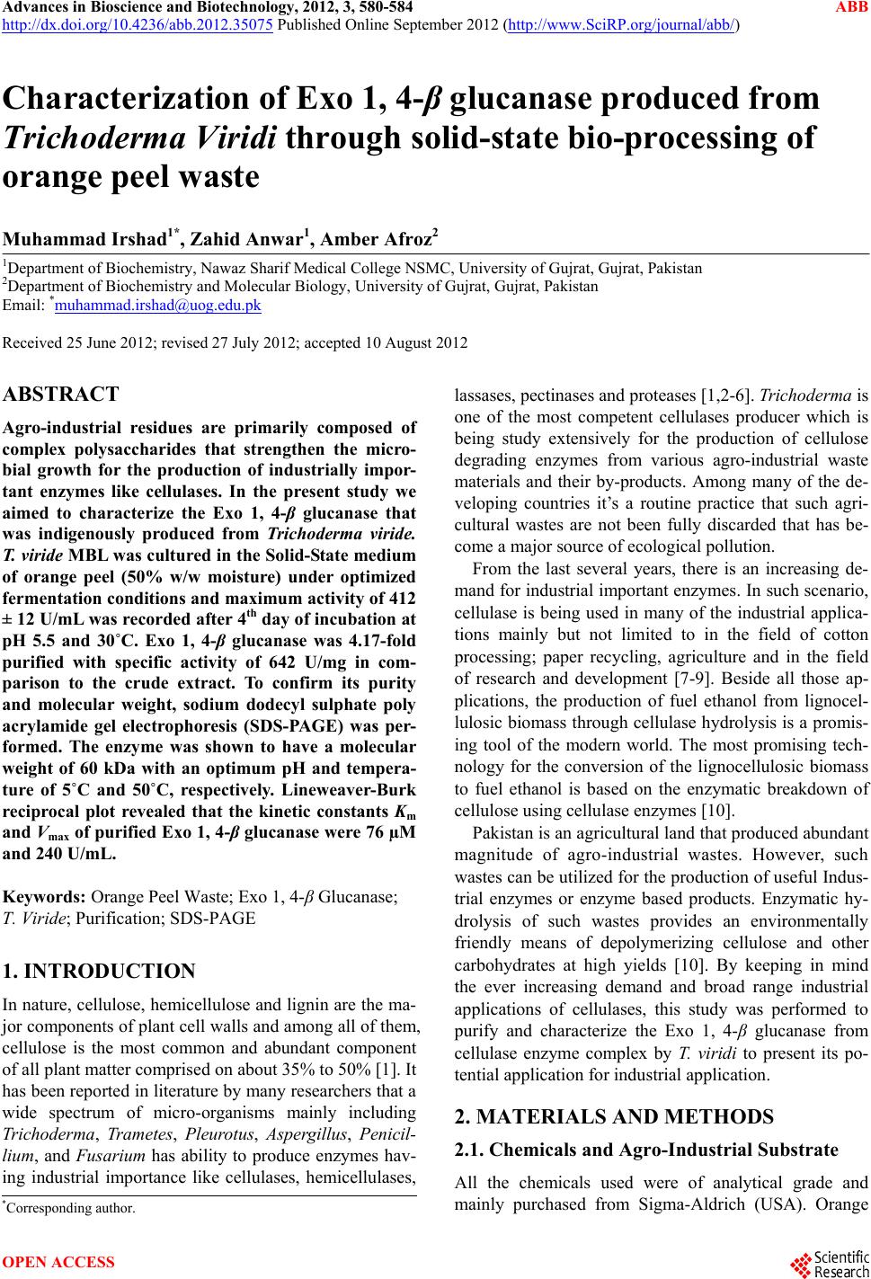
2.7. Purification of Exo 1, 4-β Glucanase
To purify the crude extract of Exo 1, 4-β glucanase ob-
tained from T. viridi ammonium sulfate fractionation fol-
lowed by the Sephadex-G-100 (Sigma, USA) column
(120 × 2 cm) gel filtration chromatographic technique
was adopted as described by Iqbal et al. [1], for purifica-
tion purposes. Total proteins and activity of partially pu-
rified Exo 1, 4-β glucanase were determined before and
after each purification step as described earlier.
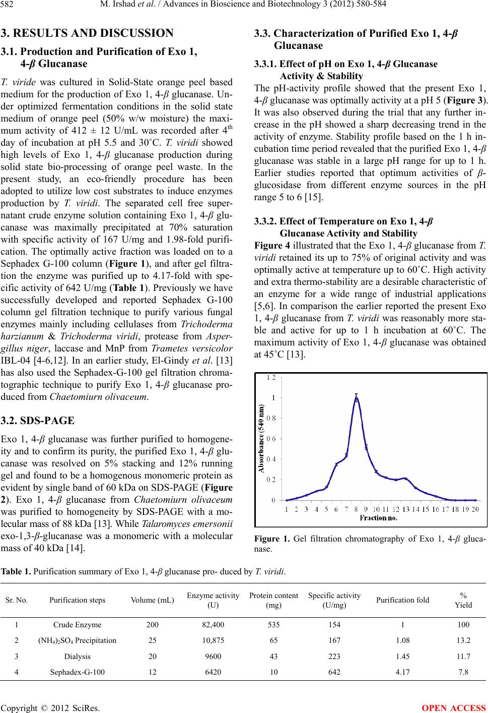
2.8. SDS-PAGE
To determine the molecular weight of purified Exo 1, 4-β
glucanase sodium dodecyl sulphate poly acrylamide gel
electrophoresis (SDS-PAGE) was performed on a 5%
stacking and a 12% resolving gel according to the meth-
odology, as described previously [1].
2.9. Characterization of Purified Exo 1, 4-β
Glucanase
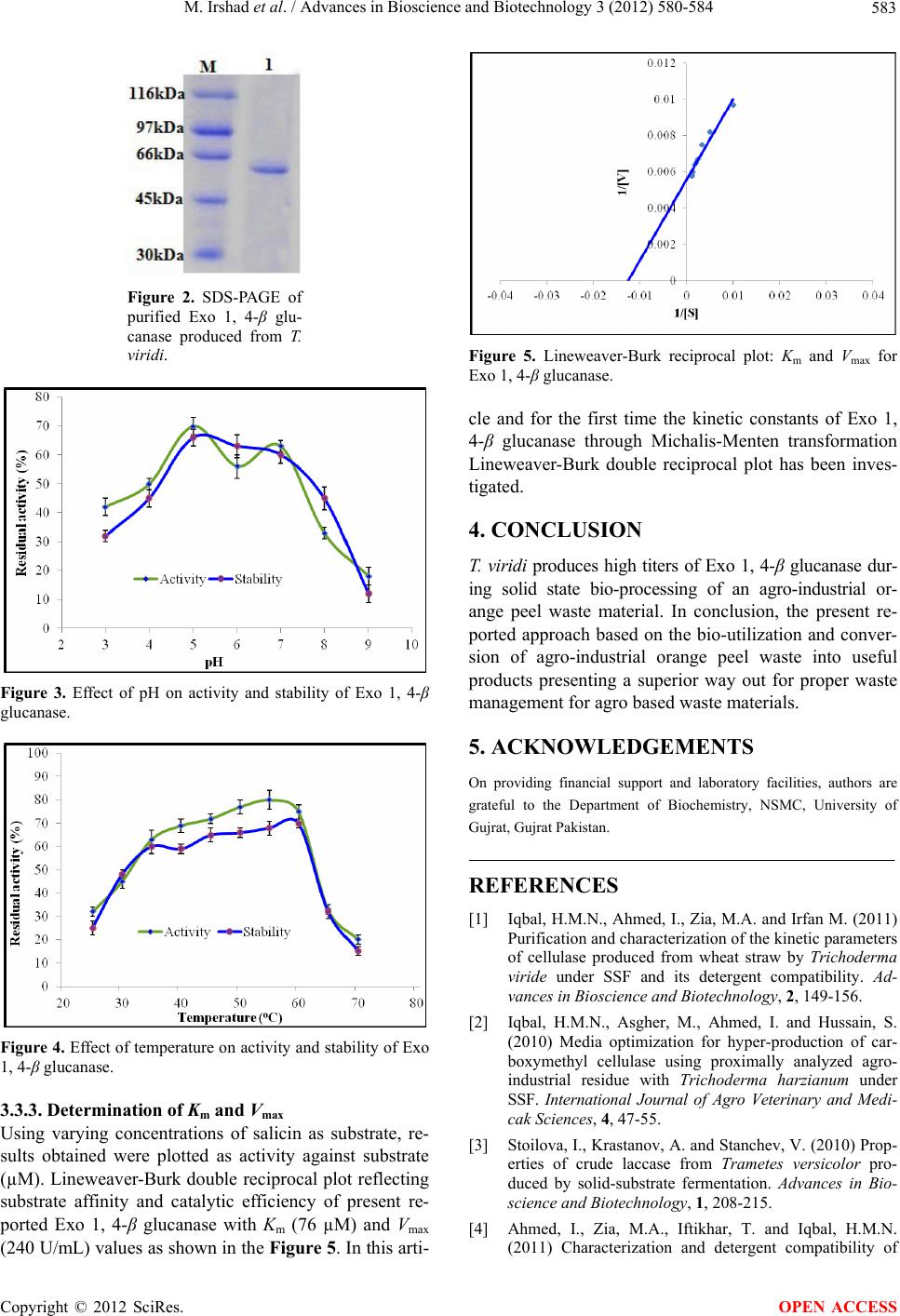
Characterization of purified Exo 1, 4-β glucanase was
done by studying the effect of various kinetic parameters
including pH, temperature and substrate concentration on
the Exo 1, 4-β glucanase activity. To investigate the ef-
fect of pH Exo 1, 4-β glucanase was incubated in buffers
of different pH (2 - 10), followed by standard assay pro-
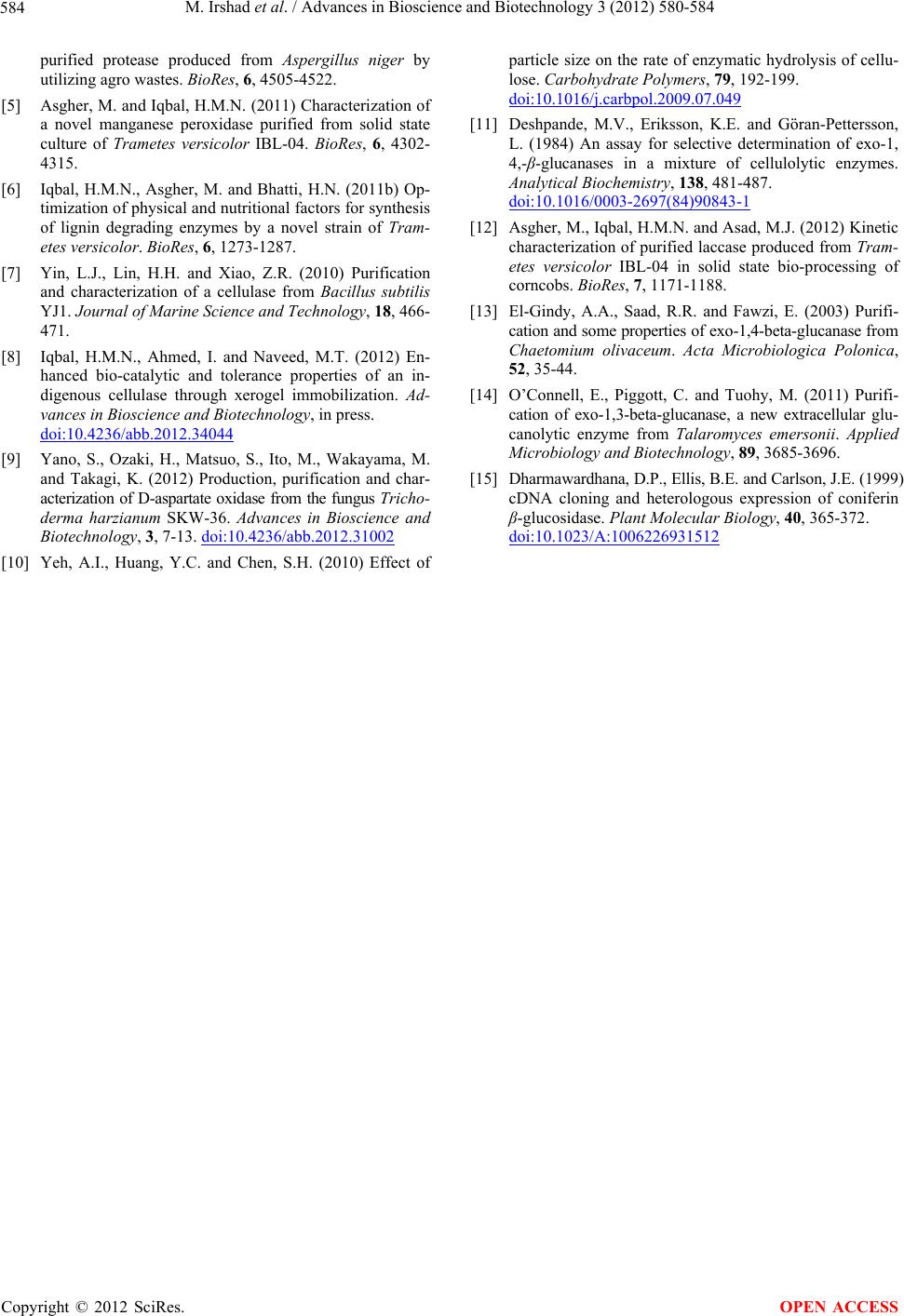
tocol. To determine the thermal features Exo 1, 4-β glu-
canase was incubated without substrate under different
temperatures ranging from 25˚C to 70˚C for 1 h time
period followed by normal assay protocol as previously
described. The Michalis-Menten kinetic constants Km
and Vmax for Exo 1, 4-β glucanase were calculated from
Lineweaver-Burk reciprocal plots using varying concen-
trations of salicin as substrate.
2.10. Statistical Analysis
All the experimental data was conducted in triplicate and
presented as mean ± standard error (SE) and SE are
showed in figures as Y-error bars.
Copyright © 2012 SciRes. OPEN ACCESS