Journal of Cosmetics, Dermatological Sciences and Applications, 2012, 2, 174-178
http://dx.doi.org/10.4236/jcdsa.2012.23033 Published Online September 2012 (http://www.SciRP.org/journal/jcdsa)
Re-Evaluation of Acquired Epidermodysplasia
Verruciformis in Kidney Transplant Recipients*
Khalifa E. Sharquie1#, Adil A. Noaimi2, Ali A. Al-Jobori3
1Chairman of Scientific Council of Dermatology and Venereology—Iraqi Board for Medical Specializations, Baghdad, Iraq; 2Department
of Dermatology and Venereology, College of Medicine, University of Baghdad, Baghdad, Iraq; 3Department of Dermatology and
Venereology, Baghdad Teaching Hospital, Medical City, Baghdad, Iraq.
Email: #ksharquie@yahoo.co.uk, adilnoaimi@yahoo.com, ali60aljobori@yahoo.com
Received March 2nd, 2012; revised April 5th, 2012; accepted April 16th, 2012
ABSTRACT
Background: Epidermodysplasia verruciformis (EV) is a skin disease associated with numerous early onset viral warts
on the sun exposed parts of the body that usually progress into different skin malignancies. Kidney transplant patients
are also commonly associated with viral warts that might change into skin cancers. Objective: To re-evaluate all fea-
tures of the acquired epidermodysplasia verruciformis in kidney transplant patients in comparison with healthy indi-
viduals. Patients and Methods: This case controlled study that included 100 patients with kidney transplantation (87
males, 13 females) their ages ranged from 14 - 70 (46.65 ± 4.74) years and was done in kidney Transplantation Centers
in Baghdad and Al-Karma Teaching Hospitals from June 2009 - August 2010. Patients included were receiving multiple
immunosuppressive drugs like azathioprine, prednisolone, cyclosporine, mycophenolate mofetil ranged from 6 months -
25 years. One hundred healthy individuals (70 males, 30 females), there ages ranged from 14 - 73 (41.63 ± 9.55) years,
had been examined as a control for the presence of viral warts and tumors. These control cases had been taken randomly
from general population in multiple regions in Baghdad. Results: Forty (40%) patients out of one hundred kidney trans-
plant patients had viral warts and were seen in 9 (9%) of control group and there was statistically significant difference
between patients and control cases (P value < 0.00001). Most of the viral warts were multiple and of the verrucae vul-
garis type and were mainly located on the exposed areas of the body, mostly on the face and dorsa of the hands. Solar
keratosis admixed with viral warts were found in 14 out of 100 kidney transplant patients. Skin malignancies in these
fourteenth patients were observed in 6 (42.14%) cases: 2 squamous cell carcinoma (SCC) and 4 basal cell carcinoma
(BCC) and these were mainly located on the exposed areas of the body, mostly on the face and dorsa of the hands. No
skin malignancies were observed in control group. Conclusions: Kidney transplant recipients had all the features that
simulate the inherited epidermodysplasia verruciformis.
Keywords: Acquired Epidermodysplasia Verruciformisin; Kidney Transplant; Tumors
1. Introduction
Epidermodysplasia verruciformis (EV) is an inherited
autosomal recessive gene disorder in which there is early
onset, numerous, widespread, persistent, and refractory
infection with human papilloma virus (HPV) [1-4]..The
individual lesions typically have either the appearance of
that warts or flat scaly red-brown macules, resemble le-
sions of pityriasis versicolor or pityriasis rosea. The first
type of lesion is usually caused by the same HPV types
as those found in flat warts in the general population (e.g.
HPV-3 & 10),while the second one is usually caused by
EV HPV types (e.g. HPV-5, 8, 9, 12, 14, 15, 17, 19-25,
28, 29, 36-38, 47, 49 and 50) [2-7]. There may be more
than one HPV type in the same patient [5,6]. The patho-
genesis of this syndrome is unknown but is felt to be a
specific defect of cell mediated immunity [2-4,8].
Some EV patients are at high risk of developing cuta-
neous squamous cell carcinoma (SCC) [1-4,9]. Although,
pityriasis like lesions caused by any EV type may be at
increased risk of becoming malignant, the risk appears to
be greatest for those caused by HPV 5, 8 and 47 types
[1-4,6,9]. The lesions of individuals with EV who have
only flat warts caused by non-EV HPV types do not
seem to be at increased risk of becoming malignant. Squa-
mous cell carcinomas develop in 30% - 60% patients.
Most often, skin cancers appear on sun exposed sur-
faces, but they can appear on any parts of the body.
They begin to appear at ages 20 - 40 years. HPV 5, 8 and
47 are found in more than 90% of EV skin cancers. The
*This study was an independent study and not funded by any of the
drug companies.
#Corresponding author.
Copyright © 2012 SciRes. JCDSA
Re-Evaluation of Acquired Epidermodysplasia Verruciformis in Kidney Transplant Recipients 175
squamous cell carcinoma may appear de-novo but usu-
ally appear on the background of numerous actinic kera-
tosis and lesions of Bowen’s disease, locally and usually
aggressive [1-4].
The aim of the present work is to re-evaluate the cli-
nical features of the acquired epidermodysplasia verru-
ciformisin in kidney transplant recipients.
2. Patients and Methods
This case controlled study that included 100 patients with
kidney transplantation (87 males, 13 females) their ages
ranged from 14 - 70 years with a mean ± SD of 46.65 ±
4.74 years.This was carried out in kidney Transplanta-
tion Centers in Baghdad and Al-Karma Teaching Hospi-
tals from June 2009 - August 2010. All patients with kid-
ney transplant were receiving multiple immunosuppres-
sive drugs like azathioprine, prednisolone, cyclosporine,
mycophenolate mofetil ranged from 6 months - 25 years.
They consisted of rapidly decreasing doses of intrave-
nous methylprednisolone succinate for the first 3 days
followed by oral prednisolone 60 mg/day tapered slowly
to a maintenance dose of 10 - 20 mg/day every other day.
While azathioprine was given at doses were ranging from
1 - 3 mg/kg/day according to the white blood cell count.
Cyclosporine, in combination with azathioprine was also
given orally starting with 8 - 10 mg/kg/day, decreased
every two weeks by 2 mg/kg/day to be continued in a
maintenance dose of 5 mg/kg/day. Mycophenolate mofe-
tal was added with other immunosuppression in a dose 1 -
2 gm/day. History was taken from all patients including
all relevant points. These patients were divided into 3
groups according to the duration of immunosuppressive
therapy: Group A 1 - 10 years, Group B 11 - 20 years
and Group C more than 20 years. Biopsies were taken
from the viral warts lesions, solar keratosis and associ-
ated skin malignancies.
One 100 healthy individuals, 70 males, 30 females,
their ages ranged from 14 - 73 years, a mean ± SD of was
41.63 ± 9.55 years, had been examined as a control for
the presence of viral warts and tumors. This control group
had been collected from general population in multiple
regions in Baghdad City.
Formal consent was taken from each patient after full
explanation about the goal and nature of the present study.
Also, ethical approval was performed by the Scientific
Council of Dermatology and Venereology-Iraqi Board for
Medical Specializations.
Statistical analysis were done using EPI-info version 6
by estimation of both descriptive and analytic statistics. P
value equal or less than 0.05 was considered as significant.
3. Results
Forty patients out of one hundred kidney transplant pa-
tients had viral warts, their ages ranged from 22 - 70
years with a mean ± SD of 39.27 ± 10.62 years, 34 males
and 6 females, and the duration of the warts ranged from
0.3 - 15 years with a mean ± SD of 4.64 ± 3.66 years.
The distribution of patients with viral warts among the
groups was as follows: Group A 28 (70%), Group B 7
(17.5%), Group C 5 (12.5%) cases. While viral warts
seen only in 9 (9%) individuals of control group. There
was statistically significant difference between patients
and control cases (P value < 0.00001). Most of the viral
warts in patients with kidney transplantation were nu-
merous, larger in size and of the verrucae vulgaris type.
They were mainly located on the exposed areas of the
body, mostly on the face and dorsa of hands.
Viral warts admixed up with solar keratosis and skin
malignancies were observed in 14 out of 100 kidney trans-
plant patients: their ages ranged from 35 - 70 years with a
mean ± SD of 54.28 ± 9.35 years and the duration of
solar keratosis ranged from 0.5 - 25 years with a mean
5.54 ± 3.34 years.
The distribution of patients with solar keratosis among
the groups were as follow: Group A 4 (28.58%), Group B
5 (35.71%), Group C 5 (35.71%) cases. While solar kera-
tosis seen in 1 (1%) individual in a control group. Skin
malignancies in these fourteenth patients with combined
viral warts and solar keratosis were observed in 6 (42.14%)
cases: 2 squamous cell carcinoma and 4 basal cell carci-
noma. These skin malignancies were mainly located on
the exposed areas of the body, mostly on the face and
dorsa of the hands. No cases were seen in Group A, while
three (60%) cancer cases in Group B and three (60%)
patients in Group C. No skin malignancies were ob-
served in control group (Figures 1 and 2).
4. Discussion
Kidney transplant recipients receive multiple immuno-
Figure 1. Sixty-seven years old male patient with kidney
transplant showing multiple solar keratosis with big squa-
mous cell carcinomaon the left cheek.
Copyright © 2012 SciRes. JCDSA
Re-Evaluation of Acquired Epidermodysplasia Verruciformis in Kidney Transplant Recipients
176
Figure 2. Sixty five years old patient with kidney transplant
showing numerous solar keratosis admixed up with many
basal cell carcinomas.
suppressive drugs that are used to prevent organ rejec-
tions [10,11]. This immunosuppression usually ends with
increase tendency for infections especially viral warts
[11].
The present work showed that viral warts had been
found in 40% of kidney transplant patients, while viral
warts admixed up with solar keratosis found in 14 (14%)
of cases and sometimes it is not easy to differentiate be-
tween viral warts and solar keratosis as they could be
similar and mixed up with each other’s especially in late
cases. Skin malignancies mainly squamous cell carcino-
ma and basal cell carcinoma were seen in 6 (6%) cases of
kidney transplant patients in combination with viral warts
and solar keratosis. In all cases, these lesions were ob-
served on the sun exposed areas mainly face. This in-
crease in rate of malignancies among patients with kid-
ney transplantations was similar to what has been pub-
lished [4,12-15].
In a previous Iraqi study, viral warts were reported in
50% of patients with kidney transplant while skin ma-
lignnancies was seen in 4.6% of cases (six cases: one
BCC and five SCC) in comparison with the present work
6 (42.14%) of cases (four BCC, two SCC). The present
study showed very important observation as over years
of immunosuppressive therapies, the number of viral
warts decreased while the number of solar keratosis to-
gether with skin malignancies were increased. This was
almost similarly reported [10].
Inherited EV is usually autosomal recessive disease,
that associated with early onset of numerous viral warts
similar to pigmented pityrasis rosea or tinea versicoler
like lesions [1-4] with high tendency to develop skin ma-
lignancies especially squamous cell carcinoma in 30% -
60% of cases. They begin to appear at ages 20 - 40 years
[4]. Most of the malignant tumors remains local, but re-
gional & distant metastasis may occur [1-3]. The sero-
types of HPV that responsible for this oncogensity are 5,
8 and 47 types [1-4,6,9].
It is well known that EV associated with skin malig-
nancies like squamous cell carcinoma and basal cell car-
cinoma and HPV types 5, 8 and 47 have been incrimi-
nated in their etiopathogenesis [1-4,6,9]. Most recently, it
has been observed that HPV had contributed in the de-
velopment of squamous cell carcinoma even in patients
without viral warts [16,17]. So, HPV in patients with kid-
ney transplant could act as a co-factor in the tumor de-
velopment along with immunosuppresion, solar radiation
and other environmental factors [1-4,14].
In reviewing, the clinical picture of the inherited EV in
comparison with that of the clinical features in patients
with kidney transplant, we found the following similar
observations in both conditions: There is immunosup-
pression because of impaired cell mediated immunity,
the rash occurs in young patients, there are numerous
skin lesions, they are recalcitrant to therapy, viral warts
and skin cancers occur on any parts of the body espe-
cially in sun exposed areas of the body and skin malign-
nancies is a common feature [1-10,15,17-20]. The se-
rotypes of human papilloma virus of viral warts and
malignancies in patients with kidney transplant is simi-
lar to serotypes of HPV found in the inherited epider-
modysplasia verruciformis, these HPV types include HPV-
3, 5, 8, 9, 10, 12, 14, 15, 17, 19, 25 and 36 through 38
[17,21,22].
So, the infective agents, the immune defect, clinical
pictures of the skin rash and the course of the disease in
kidney transplant recipients having many similarities with
that of the inherited form of EV [13-20]. These clinical
pictures in patients with kidney transplant have been de-
scribed as acquired epidermodysplasia verruciformis.
The present work had supported the features of this new
entity so called acquired epidermodysplasia verruciforms
[10].
Cytotoxic drugs can induce skin malignancies in pa-
tients with solid organ transplants especially in patients
with kidney transplant and this in collaboration with
other factors like fair skin, UV light exposure, and HPV
infections [10,23-28]. Accordingly these patients should
be protected from sun light exposure and even could be
Copyright © 2012 SciRes. JCDSA
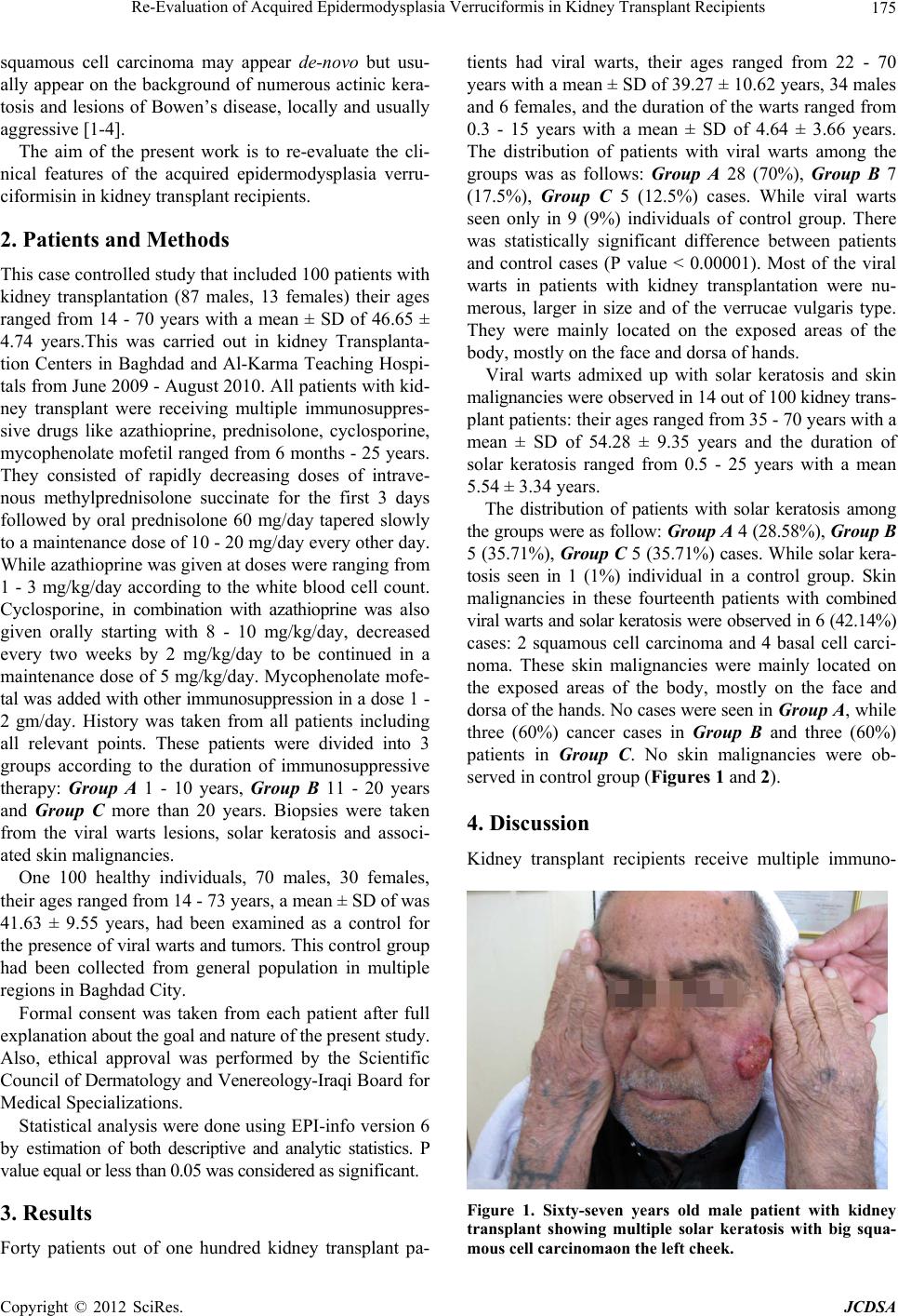
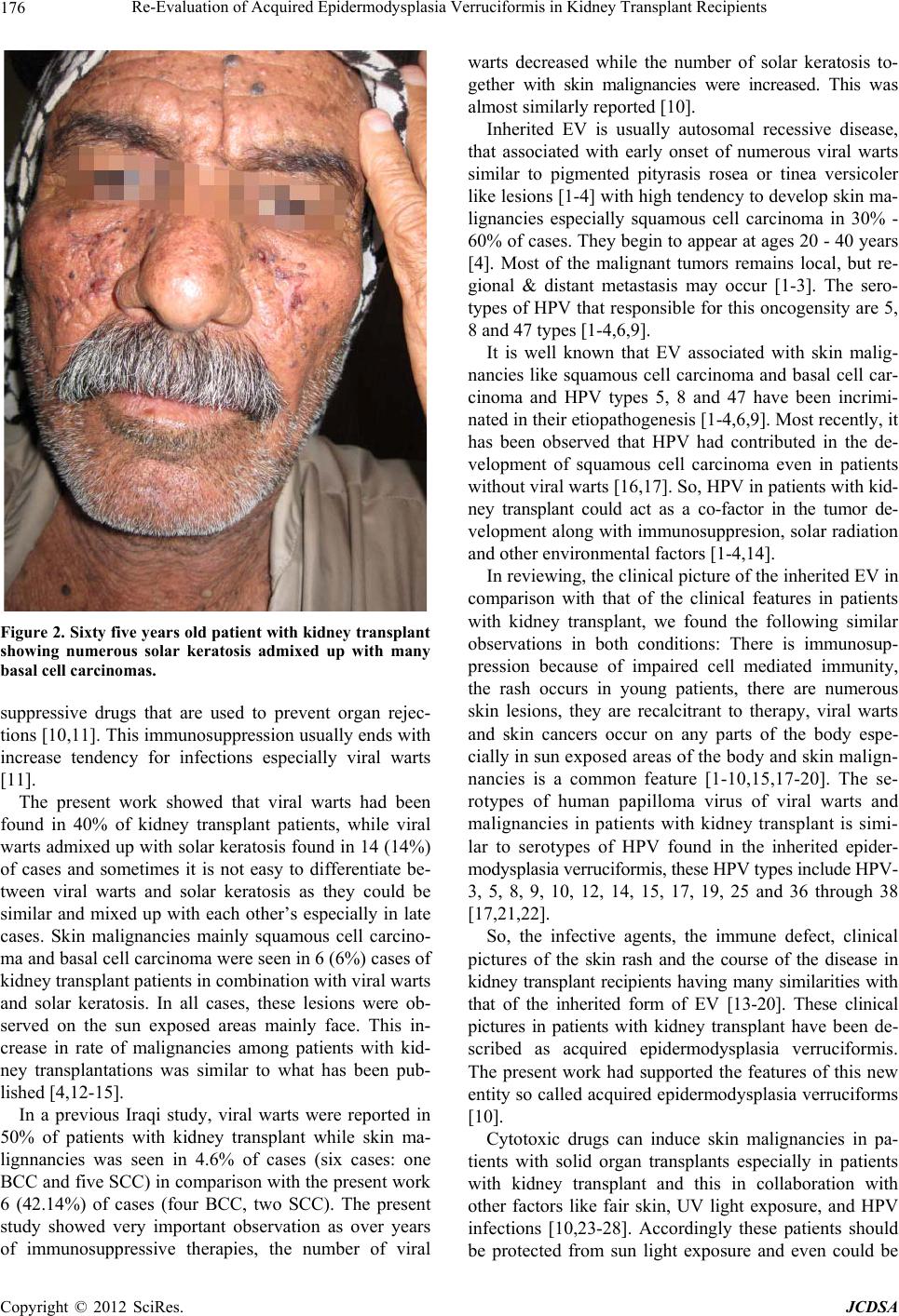
Re-Evaluation of Acquired Epidermodysplasia Verruciformis in Kidney Transplant Recipients 177
immunized against HPV infections before doing organ
transplant and giving cytotoxic drugs for prolonged course.
In conclusion, the features of viral warts, solar kerato-
sis and skin malignancy were recorded in patients with
kidney transplant and deserved the term acquired epi-
dermodysplasia verruciforms as has been reported [10].
REFERENCES
[1] J. C. Sterling and J. B. Kurtz, “Viral Infections,” In: R. H.
Champion, J. L. Burton and D. A. Burn, Eds., Rooks
Textbook of Dermatology, Blackwell Science Ltd. Edito-
rial Office, Oxford, 1998, pp. 995-1095.
[2] N. Penneys, “Diseases Caused by Viruses,” In: D. Elder,
R. Elenitsas, C. Jaworsky and B. Johnson Jr., Eds., Le-
vers Histopathology of the Skin, W.B. Saunders Com-
pany, Philadelphia, 1997, pp. 569-589.
[3] D. R. Lowy and E. J. Androphy, “Warts,” In: I. M. Freed-
berg, A. Z. Eisen, K. Wolff, K. F. Austen, L. A. Gold-
smith and S. I. Katz, Eds., Fitzpatricks Dermatology in
General Medicine, The McGraw-Hill Companies, New
York City, 2003, pp. 2119-2131.
[4] R. B. Odom, W. D. James and T. G. Berger, “Viral, Dis-
eases,” In: Andrews Diseases of the Skin: Clinical Der-
matology, W.B. Saunders Company, Philadelphia, 2000,
pp. 473-525.
[5] R. S. Ostrow, D. Manias, A. J. Mitchell, L. Stawowy and
A. J. Faras, “Epidermodysplasia Verruciformis. A Case
Associated with Primary Lymphatic Dysplasia, Depressed
Cell-Mediated Immunity, and Bowen’s Disease Contain-
ing Human Papillomavirus 16 DNA,” Archives of Der-
matology, Vol. 123, No. 11, 1987, pp. 1511-1516.
[6] H. Pfister, “Human Papillomaviruse and Impaired Immu-
nity vs Epidermodysplasia Veruciformis,” Archives of
Dermatology, Vol. 123, No. 11, 1987, pp. 1469-1470.
doi:10.1001/archderm.1987.01660350069015
[7] M. Yutsudo, T. Tanigaki, R. Kanada, T. Sasagawa, T.
Inoue, P. Jing, I. I. H. Yong and A. Hakura, “Involvement
of the Human Papilloma Viruse Verruciformis Skin Car-
cinogenesis,” Journal of Clinical Microbiology, Vol. 32,
No. 4, 1994, pp. 1076-1078.
[8] S. Majewski, E. Skopinska-Rozewska, S. Jablonska, M.
Wasik and J. Misiewicz, “Partial Defects of Cell-Me-
diated Immunity in Patients with Epidermodysplasia Ver-
ruciformis,” Journal of the American Academy of Der-
matology, Vol. 15, No. 5, 1986, pp. 966-973.
doi:10.1016/S0190-9622(86)70258-2
[9] G. Orth, “Epidermodysplasia Verruaformis: A Model for
Understanding the Oncogensity of Human Papilloma Vi-
ruses,” Ciba Foundation Symposium, Vol. 120, 1986, pp.
157-174.
[10] K. E. Sharquie, S. A. Al-Mashhadani, A. A. Noaimi and
M. Y. Abbas, “Acquired Epidermodysplasia Verrucifor-
mis in Kidney Transplant Patients,” Journal of the Saudi
Society of Dermatology & Dermatologic Surgery, Vol. 15,
No. 2, 2011, pp. 53-56.
[11] D. Hamilton, “A Kidney Transplantation: A History,” In:
P. J. Morris, Ed., Kidney Transplantation Principles and
Practice, W.B. Saunders Company, Philadelphia, 1988,
pp. 1-11.
[12] G. Rowan, R. G. Walker and J. F. Antony, “A 2 Azthio-
prine and Steroids,” In: P. J. Morris, Ed., Kidney Trans-
plantation, Principles and Practice, W.B. Saunders Com-
pany, Philadelphia, 1988, pp. 319-399.
[13] R. Rudlinger, I. W. Smith, M. H. Bunney and J. A. Hunter,
“13-Human Papilloma Virus Infections in a Group of Re-
nal Transplant Recipients,” British Journal of Dermatol-
ogy, Vol. 115, No. 6, 1986, pp. 681-692.
doi:10.1111/j.1365-2133.1986.tb06649.x
[14] I. M. Leigh and M. T. Glover, “Skin Cancer and Warts in
Immuno Suppressed Renal Transplant Recipients,” Re-
cent Results in Cancer Research, Vol. 139, 1995, pp. 69-
86. doi:10.1007/978-3-642-78771-3_6
[15] I. M. Leigh and M. T. Glover, “Cutaneous Warts and
Tumors Inimmunosuppressed Patients,” Journal of the
Royal Society of Medicine, Vol. 88, No. 2, 1995, pp. 61-
62.
[16] K. E. Sharquie and S. A. Al-Sadawi, “Cutaneous Mani-
festations in Renal Transplant Recipients among Iraqi Pa-
tients,” Iraqi Journal of Community Medicine, Vol. 14,
No. 1, 2001, pp. 1-4.
[17] K. Murao, Y. Kubo, K. Fukubara, K. Mastsumoto and S.
Arape, “Three Cases of Bowen’s Disease on the Lower
Abdomen with High Risk Types 16, 33 and 59 of HPVs,”
Journal of the American Academy of Dermatology, Vol.
52, No. 4, 2004, pp. 723-724.
[18] L. M. De Jong-Tieben, R. J. Berkhout, H. L. Smits, J. N.
Bouwes Bavinck, B. J. Vermeer, F. J. van der Woude and
J. ter Schegget, “High Frequency of Detection of Epi-
dermodysplasia Verruciformis Associated Human Papil-
loma Viruses DNA Detection in Biopsies from Malignant
and Pre Malignant Lesions from Renal Transplant Re-
cipient,” Journal of Investigative Dermatology, Vol. 105,
No. 3, 1995, pp. 367-371.
[19] A. Annelies, A. Peter and B. Martina, “Skin Changes and
Tumors after Renal Transplantation,” Clinical Issues in
Nephrology, Vol. 91, No. 2, 2002, pp. 188-196.
doi:10.1159/000058391
[20] R. Jeffrey, L. Edgar and A. Reza, “Cutaneous Neoplasm
in Renal Transplant Recipients,” European Journal of
Dermatology, Vol. 12, No. 6, 2000, pp. 532-535.
[21] V. Shamanin, M. Glover, C. Rausche, C. Proby, I. M.
Leigh, H. zur-Hausan and E. M. de-Villiers, “Specific
Types of Human Papillomavirus Found in Benign Prolif-
eration and Carcinoma of the Skin in Immunosuppressed
Patients,” Cancer Research, Vol. 54, No. 17, 1994, pp. 4610-
4613.
[22] J. Doorbar, “Specific Interaction between HPV-16 EI-E4
and Cytokeratins Results in Collapse of the Epithelial
Cell Intermediate Filament Network,” Nature, Vol. 352,
1991, p. 824. doi:10.1038/352824a0
[23] L. M. Tieben, R. J. Berkhout, H. L. Smits, et al., “Detec-
tion of Epidermodysplasis Verruciformis-Like Human Pa-
pilloma Virus Types in Malignant and Premalignant Skin
Lesions of Renal Transplant Recipients,” British Journal
Copyright © 2012 SciRes. JCDSA
Re-Evaluation of Acquired Epidermodysplasia Verruciformis in Kidney Transplant Recipients
Copyright © 2012 SciRes. JCDSA
178
of Dermatology, Vol. 131, No. 2, 1994, pp. 226-230.
[24] I. Penn, “Cancers in Cyclosporine Treated vs. Azathio-
prine Treated Patients,” Transplantation Proceedings,
Vol. 28, No. 2, 1996, pp. 876-878.
[25] A. E. Taylor and S. Shuster, “Skin Cancer after Renal
Transplantation: The Causal Role of Azathioprine,” Acta
Dermato-Venereologica, Vol. 72, No. 2, 1992, pp. 115-
119.
[26] C. M. Perrette, S. L. Walker, P. O’Donovan, J. Warwick,
C. A. Harwood, P. Karran and J. M. McGregor, “Azathio-
prine Treatment Photosensitive Human Skin to Ultravio-
let a Radiation,” British Journal of Dermatology, Vol.
159, No. 1, 2008, pp. 198-204.
doi:10.1111/j.1365-2133.2008.08610.x
[27] L. Peyrin-Biroulet, K. Khosrotehrani, F. Carrat, A. M.
Bouvier, J. B. Chevaux, T. Smon, F. Carbonnel, J. F. Co-
lombel, J. L. Dupas, P. Godeberege, J. P. Hugot, M.
Lemann, S. Nahon, J. M. Sabate, G. Tucat, L. Beaugerie
and Cesame Study Grpup, “Increased Risk for Nonmela-
noma Skin Cancers in Patients Who Receive Thiopurines
for Inflammatory Bowel Disease,” Gastroenterology, Vol.
141, No. 5, 2011, pp. 1621-1628.
[28] K. E. Sharquie, A. A. Noaimi and S. Y. Mohsin, “Fre-
quency of Autoimmune Diseases and Estimation of P53
among Vitiligo Patients,” Thesis for Fellowship of Iraqi
Board for Medical Specializations in Dermatology and
Venerevlogy, Iraqi Board for Medical Specializations,
Baghdad, 2011.