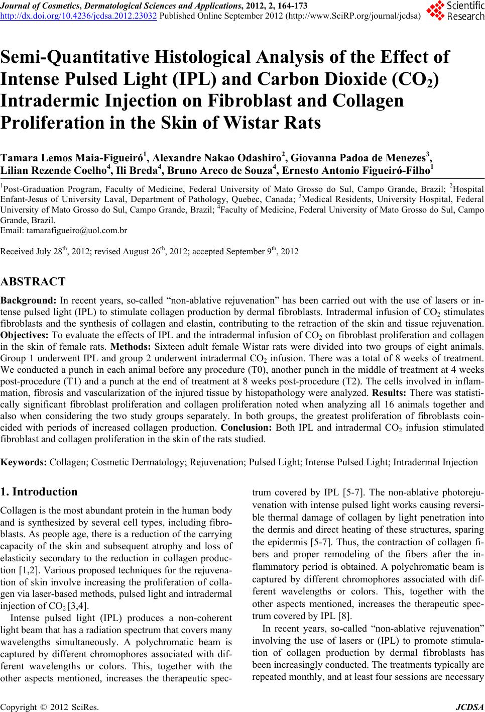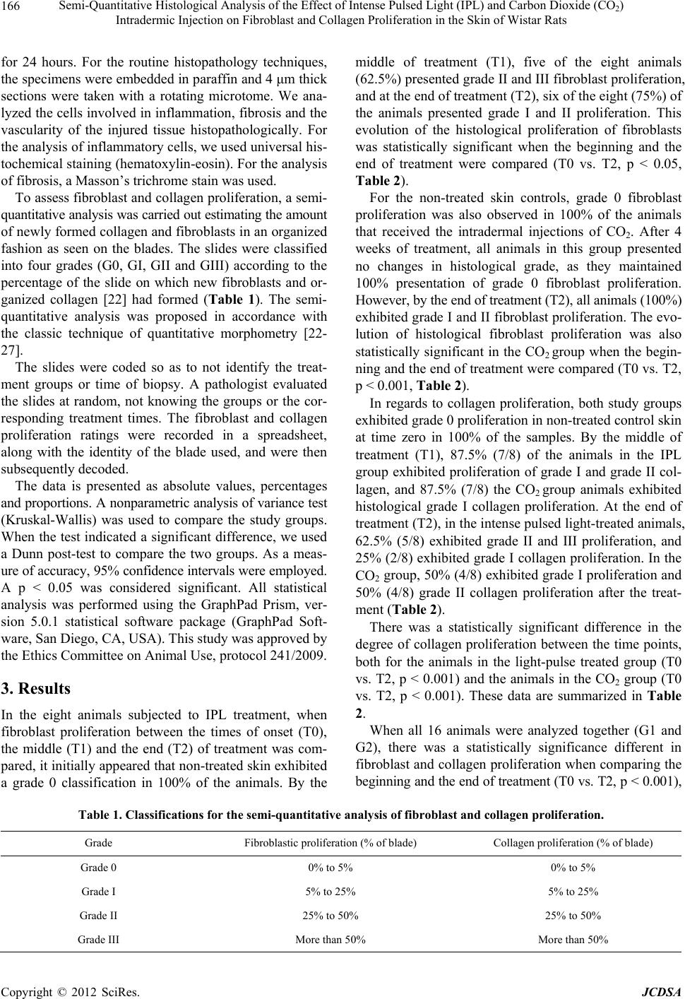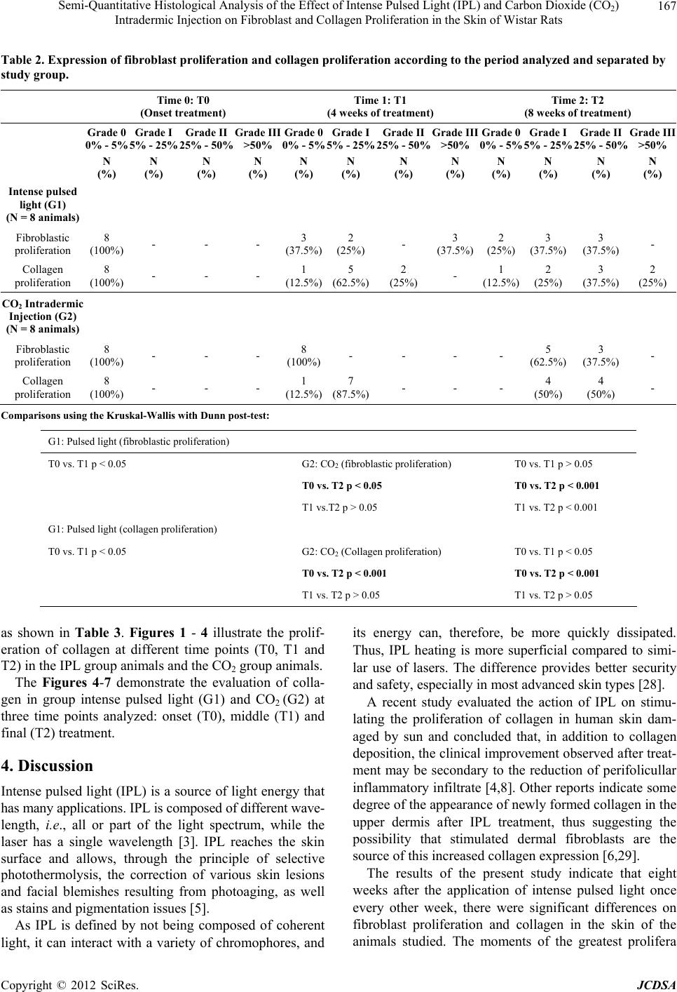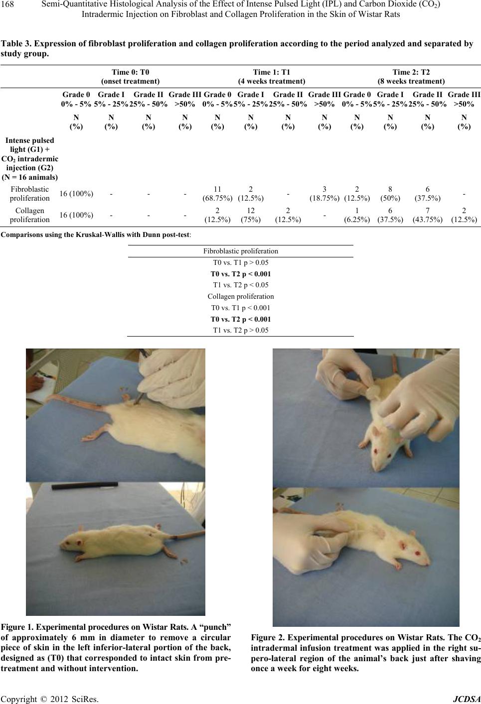
Semi-Quantitative Histological Analysis of the Effect of Intense Pulsed Light (IPL) and Carbon Dioxide (CO2)
Intradermic Injection on Fibroblast and Collagen Proliferation in the Skin of Wistar Rats
172
[3] M. G. Catorze, “Laser: Bases and Use in Dermatology,”
Medicina Cutánea Ibero-Latino-Americana, Vol. 37, No.
1, 2009, pp. 5-27.
[4] V. G. Prieto, A. H. Diwan, C. R. Shea, P. Zhang and N. S.
Sadick, “Effects of Intense Pulsed Light and the 1064 nm
Nd:YAG Laser on Sun-Damaged Human Skin: Histolo-
gic and Immunohistochemical Analysis,” Dermatologic
Surgery, Vol. 31, No. 5, 2005, pp. 522-525.
doi:10.1111/j.1524-4725.2005.31154
[5] R. C. R. Patriota, C. J. Rodrigues and L. C. Cucé, “Intense
Pulsed Light in Photoaging: Clinical, Histopathological
and Immunohistochemical Evaluation,” Anais Brasileiros
de Dermatologia, Vol. 86, No. 6, 2011, pp. 1129-1133.
doi:10.1590/S0365-05962011000600010
[6] D. J. Goldberg, “New Collagen Formation after Dermal
Remodeling with an Intense Pulsed Light Source,” Jour-
nal of Cutaneous Laser Therapy, Vol. 2, No. 2, 2000, pp.
59-61.
[7] D. J. Goldberg and K. B. Cutler, “Nonablative Treatment
of Rhytids with Intense Pulsed Light,” Lasers in Surgery
and Medicine, Vol. 26, No. 2, 2000, pp. 196-200.
doi:10.1002/(SICI)1096-9101(2000)26:2<196::AID-LSM
10>3.0.CO;2-9
[8] V. G. Prieto, P. S. Zhang and N. S. Sadick, “Evaluation of
Pulsed Light and Radiofrequency Combined for the Treat-
ment of Acne Vulgaris with Histologic Analysis of Facial
Skin Biopsies,” Journal of Cosmetic and Laser Therapy,
Vol. 7, No. 2, 2005, pp. 63-68.
[9] C. Brandi, C. D’Aniello, L. Grimaldi, B. Bosi, I. Dei, P.
Lattarulo, et al., “Carbon Dioxide Therapy in the Treat-
ment of Localized Adiposities: Clinical Study and Histo-
pathological Correlations,” Aesthetic Plastic Surgery, Vol.
25, No. 3, 2001, pp. 170-174.
doi:10.1007/s002660010116
[10] C. Brandi, L. Grimaldi, G. Nisi, A. Brafa, A. Campa, M.
Calabrò, et al., “The Role of Carbon Dioxide Therapy in
the Treatment of Chronic Wounds,” In Vivo, Vol. 24, No.
2, 2010, pp. 223-226.
[11] T. Brockow, A. Dillner, A. Franke and K. L. Resch, “An-
algesic Effectiveness of Subcutaneous Carbon-Dioxide
Insufflations as an Adjunct Treatment in Patients with
Non-Specific Neck or Low Back Pain,” Complementary
Therapies in Medicine, Vol. 9, No. 2, 2001, pp. 68-76.
doi:10.1054/ctim.2001.0434
[12] A. Sönmez, M. Yaman, O. Yalçin, B. Ersoy, M. Serin and
A. Sav, “Carbon Dioxide Therapy Increases Capillary
Formation on Random Pedicled Skin Flaps in the Rat,”
Journal of Plastic, Reconstructive & Aesthetic Surgery,
Vol. 62, No. 7, 2009, pp. e236-e237.
doi:10.1016/j.bjps.2009.01.067
[13] D. Kummer-Kloess, W. Kloess, N. Marienhoff, R. M.
Schütz, M. Zwaan and H. D. Weiss, “Angiography during
Interventional Procedures with Carbon Dioxide (CO2)
(Carbo-Angiography) in Patients with Increased Contrast
Media Risk,” Zentralblatt für Chirurgie, Vol. 122, 1997,
pp. 725-729.
[14] M. Zwaan, W. Kloess, C. Kagel, D. Kummer-Kloess, S.
Matthies-Zwaan, R. M. Schütz, et al., “Carbon Dioxide as
an Alternative Contrast Medium in Peripheral Angiogra-
phy,” Rofo, Vol. 164, No. 5, 1996, pp. 445-448.
doi:10.1055/s-2007-1015687
[15] M. Zwaan, J. Steinhoff, L. Fricke, C. Kagel, H. Lorch and
H. D. Weiss, “The Angiography of Kidney Transplant
Arteries Using Carbon Dioxide,” Dtsch Med Wochenschr,
Vol. 122, 1997, pp. 1133-1136.
doi:10.1055/s-2008-1047738
[16] B. R. Hartmann, E. Bassenge and M. Pittler, “Effect of
Carbon Dioxide-Enriched Water and Fresh Water on the
Cutaneous Microcirculation and Oxygen Tension in the
Skin of the Foot,” Angiology, Vol. 48, No. 4, 1997, pp.
337-343. doi:10.1177/000331979704800406
[17] B. R. Hartmann, E. Bassenge and M. Hartmann, “Effects
of Serial Percutaneous Application of Carbon Dioxide in
Intermittent Claudication: Results of a Controlled Trial,”
Angiology, Vol. 48, No. 11, 1997, pp. 957-963.
doi:10.1177/000331979704801104
[18] K. Field, M. Bailey, L. L. Foresman, R. L. Harris, S. L.
Motzel, R. A. Rockar, et al., “Medical Records for Ani-
mals Used in Research, Teaching, and Testing: Public
Statement from the American College of Laboratory Ani-
mal Medicine,” Institute of Laboratory Animal Resources
Journal, Vol. 48, 2007, pp. 37-41.
[19] D. F. Kohn, T. E. Martin, P. L. Foley, T. H. Morris, M. M.
Swindle, G. A. Vogler, et al., “Public Statement: Guide-
lines for the Assessment and Management of Pain in Ro-
dents and Rabbits,” Journal of American Association for
Laboratory Animal Science, Vol. 46, No. 2, 2007, pp. 97-
108.
[20] ACLAM, “ACLAM Position Statement on Animal Ex-
perimentation,” Comparative Medicine, Vol. 53, No. 5,
2003, p. 472.
[21] J. Artwohl, P. Brown, B. Corning, S. Stein and A. T.
Force, “Report of the ACLAM Task Force on Rodent
Euthanasia,” Journal of American Association for Labo-
ratory Animal Science, Vol. 45, No. 1, 2006, pp. 98-105.
[22] A. L. Domingos, S. Tucci, S. B. Garcia, J. de Bessa, A. J.
Cologna and A. C. Martins, “Use of a Latex Biomem-
brane for Bladder Augmentation in a Rabbit Model: Bio-
compatibility, Clinical and Histological Outcomes,” In-
ternational Journal of the Brazilian Society of Urology,
Vol. 35, No. 2, 2009, pp. 217-224.
[23] J. J. S. Gonçalves, L. E. V. Leão, R. G. Ferreira, R. Olivei-
ra, L. H. Ota and R. S. D. Santos, “Semiquantitative Ana-
lysis of Surgical Biopsies of Different Lung Lobes of Pa-
tients with Usual Interstitial Pneumonia/Idiopathic Pul-
monary Fibrosis,” Jornal Brasileiro de Pneumologia, Vol.
35, No. 7, 2009, pp. 676-682.
[24] D. M. Hyde, T. E. King, T. McDermott, J. A. Waldron, T.
V. Colby, W. M. Thurlbeck, et al., “Idiopathic Pulmonary
Fibrosis. Quantitative Assessment of Lung Pathology. Com-
parison of a Semiquantitative and a Morphometric Histo-
pathologic Scoring System,” American Review of Respi-
ratory Disease, Vol. 146, No. 4, 1992, pp. 1042-1047.
[25] M. F. Y. Maeda, C. D. Silva, L. S. Harima, L. F. F. Silva,
B. Ctenas and V. A. F. Alves, “Vascularization in Hepatic
Cirrhosisbased on Autopsies,” Archives of Gastroentero-
Copyright © 2012 SciRes. JCDSA