Journal of Cosmetics, Dermatological Sciences and Applications, 2012, 2, 136-140
http://dx.doi.org/10.4236/jcdsa.2012.23027 Published Online September 2012 (http://www.SciRP.org/journal/jcdsa)
A Unique Case? Darier’s Disease Presented as
Porcupine-Like Appearance and the Observation on
Acitretin Treatment*
Xi-Bao Zhang1#, Chang-Xing Li2, Xue-Mei Li1, Yu-Qing He1, Xiao Xu,1 Quan Luo1
1Department of Dermatology, Guangzhou Institute of Dermatology, Guangzhou, China; 2Department of Dermatology, Dongguan
Hospital of Chronic Disease, Dongguan, China.
Email: #lilichangxing@163.com
Received May 6th, 2012; revised June 10th, 2012; accepted June 29th, 2012
ABSTRACT
Dyskeratosis follicularis (Darier’s disease, DD) is rare autosomal dominant disease characterized by hyperkeratotic
papules that coalesce into plaques and occur primarily in seborrheic or intertriginous areas. Associated findings include
nail abnormalities. A 3-year-old boy presented with porcupine-like appearance for 2 years. The lesion from the back
was taken for light microscopy and electron microscopy. He was treated with acitretin (0.31 mg/d to 0.66 mg/d) for 8
years. Light microscopy and electron microscopy showed that the typical features of DD. The patient show good re-
spond to the treatment. During 8 years treatment, the patient had dry mouth and pruritus. The skeletal abnormalities
didn’t happen in the patient. Evaluation of the serum lipid profile, liver function and renal function were within normal
lever after treatment. Our findings showed that porcupine-like appearance is a unique pattern of DD. Acitretin may be a
useful therapeutic agent in children with DD and less likely to cause skeletal problems.
Keywords: Darier’s Disease; Acitretin
1. Introduction
The prevalence of DD has been estimated vary from one
in 36,000 to one in 100,000 [1]. Clinically, the distinctive
lesion is characterized by hyperkeratotic papules that
coalesce into plaques and occur primarily in seborrheic,
but also in intertriginous areas [1,2]. Coalescence of the
papules produces irregular warty plaques or papilloma-
tous masses, which, in the flexures, become hypertrophic,
and malodorous with painful fissured. Associated ab-
normalities include nail abnormalities characterized by
nail fragility, red and white longitudinal stripes and v-
shaped notches at the free margin of the nails [2]. In this
article, we describe a case of DD with porcupine-like ap-
pearance. The patient showed good respond to the treat-
ment of acitretin therapy.
2. Case Report
A 3-year-old boy had healthy skin at birth. At the age of
1 year, a brownish-red, keratotic papules and plaques
appeared in his scalp, ears, perianal and genital areas. A
biospy taken from his lesion revealed DD in the local
hospital. He showed no signifficant respond to the treat-
ment of 0.025% retinoc acid cream or 0.1% mometasone
furoate cream. The keratotic skin lesions worsened gra-
dually as she grew. At 3 years of age, roughening, thick-
ening and dryness of the keratotic skin lesions increased
progressively to most parts of the body. Mentally the
patient was normal and he had done well in school. His
general health had been good. No other family members
were affected.
On physical examination, the erythematous, keratotic,
follicular papules were over his scalp, face and ear (Fig-
ure 1(a)). The disseminated heavy papillomatous hy-
perkeratosis was dark gray-brown and thickest on the
legs, arms and trunk (Figures 1(b) and (c)). Nail involve-
ment was subungual hyperkeratosis, dystrophy of the
free margin of the nail and v-shaped notches at the free
margin of the nails (Figure 1(d)).
3. Materials and Methods
Biopsy specimens were taken from affected skin on his
back before treatment. The tissue specimens were studied
under the light microscope as paraffin sections stained
with hematoxylin-eosin or as l μm epoxy resin-fixed sec-
tions. For electron microscopy, the tissue was fixed 2
*Authors Xi-Bao Zhang and Chang-Xing Li contributed equally to this
work.
#Corresponding author.
Copyright © 2012 SciRes. JCDSA
A Unique Case? Darier’s Disease Presented as Porcupine-Like Appearance and the Observation on Acitretin Treatment 137
Figure 1. (a) The erythematous, keratotic, follicular papules were over his scalp, face and ear; (b) and (c) The disseminated
heavy papillomatous hyperkeratosis was dark gray-brown and thickest on the legs, arms and trunk; (d) Nail involvement was
subungual hyperkeratosis, dystrophy of the free margin of the nail and v-shaped notches at the free margin of the nails; (e)
and (f) The patient’s condition showed great improvement.
hours to 4 hours in 3% glutaraldehyde and postfixed in
1% osmium tetroxide. The specimens were stained en
bloc with uranyl acetate-lead citrate, dehydrated in a
graded alcohol series, and embedded in epoxy resin
(Epon812). Ultrathin sections were cut on an ultramicro-
tome, stained with uranyl acetate-lead citrate solutions,
and examined with an electron microscope.
Assessment of efficacy:
The definitions of efficacy used in the present investi-
gation were: 1) Cured in clinically. The improvement of
skin lesions comes to over 90% been cured; the kerato-
derma is totally desquamated or light coarse remains on
the skin. The patients can normally study and live in the
society without any handicap; the life quality was im-
proved completely; 2) Marked improvement. The im-
provement of skin lesions comes to 60% - 90% and the
patients can normally study and live, the life quality was
improved remarkable; 3) Improvement. The improve-
ment of skin lesions comes to 30% - 60% and the pa-
tients can nearly study and live as normal the life quality
was improved; 4) Failure. The skin improvement is less
than 30% and the patients can’t live in a normal life, the
life quality was improved in a little.
Safety evaluation: Evaluate the safety according to the
clinical side effects, changes in body weight and height,
examination results of routine blood tests, liver and renal
function, blood lipids and selective X-ray check for those
cases been treated more than one year.
4. Results
Biopsies taken from back showed hyperkeratosis, para-
keratosis, follicular plugging, irregular acanthosis, acan-
tholytic keratinocytes forming corp ronds and corp grains
(Figure 2), and an upward proliferation of papillae into
the clefts. The direct immunofluorescence was negative.
Electron microscopic studies have demonstrated cell
membrane damage, cytoplasmic vacuolization, dissolu-
tion of desmosome-keratin filament complexes and ag-
gregation of keratin filaments around cell nucle. In the
granular layer, the abnormality is cytoplasmic vacuoliza-
tion. The affected cells display a spectrum of vacuoliza-
Copyright © 2012 SciRes. JCDSA
A Unique Case? Darier’s Disease Presented as Porcupine-Like Appearance and the Observation on Acitretin Treatment
138
tion ranging from a predominantly perinuclear distribu-
tion giving a foamy or cytolytic appearance. The tono-
filaments and keratohyaline granules appear compressed
into dense rings without attachments with neighboring
cells (Figure 3(a)). Cells of the prickle layer, which are
within lacunae, are separated from each other, having
smooth rounded plasma membranes devoid of microvilli
and desmosome attachments, and increasing aggregation of
tonofilaments with perinuclear ditriution (Figures 3(b) - (d)).
Acitretin was used in the treatment of the case. The
start treatment dose was 5 mg/day (0.38 mg/kg/d). The
response was good and the patient’s condition showed
great improvement within 1 year. After 1 year, the dose
of acitretin was increased to 10 mg/day (0.66 mg/kg/d),
and the skin remained in good condition. After 2 years,
the dose of acitretin was reduced to 6 mg/day (0.31 mg/
kg/d), and the skin remained in good condition within 2
years. After 4 years, the dose of acitretin was reduced to
5 mg/day (0.31 mg/kg/d), and the skin remained in good
condition (Figures 1(e)-(f)) (Table 1).
During 8 years treatment, our patient had dry mouth
and pruritus. The skeletal abnormalities didn’t happen in
the patient. Evaluation of the serum lipid profile, liver
function and renal function were within normal lever
after treatment.
5. Discussion
The clinical expression of IH (Curth-Macklin) varies from
palmoplantar keratoderma to a severe generalized involve-
ment [3]. Histopathology shows granular cells with peri-
nuclear oedema and shell formation, and binuclear cells,
and the most prominent feature ultrastructurally are
Figure 2. Histology showed hyperkeratosis, parakeratosis,
follicular plugging, irregular acanthosis, acantholytic kera-
tinocytes forming corp ronds and corp grains.
(a)
(b)
(c)
(d)
Figure 3. (a) The tonofilaments and keratohyaline granules appear compressed into dense rings without attachments with
neighboring cells; (b-d) Cells of the prickle layer, which are within lacunae, are separated from each other, having smooth
rounded plasma membranes devoid of microvilli and desmosome attachments, and increasing aggregation of tonofilaments
with perinuclear ditriution.
Copyright © 2012 SciRes. JCDSA
A Unique Case? Darier’s Disease Presented as Porcupine-Like Appearance and the Observation on Acitretin Treatment 139
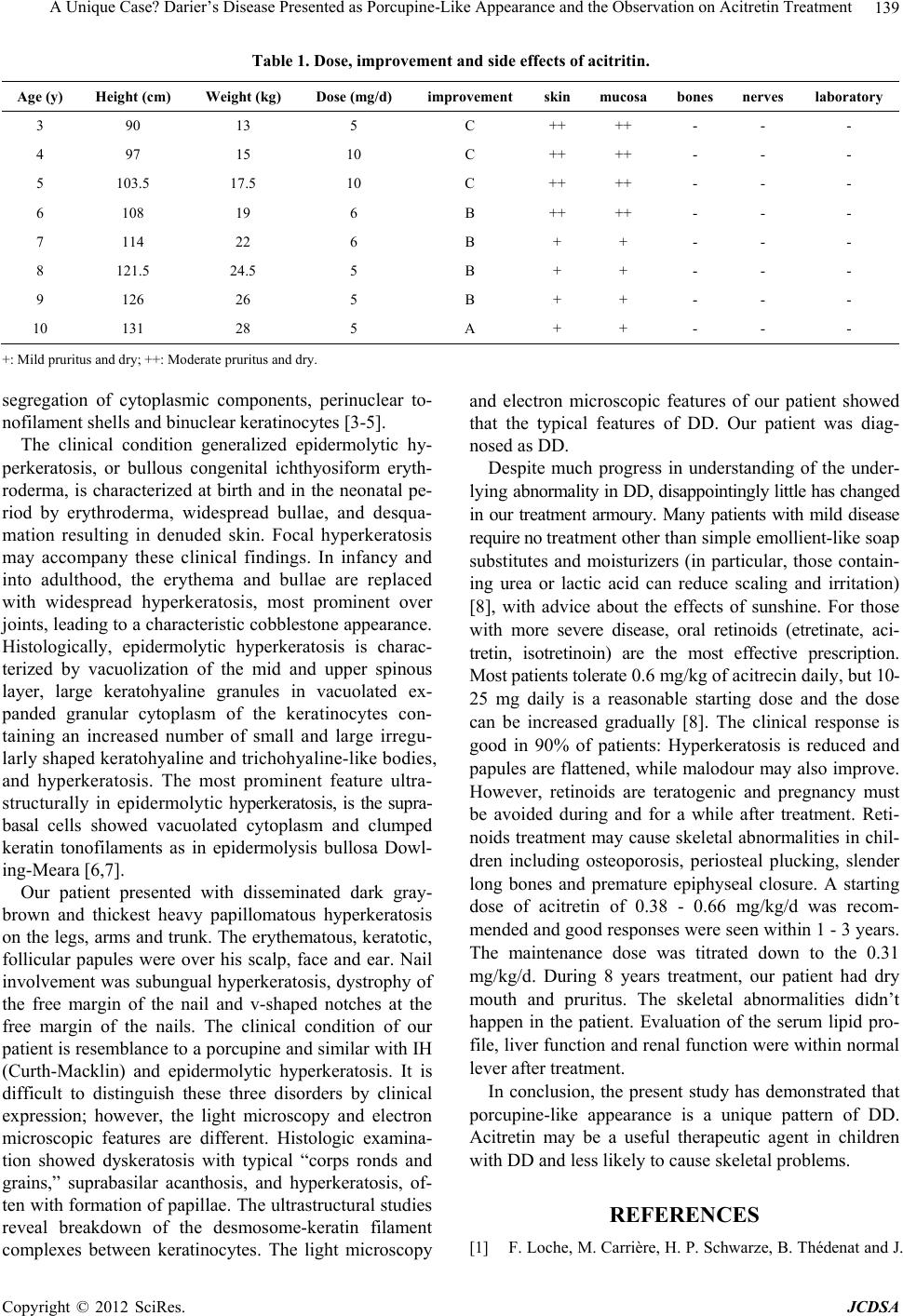
Table 1. Dose, improvement and side effects of acitritin.
Age (y) Height (cm) Weight (kg) Dose (mg/d) improvement skinmucosa bones nerves laboratory
3 90 13 5 C ++ ++ - - -
4 97 15 10 C ++ ++ - - -
5 103.5 17.5 10 C ++ ++ - - -
6 108 19 6 B ++ ++ - - -
7 114 22 6 B + + - - -
8 121.5 24.5 5 B + + - - -
9 126 26 5 B + + - - -
10 131 28 5 A + + - - -
+: Mild pruritus and dry; ++: Moderate pruritus and dry.
segregation of cytoplasmic components, perinuclear to-
nofilament shells and binuclear keratinocytes [3-5].
The clinical condition generalized epidermolytic hy-
perkeratosis, or bullous congenital ichthyosiform eryth-
roderma, is characterized at birth and in the neonatal pe-
riod by erythroderma, widespread bullae, and desqua-
mation resulting in denuded skin. Focal hyperkeratosis
may accompany these clinical findings. In infancy and
into adulthood, the erythema and bullae are replaced
with widespread hyperkeratosis, most prominent over
joints, leading to a characteristic cobblestone appearance.
Histologically, epidermolytic hyperkeratosis is charac-
terized by vacuolization of the mid and upper spinous
layer, large keratohyaline granules in vacuolated ex-
panded granular cytoplasm of the keratinocytes con-
taining an increased number of small and large irregu-
larly shaped keratohyaline and trichohyaline-like bodies,
and hyperkeratosis. The most prominent feature ultra-
structurally in epidermolytic hyperkeratosis, is the supra-
basal cells showed vacuolated cytoplasm and clumped
keratin tonofilaments as in epidermolysis bullosa Dowl-
ing-Meara [6,7].
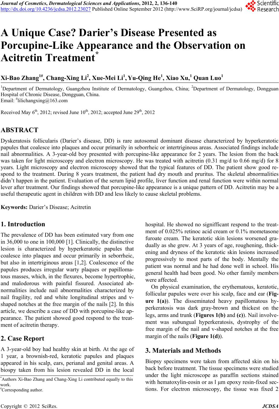
Our patient presented with disseminated dark gray-
brown and thickest heavy papillomatous hyperkeratosis
on the legs, arms and trunk. The erythematous, keratotic,
follicular papules were over his scalp, face and ear. Nail
involvement was subungual hyperkeratosis, dystrophy of
the free margin of the nail and v-shaped notches at the
free margin of the nails. The clinical condition of our
patient is resemblance to a porcupine and similar with IH
(Curth-Macklin) and epidermolytic hyperkeratosis. It is
difficult to distinguish these three disorders by clinical
expression; however, the light microscopy and electron
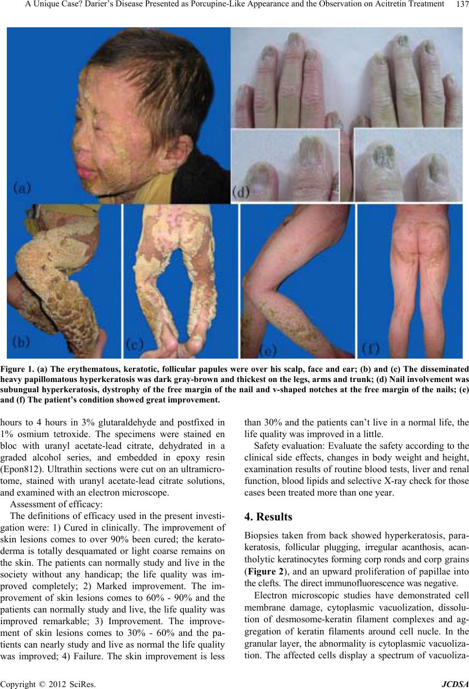
microscopic features are different. Histologic examina-
tion showed dyskeratosis with typical “corps ronds and
grains,” suprabasilar acanthosis, and hyperkeratosis, of-
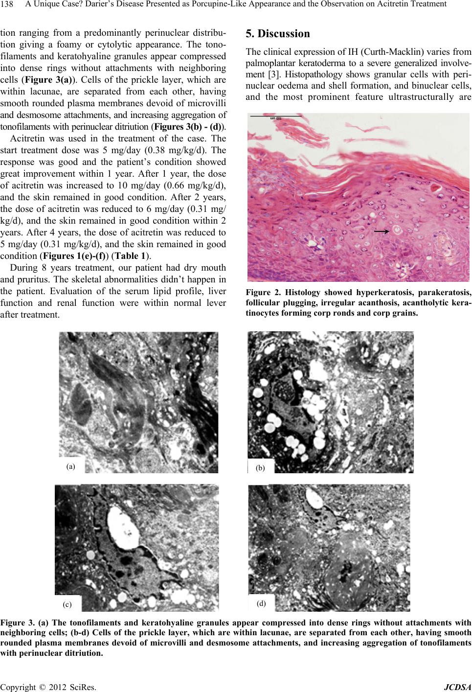
ten with formation of papillae. The ultrastructural studies
reveal breakdown of the desmosome-keratin filament
complexes between keratinocytes. The light microscopy
and electron microscopic features of our patient showed
that the typical features of DD. Our patient was diag-
nosed as DD.
Despite much progress in understanding of the under-
lying abnormality in DD, disappointingly little has changed
in our treatment armoury. Many patients with mild disease
require no treatment other than simple emollient-like soap
substitutes and moisturizers (in particular, those contain-
ing urea or lactic acid can reduce scaling and irritation)
[8], with advice about the effects of sunshine. For those
with more severe disease, oral retinoids (etretinate, aci-
tretin, isotretinoin) are the most effective prescription.
Most patients tolerate 0.6 mg/kg of acitrecin daily, but 10-
25 mg daily is a reasonable starting dose and the dose
can be increased gradually [8]. The clinical response is
good in 90% of patients: Hyperkeratosis is reduced and
papules are flattened, while malodour may also improve.
However, retinoids are teratogenic and pregnancy must
be avoided during and for a while after treatment. Reti-
noids treatment may cause skeletal abnormalities in chil-
dren including osteoporosis, periosteal plucking, slender
long bones and premature epiphyseal closure. A starting
dose of acitretin of 0.38 - 0.66 mg/kg/d was recom-
mended and good responses were seen within 1 - 3 years.
The maintenance dose was titrated down to the 0.31
mg/kg/d. During 8 years treatment, our patient had dry
mouth and pruritus. The skeletal abnormalities didn’t
happen in the patient. Evaluation of the serum lipid pro-
file, liver function and renal function were within normal
lever after treatment.
In conclusion, the present study has demonstrated that
porcupine-like appearance is a unique pattern of DD.
Acitretin may be a useful therapeutic agent in children
with DD and less likely to cause skeletal problems.
REFERENCES
[1] F. Loche, M. Carrière, H. P. Schwarze, B. Thédenat and J.
Copyright © 2012 SciRes. JCDSA
A Unique Case? Darier’s Disease Presented as Porcupine-Like Appearance and the Observation on Acitretin Treatment
140
Bazex, “Darier White Disease and Dermatofibrosarcoma
Protuberans,” Dermatology, Vol. 199, No. 3, 1999, p. 279.
doi:10.1159/000018268
[2] S. Tavadia, E. Mortimer and C. S. Munro, “Genetic Epi-
demiology of Darier’s Disease: A Population Study in the
West of Scotland,” British Journal of Dermatology, Vol.
146, No. 1, 2002, pp. 107-109.
doi:10.1046/j.1365-2133.2002.04559.x
[3] H. O. Curth, F. H. J. Allen, U. W. Schnyder and I. An-
ton-Lamprecht, “Follow-Up of a Family Group Suffering
from Ichthyosis Hystrix Type Curth-Macklin,” Human-
genetik, Vol. 17, No. 1, 1972, pp. 37-48.
doi:10.1007/BF01789598
[4] L. Kanerva, J. Karvonen, A. Oikarinen, J. Lauharanta, A.
Ruokonen and K. M. Niemi, “Ichthyosis Hystrix (Curth-
Macklin). Light and Electron Microscopic Studies Per-
formed before and after Etretinate Treatment,” Archives
of Dermatology, Vol. 120, No. 9, 1984, pp. 1218-1223.
doi:10.1001/archderm.1984.01650450100030
[5] K. M. Niemi, I. Virtanen, L. Kanerva and M. Muttilainen,
“Altered Keratin Expression in Ichthyosis Hystrix Curth-
Macklin. A Light and Electron Microscopic Study,” Ar-
chives of Dermatological Research, Vol. 282, No. 4, 1990,
pp. 227-233. doi:10.1007/BF00371641
[6] A. L. Ingrun, “Ultrastructural Identification of Basic Ab-
normalities as Clues to Genetic Disorders of the Epider-
mis,” Journal of Investigative Dermatology, Vol. 103, No.
5, 1994, pp. 6-12. doi:10.1111/1523-1747.ep12398887
[7] A. B. Ackerman and G. Goldman, “Combined Epidermo-
lytic Hyperkeratosis and Focal Acantholytic Dyskerato-
sis,” Archives of Dermatology, Vol. 109, No. 3, 1974, pp.
385-386. doi:10.1001/archderm.1974.01630030043010
[8] S. Bruge, “Management of Darier’s Disease,” Clinical
and Experimental Dermatology, Vol. 24, No. 2, 1999, pp.
53-56. doi:10.1046/j.1365-2230.1999.00416.x
Copyright © 2012 SciRes. JCDSA