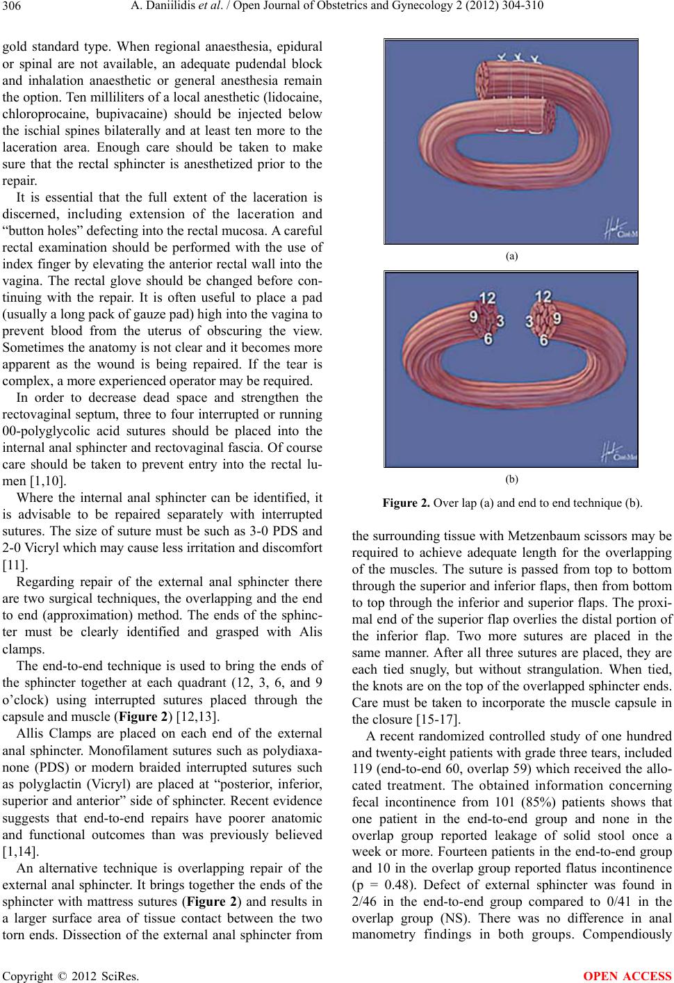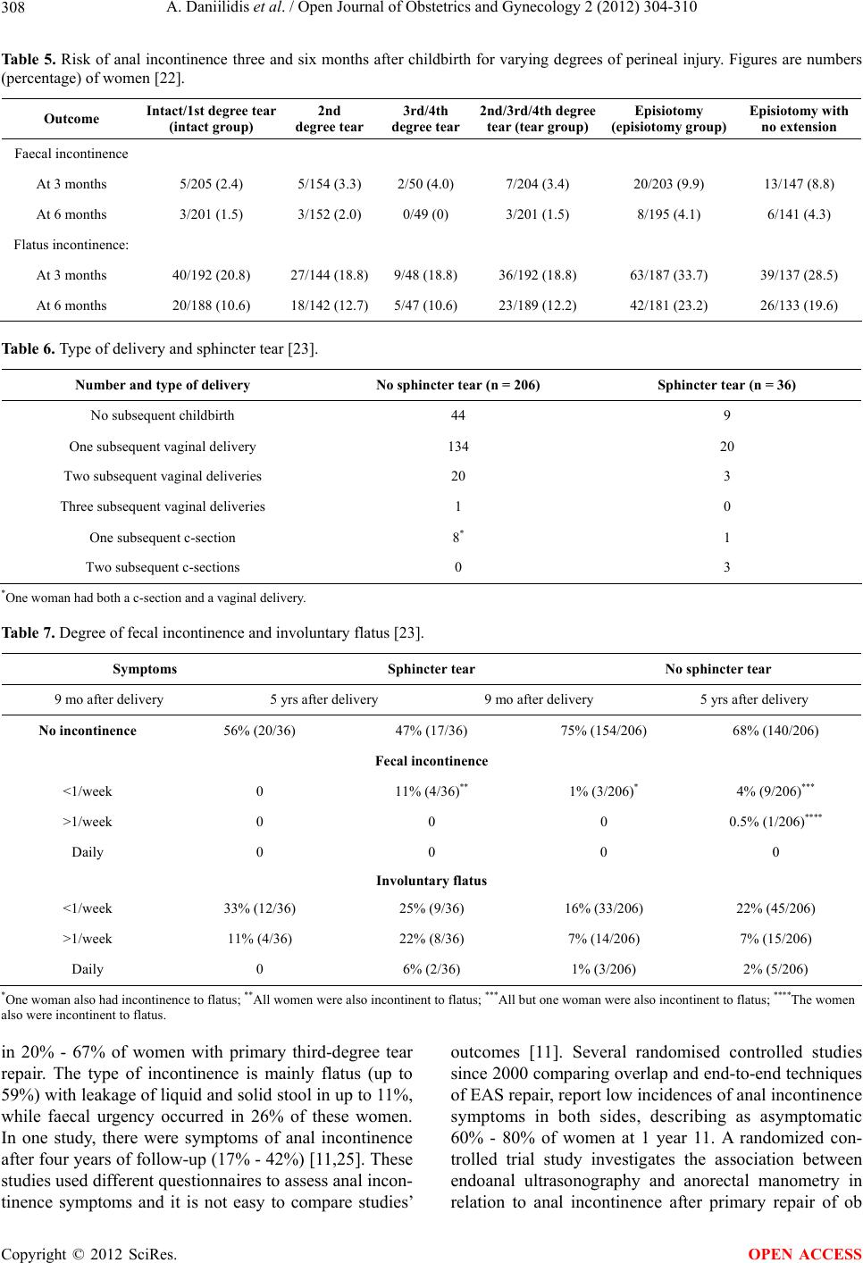
A. Daniilidis et al. / Open Journal of Obstetrics and Gynecology 2 (2012) 304-310
310
doi:10.1111/j.1471-0528.1992.tb13695.x
[7] De Leew, J.W., Struijk, P.C., Vierhout, M.E. and Wallen-
burg, H.C. (2001) Risk factors for third degree perineal
ruptures during delivery. British Journal of Obstetrics
and Gynaecology, 108, 383-387.
doi:10.1016/S0306-5456(00)00090-5
[8] Williams, A. (2003) Third-degree perineal tears: Risk
factors and outcomes after primary repair. American
Journal of Obstetrics & Gynecology, 23, 611-614.
doi:10.1080/01443610310001604358
[9] Bodner, K., Bodner-Adler, B., Wagenbichler, P., Kaider,
A., Leodolter, S., Husslein, P. and Mayerhofer, K. (2001)
Perineal lacerations during spontaneous vaginal delivery.
Wiener Klinische Wochenschrift, 11 3, 743-746.
[10] Peleg, D., Kennedy, C.M., Merrill, D. and Zlatnik, F.J.
(1999) Risk of repetition of a severe perineal laceration.
Obstetrics & Gynecology, 93, 1021-1024.
doi:10.1016/S0029-7844(98)00556-0
[11] Royal College of Obstetricians and Gynecologists (2007)
The management of third- and fourth-degree perineal
tears. Green-Top Guideline.
[12] Sultan, A.H., Kamm, M.A., Hudson, C.N. and Bartram,
C.I. (1994) Third degree obstetric anal sphincter tears:
Risk factors and outcome of primary repair. British Me-
dical Journal, 308, 877-891.
doi:10.1136/bmj.308.6933.887
[13] Kammerer-Doak, D.N., Wesol, A.B., Rogers, R.G., Do-
minguez, C.E. and Dorin, M.H. (1999) A prospective co-
hort study of women after primary repair of obstetric anal
sphincter laceration. American Journal of Obstetrics &
Gynecology, 181, 1317-1322.
doi:10.1016/S0002-9378(99)70370-4
[14] Williams, A., Adams, E.J., Tincello, D.G., Alfirevic, Z.,
Walkinshaw, S.A. and Richmond, D.H. (2006) How to
repair an anal sphincter injury after vaginal delivery: Re-
sults of a randomised controlled trial. An International
Journal of Obstetrics & Gynaecology, 113, 201-107.
doi:10.1111/j.1471-0528.2006.00806.x
[15] Fitzpatrick, M., Behan, M., O’Connell, P.R. and O’Her-
lihy, C. (2000) A randomized clinical trial comparing
primary overlap with approximation repair of third-de-
gree obstetric tears. American Journal of Obstetrics &
Gynecology, 183, 1220-1224.
doi:10.1067/mob.2000.108880
[16] Homsi, R., Daikoku, N.H., Littlejohn, J. and Wheeless,
C.R. (1994) Episiotomy: Risks of dehiscence and rec-
tovaginal fistula. Obstetrical & Gynecological Survey, 49,
803-808. doi:10.1097/00006254-199412000-00002
[17] Sultan, A.H., Monga, A.K., Kumar, D. and Stanton, S.L.
(1999) Primary repair of obstetric anal sphincter rupture
using the overlap technique. British Journal of Obstetrics
and Gynaecology, 106, 318-323.
doi:10.1111/j.1471-0528.1999.tb08268.x
[18] Rygh, A.B., et al. (2010) The overlap technique versus
end-to-end approximation technique for primary repair of
obstetric anal sphincter rupture: A randomized controlled
study. Acta Obstetricia et Gynecologica Scandinavica, 89,
1256-1262. doi:10.3109/00016349.2010.512073
[19] Kalis, V., Bednarova, B., et al. (2010) Repair of the 3rd
and 4th degree obstetric perineal tear. Ceská Gynekologie,
75, 284-291.
[20] Legino, L.J., Woods, M.P., Rayburn, W.F. and McGoogan,
L.S. (1988) Third- and fourth-degree perineal tears—50
years’ experience at a university hospital. The Journal of
Reproductive Medicine, 33, 423-426.
[21] Abdul, H., et al. (1999) Primary repair of obstetric anal
sphincter rupture using the overlap technique. British
Journal of Obstetrics and Gynecology, 106, 318-323.
[22] Lisa, B. et al. (2000) Midline episiotomy and anal incon-
tinence: Retrospective cohort study. British Medical Jour-
nal, 320, 86-90.
[23] Nordenstam, J. (2010) Anal Incontinence and Obstetric
Anal Sphincter Tears. Karolinska Institutet, Stockholm.
[24] Fernando, R., Sultan, A.H., Kettle, C., Thakar, R. and
Radley, S. (2006) Methods of repair for obstetric anal
sphincter injury. Cochrane Database of Systematic Re-
views, 19, CD002866.
[25] Poen, A.C., Felt-Bersma, R.J., Dekker, G.A., Deville, W.,
Cuesta, M.A. and Meuwissen, S.G. (1997) Third degree
obstetric perineal tears: Risk factors and the preventative
role of mediolateral episiotomy. An International Journal
of Obstetrics & Gynaecology, 104, 563-566.
doi:10.1111/j.1471-0528.1997.tb11533.x
[26] Nordenstam, J.F., et al. (2010) Impaired rectal sensation
at anal manometry is associated with anal incontinence
one year after primary sphincter repair in primiparous
women. Diseases of the Colon & Rectum, 53, 1409-1414.
doi:10.1007/DCR.0b013e3181eb9f01
Copyright © 2012 SciRes. OPEN ACCESS