Psychology
2012. Vol.3, No.8, 578-582
Published Online August 2012 in SciRes (http://www.SciRP.org/journal/psych) http://dx.doi.org/10.4236/psych.2012.38086
Copyright © 2012 SciRes.
578
Heart Rate Variability Predicts Emotional Flexibility in Response
to Positive Stimuli
To momi Fujimura1,2, Kazuo Okanoya1,2,3*
1Japan Science Technology Agency, ERATO, Okanoya Emotional Inform ati on Project, Saitama, Japan
2RIKEN Brain Science Institute, Saitama, Japan
3Department of Life Sciences, Graduate School of Arts and Sc ien ces , The University of Tokyo, T okyo, Japan
Email: *kazuookanoya@gmail.com
Received May 7th, 2012; revised June 7th, 2012; accepted July 10th, 2012
Flexible adaptation to constantly changing environments is linked to mental health and psychological
functioning. Heart rate variability (HRV), an index of autonomic flexibility, has been implicated in emo-
tional flexibility, the ability to generate contextually dependent emotional responses in accordance with
situational demands. The current study investigated whether HRV during rest is associated with experi-
enced emotion, one of the measures of emotional flexibility. To assess experienced emotion in response to
changing events, three types of stimuli sets were created by presenting two stimuli successively. First, two
stimuli represented the same valence (i.e., negative/negative or positive/positive). Second, two stimuli
represented opposite valences (i.e., negative/positive or positive/negative). Third, a neutral stimulus was
followed by negative or positive stimulus (i.e., neutral/negative or neutral/positive). Psychological ratings
for experienced emotion to the second stimulus were collected with regard to valence and arousal. The
results showed that subjects with lower resting HRV experienced more aroused states in response to suc-
cessive positive stimuli. Resting HRV may be a proxy of emotional flexibility indexed by subjective
arousal states to positive events.
Keywords: Heart Rate Variability; Emotional Flexibility; Psychological Ratings
Introduction
Life involves constantly changing events in the external en-
vironment. Rapid and adequate adaptation to these changes is
vital for organisms to survive. Recent research has focused on
emotional flexibility, the ability to flexibly respond to positive
and negative events and to regulate emotion effectively (Waugh,
Thompson, & Gotlib, 2011; Westphal, Seivert, & Bonanno,
2010). Individuals with greater emotional flexibility have posi-
tive reactivity to positive events and negative reactivity to
negative events, in accordance with situational demands. These
context-dependent emotional responses are characterized by
flexible autonomic, somatic, and psychological responses to
changing events.
Heart rate variability (HRV), an index of autonomic flexibil-
ity, is positively associated with good psychological and
physiological functioning (Thayer & Lane, 2000). HRV is cal-
culated by R-R wave intervals produced by electrocardio-
graphic data. In healthy individuals, heart rate fluctuates during
resting states due to the different frequency characteristics of
sympathetic and parasympathetic neural modulations of heart
rate. Therefore, HRV is indicative of autonomic balance be-
tween sympathetic and parasympathetic activity. Low HRV is
associated with a number of psychological disorders character-
ized by poor emotion regulation and behavioral inflexibility
(see Thayer & Brosschot, 2005 for a review). Given that emo-
tional flexibility can be defined by autonomic and behavioral
reactivity, it is possible that HRV assesses an individual’s emo-
tional flexibility, or how well an individual responds to emo-
tional events and generates contextually dependent emotional
responses.
Individual differences in regulated emotional response can be
predicted by baseline HRV (see Appelhans & Luecken, 2006
for a review). For example, subjects with low resting HRV
showed a more exaggerated startle reflex potentiated by the
threat of shock compared with subjects with high resting HRV
(Melzig, Weike, Hamm, & Thayer, 2009). Furthermore, the
emotion-modulated startle reflex was higher in subjects with
low HRV than in those with high HRV and subjects with low
HRV showed no difference in startle magnitude during negative,
positive, and neutral foregrounds (Ruiz-Padial, Sollers, Vila, &
Thayer, 2003). This evidence suggests that individuals with low
resting HRV have a tendency to produce an exaggerated startle
reflex regardless of valence, reflecting an inappropriate re-
sponse to situational demands.
Attention allocation, which involves the selection of mean-
ingful information from the external environment, is also asso-
ciated with resting HRV (Thayer & Brosschot, 2005). A previ-
ous study reported that individuals with low HRV could not
shift their attention away from negative stimuli, resulting in
prolonged response initiation after the presentation of negative
stimuli (Krypotos, Jahfari, van Ast, Kindt, & Forstmann, 2011).
In relation to this, Johnsen, Thayer, Laberg, Wormnes, Raadal,
Skaret, Kvale, and Berg (2003) found increased attentional bias
to threat-related words (i.e., dental-related words for dental
phobics) in subjects with low HRV. This evidence indicates
that HRV during rest is associated with sensitivity to threat
signals. Because attentional control is important for adapting to
a constantly changing environment, excessive sensitivity to
*Corresponding author.
T. FUJIMURA, K. OKANOYA
threat signals interferes with the processing of subsequent
events, leading to emotional inflexibility.
The aforementioned studies have revealed that resting HRV
predicts individual differences in emotional flexibility by mea-
suring reflexes and attention to emotional stimuli directly or
indirectly. However, it remains unknown whether subjective
emotions measured by psychological ratings are related to rest-
ing HRV, although subjective emotion is one of the indices to
assess emotional flexibility. Waugh et al. (2011) demonstrated
that high-resilient people who exhibited emotional flexibility,
compared to low-resilient people, showed a greater difference
in self-reported affect between current negative and positive
pictures. Given that emotional flexibility is characterized by
context-dependent experienced emotion, HRV, an index of
flexibility, is associated with experienced emotion as measured
by psychological ratings. That is, people with low HRV may
show poor emotional experiences to positive and negative
stimuli.
The present study aimed to examine whether resting HRV
predicts individual differences in the experience of subjective
emotion. To assess emotional flexibility, experienced emotion
was evaluated in response to target stimuli followed by three
types of prime stimuli. In the first case, the prime and target
stimuli represented the same valence (i.e., negative/negative or
positive/positive). In the second case, the prime and target
stimuli represented opposite valences (i.e., negative/positive or
positive/negative). In the third case, a neutral prime stimulus
was followed by negative or positive target stimuli (i.e., neu-
tral/negative or neutral/positive). Participants were asked to rate
their experienced emotion when viewing the picture that was
presented second (i.e., the target picture). We hypothesized that
subjects with high resting HRV, in comparison with subjects
with low resting HRV, would have stronger experiences of
pleasantness or unpleasantness in response to the target stimu-
lus, regardless of which stimulus was presented first. In addi-
tion to valence ratings, arousal ratings for experienced emotion
were collected. Individuals with low resting HRV are charac-
terized by chronic autonomic hyperarousal and diminished
response habituation (Friedman & Thayer, 1998). Accordingly,
experienced arousal may be exaggerated in individuals with
low resting HRV. Furthermore, this tendency may be promi-
nent in the congruent condition in which same-valence stimuli
were presented successively because individuals with low HRV
are unlikely to habituate these emotional stimuli. In the present
study, we expected that individual difference in resting HRV
would translate into positive and negative reactivity, assessed
by experienced emotion.
Method
Participants
Forty-three adults (24 men and 19 women; mean ± SD age,
25.02 ± 6.74 years) participated in the study. The participants
were recruited by advertisements placed with an intermediary
company, and their occupational backgrounds varied widely.
They received compensation for participating in the experiment.
All of the participants had normal or corrected-to-normal vi-
sion.
Stimulus Material
The stimuli were 35 pictures selected from the International
Affective Picture System (IAPS: Lang, Bradley, & Cuthbert,
2008), consisting of 10 pleasant, 5 neutral, and 10 unpleasant
pictures. The mean of valence/arousal ratings were: 7.63/5.10
for the pleasant pictures, 4.99/2.45 for neutral pictures, and
2.73/5.23 for unpleasant pictures. Arousal ratings were equiva-
lent for pleasant and unpleasant pictures. All pictures were
presented in full color.
Apparatus and Physiological Measurement
Experimental events were controlled by a program written in
Inquisit 3.0 (Millisecond) and were implemented on a computer
(Vostro 420, Dell) using the Microsoft Windows XP operating
system. Stimuli were presented on a 19-inch LCD monitor
(E1902S, Iiyama; 1024 × 768 pixels, 75 Hz refresh rate) and
subtended a visual angle of abou t 20.8˚ × 28.1˚.
The electrocardiogram (ECG) was recorded with a data ac-
quisition system (MP150 system; BIOPAC sy stems Inc., Goleta,
CA) with electrodes placed in a Lead II configuration. The
ECG signals were .5 to 35 Hz bandpass filtered and amplified
using a BIOPAC amplifier (ECG100C; BIOPAC systems Inc.,
Goleta, CA) and were digitized with a sampling rate of 1000
Hz.
Procedure
Experiments were conducted individually in an electronically
shielded and sound-attenuated room. Upon arrival, participants
were told that the electrodes were harmless and that they could
withdraw from the experiment at any time. All participants
completed the informed consent form and successfully partici-
pated in the experiment. After all sensors were attached, task
instructions were provided.
To assess baseline values of HRV in a resting period, ECG
was measured for 5 min during a resting state. Participants were
asked to relax into a chair but not to close their eyes to avoid
falling into sleep.
After the measurement of resting HRV, a picture evaluation
task was conducted. For each trial, two pictures were consecu-
tively displayed. To assess experienced emotion elicited by the
second picture (i.e., the target stimulus) preceded by the first
picture (i.e., the prime stimulus), three types of conditions in-
cluding pairings of pictures were produced. The first pairing
consisted of negative/negative or positive/positive stimuli. The
second pairing consisted of negative/positive or positive/ negative
stimuli. The third pairing consisted of neutral/negative or neu-
tral/positive stimuli. These six pairings were arranged into three
blocks according to the hedonic content of the prime stimuli
(positive, neutral, and negative blocks). Each prime stimulus
was presented two times within a block. In each block, 10 pair-
ings of pictures were presented, including five positive pictures
and five negative pictures as target stimuli. The target stimuli
were identical across the three blocks. The three blocks were
randomly conducted across participants.
A trial began with a 2-s fixation point followed by two suc-
cessive pictures presented for 6 s each. After a 6-s blank screen,
the rating screen was displayed until the participant responded.
The inter-trial interval was 6 s. The psychological rating task
was the 9 × 9 Affect Grid, which assessed affect along the di-
mensions of valence and arousal (Russell, Weiss, & Mendel-
sohn, 1989). Participants were asked to rate how they felt when
Copyright © 2012 SciRes. 579
T. FUJIMURA, K. OKANOYA
Copyright © 2012 SciRes.
580
Figure 1.
Scatter plots for functions of psychological ratings and RMSSD and correlation coefficients for each relation: (a) Valence ratings and RMSSD; ( b)
Arousal ratings and RMSS D.
T. FUJIMURA, K. OKANOYA
they viewed the picture that was presented second by indicating
a square on a two-dimensional emotional space using a com-
puter mouse. All the participants used the right hand to rate the
feeling.
Data analysis
To evaluate HRV, inter-beat intervals (IBI) were derived
from ECG signals using software for analysis (Acknowledge
4.1; BIOPAC systems Inc., Goleta, CA). First, IBI were
checked in a tachograph and corrected if the R-wave triggers
were misplaced. To evaluate a time domain of HRV, the root
mean square successive difference (RMSSD) (see the Task
Force of the European Society of Cardiology and the North
American Society of Pacing Electrophysiology, 1996) was
calculated from IBI for a 5-min resting period for each partici-
pant.
For the psychological ratings, valence and arousal ratings on
a 9-point scale were collected for each trial.
Results
The mean of the RMSSD calculated by IBI during the 5-min
resting period across participants was 40.84 ms (SD = 20.53).
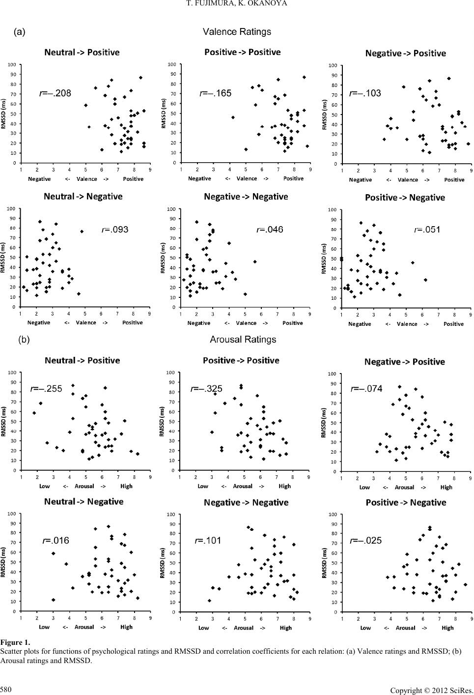
Figure 1 shows scatter plots by function of valence or arousal
ratings and RMSSD. To determine the relationship between
subjective states elicited by emotional stimuli and baseline
HRV, correlation coefficients were calculated for each condi-
tion. We found a significant negative correlation between
arousal ratings for positive stimuli preceded by positive stimuli
and RMSSD (r = –.325; t(41) = 2.20, p < .05). There were no
other significant correlations.
Discussion
In the current study, we investigated whether resting HRV is
associated with experienced emotion elicited by multiple emo-
tional events. The results showed that individuals with lower
HRV experienced more aroused states when viewing succes-
sive positive stimuli. This is the first evidence indicating an
association between resting HRV and subjective experienced
emotion by measuring psychological ratings.
Resting HRV was negatively associated only with arousal
ratings, not valence ratings, when viewing successive two posi-
tive pictures. This result showed that lower resting HRV led to
higher arousal states in response to multiple positive stimuli.
This finding is consistent with previous findings suggesting that
individuals with low HRV are in states of hyperarousal and
diminished habituation by various autonomic measures (Frie-
dman & Thayer, 1998). Furthermore, low HRV during rest has
been shown to resist habituation, even to non-threat stimuli, due
to hypervigilance (Thayer, Friedman, Borkovec, Johnsen, &
Molina, 2000). According to this evidence, subjects with low
HRV fail to adapt to successive positive stimuli, resulting in
feelings of relatively high arousal instead of pleasantness. This
finding suggests that individuals with low HRV show poor
habituation and assess their subjective emotional states as
aroused. Resting HRV may be associated with the ability to
control subjective arousal states rather than to enhance pleas-
antness or unpleasantness when confronting emotional stimuli.
Another explanation of the relationship between reduced HRV
and high arousal states is that HRV is positively related to good
emotion regulation (Porges, 2007; Porges & Byrne, 1992;
Thayer & Lane, 2000, 2009). Emotion regulation involves the
process by which people manage both negative and positive
emotions (Gross, 1998). Successful emotion regulation, by
either reappraisal or suppression, has been shown to lead to
increased vagally mediated HRV (Butler, Wilhelm, & Gross,
2006; Di Simplicio, Costoloni, Western, Hanson, Taggart, &
Harmer, 2011). Furthermore, participants with a high baseline
of vagally mediated HRV spontaneously use emotion regula-
tion strategies more often during emotional conversation than
participants with a low baseline of vagally mediated HRV
(Butler et al., 2006). Consistent with these findings, our results
suggest that high arousal states in response to positive stimuli
result from failures of emotion regulation in subjects with lower
HRV.
Notably, our findings indicated that resting HRV was related
to emotional experience elicited by positive stimuli, not nega-
tive stimuli, although most previous studies have shown an
association between resting HRV and the processing of threat-
ening or negative stimuli (Appelhans & Luecken, 2006; John-
sen et al., 2003; Krypotos et al., 2011; Melzig et al., 2009).
However, individuals with low resting HRV produced an ex-
aggerated emotion-modulated startle reflex in response to neu-
tral and positive pictures (Ruiz-Padial et al., 2003). This ten-
dency could be due to sensitivity to non-threat signals. In this
study, it is possible that the misperception of safety (i.e., posi-
tive stimuli) in individuals with low HRV caused higher arousal
states compared to individuals with high HRV. This finding
provides evidence of emotional inflexibility in response to mul-
tiple positive stimuli. Thus, resting HRV may predict emotional
flexibility indexed by the experience of high arousal to positive
events.
In summary, the current study revealed that resting HRV is a
good proxy of emotional flexibility indexed by subjective ex-
perienced emotion. Individuals with lower HRV experienced
more arousal states when faced with multiple positive stimuli.
This may be due to emotional inflexibility derived from sensi-
tivity to non-threat signals and/or failure of emotion regulation.
HRV may indicate how individuals respond to positive events
effectively and successfully. Future research should investigate
the use of resting HRV as a proxy of emotional flexibility by
measuring psychological, autonomic, and behavioral indices.
Resting HRV provides useful information to understand indi-
vidual differences in emotional flexibility.
Acknowledgements
We thank Kentaro Katahira and for assistance with ed-
iting this manuscript.
REFERENCES
Appelhans, B., & Luecken, L. (2006). Heart rate variability as an index
of regulated emotional responding. Review of General Psychology,
10, 229-240. doi:10.1037/1089-2680.10.3.229
Butler, E. A., Wilhelm, F. H., & Gross, J. J. (2006). Respiratory sinus
arrhythmia, emotion, and emotion regulation during social interac-
tion. Psychophysiology, 43, 612-622.
doi:10.1111/j.1469-8986.2006.00467.x
Di Simplicio, M., Costoloni, G., Western, D., Hanson, B., Taggart, P.,
& Harmer, C. J. (2011). Decreased heart rate variability during emo-
tion regulation in subjects at risk for psychopathology. Psychological
Medicine, 1-9.
Friedman, B. H., & Thayer, J. F. (1998). Autonomic balance revisited:
Copyright © 2012 SciRes. 581
T. FUJIMURA, K. OKANOYA
Panic anxiety and heart rate variability. Journal of Psychosomatic
Research, 44, 133-151. doi:10.1016/S0022-3999(97)00202-X
Gross, J. (1998). The emerging field of emotion regulation: An integra-
tive review. Revi ew of Gen eral Psychology, 2, 271-299.
doi:10.1037/1089-2680.2.3.271
Johnsen, B. H., Thayer, J. F., Laberg, J. C., Wormnes, B., Raadal, M.,
Skaret, E., Kvale, G., & Berg, E. (2003). Attentional and physiologi-
cal characteristics of patients with dental anxiety. Journal of Anxiety
Disorders, 17, 75-87. doi:10.1016/S0887-6185(02)00178-0
Krypotos, A., Ja h fari, S., v a n As t , V. A., Kindt, M., & Forstmann, B. U.
(2011). Individual differences in heart rate variability predict the de-
gree of slowing during response inhibition and initiation in the pres-
ence of emotional stimuli. Frontiers in Psychol o gy , 2, 1-8.
doi:10.3389/fpsyg.2011.00278
Lang, P. J., Bradley, M. M., & Cuthbert, B. N. (2008). International
affective picture system (IAPS): Affective ratings of pictures and in-
struction manual. Technical Report A-8. Gainesville, FL: University
of Florida.
Melzig, C., Weike, A., Hamm, A., & Thayer, J. (2009). Individual
differences in fear-potentiated startle as a function of resting heart
rate variability: Implications for panic disorder. International Jour-
nal of Psychophysiology, 71, 109-117.
doi:10.1016/j.ijpsycho.2008.07.013
Porges, S. W. (2007). The polyvagal perspective. Biological Psychol-
ogy, 74, 116-143. doi:10.1016/j.biopsycho.2006.06.009
Porges, S. W., & Byrne, E. A. (1992). Research methods for measure-
ment of heart rate and respiration. Biological Psychology, 34, 93-
130. doi:10.1016/0301-0511(92)90012-J
Ruiz-Padial, E., Sollers, J. J., Vila, J., & Thayer, J. F. (2003). The
rhythm of the heart in the blink of an eye: Emotion-modulated startle
magnitude covaries with heart rate variability. Psychophysiology, 40,
306-313. doi:10.1111/1469-8986.00032
Russell, J. A., Weiss, A., & Mendelsohn, G. A. (1989). Affect Grid: A
single-item scale of pleasure and arousal. Journal of Personality and
Social Psychology, 57, 493-502. doi:10.1037/0022-3514.57.3.493
Thayer, J. F., & Brosschot, J. F. (2005). Psychosomatics and psycho-
pathology: Looking up and down from the brain. Psychoneuroendo-
crinology, 30, 1050-1058. doi:10.1016/j.psyneuen.2005.04.014
Thayer, J. F., Friedman, B. H., Borkovec, T. D., Johnsen, B. H., &
Molina, S. (2000). Phasic heart period reactions to cued threat and
nonthreat stimuli in generalized anxiety disorder. Psychophysiology,
37, 361-368. doi:10.1111/1469-8986.3730361
Thayer, J. F., & Lane, R. D. (2000). A model of neurovisceral integra-
tion in emotion regulation and dysregulation. Journal of Affective
Disorders, 61, 201-216. doi:10.1016/S0165-0327(00)00338-4
Thayer, J. F., & Lane, R. D. (2009). Claude Bernard and the heart-brain
connection: Further elaboration of a model of neurovisceral integra-
tion. Neuroscience Bi obehavioral Review, 33, 81-88.
doi:10.1016/j.neubiorev.2008.08.004
Waugh, C. E., Thompson, R. J., & Gotlib, I. H. (2011). Flexible emo-
tional responsiveness in trait resilience. Emotion, 11, 1059-1067.
doi:10.1037/a0021786
Westphal, M., Seivert, N. H., & Bonanno, G. A. (2010). Expressive
flexibility. Emotion, 10, 92-100. doi:10.1037/a0018420
Copyright © 2012 SciRes.
582