Paper Menu >>
Journal Menu >>
 Journal of Min erals & Materials Characterization & Engineering , Vol. 8, No.1, pp 73-78, 2009 jmmce.org Printed in the USA. All rights reserved 73 In-Vitro Studies of Artificially Removed Human Renal Stone Minerals by Sonic Engineering Approach-I G. Kanchana, P. Sundaramoorthi*, and G.P. Jeyanthi Department of Bio-chemistry, Avinashilingam University for Women, Coimbatore, India. *Department of Physics, Thiruvalluvar Govt. Arts College, Rasipuram, Namakkal, India-637401. (Ph.No.-04287-231802 (off.) Fax- 04287-231882 , MailID-moorthi.sundara@gmail,com) ABSTRACT Ultrasonic waves are the main tools for measuring the unknown parameters in science and technological analysis of a particular biological sample. The sonic wave velocity measurement applied in the in-vitro & in-vivo studies in medical fields has been made in different renal stones removed from the human kidney. The samples were analyzed at different frequencies and the corresponding wavelength at room temperature (305K) were found. The experimental results have been used to calculate many physical constants of the samples, such as stone density, sonic velocity, acoustical wavelength, specific acoustic impedance, transmitivity, reflectivity and dynamic modulus of elasticity. The investigated results have been agreed to the reported information. Key Words: Mineral processing, Sampling, Process instrumentation, Bio-oxidation, Ion exchange 1. INTRODUCTION The ultrasonic sound velocity gives the necessary data in all the states. From the sonic velocity of a particular medium and its propagation results, one can find the texture, structure, porosity, compression, and particle size in a direction, which is kept at constant physical and chemical environments. More number of parameters can be calculated in solids [1-2] and hence it is applied to the human physiological disorders. Ultrasonic wave velocity has been used in conventional medical diagnostics therapy and surgical tools in different sensitive parts of the human body such as brain, glands, breast, heart, urinary tracts, peripheral blood vessels, reproductive organs, etc.[3-5]. Especially in a human physiological  74 G. Kanchana, P. Sundaramoorthi*, and G.P. Jeyanthi Vol.8, No.1 system, kidney is a major organ, which separate extra minerals, water, wastes etc. from the blood after digestion. In the pathological condition let the kidney failure, inhibitor disorder, and increase in mineral values in the blood or urine continuously, one can get the renal stones any where within the urinary tracts. The kidney stone formation, or renal stone diseases or mineral deposition or crystals grown in the kidney creates socio-economic disorders. To avoid this type of problem one must clearly know about the diseases to prevent the deposition either before the start or after the start. Extra corporeal shock wave lithotripsy (ESWL), Laser and UV has created large disadvantages such as tissue damage, pain etc. [6- 8] which can be overcome by ultrasonic method of treatment [9]. In the present investigation, the longitudinal sonic parameters of six different kidney stones at different frequency ranges from 0.5MHZ to 12 MHZ at 305K were used. 2. MATERIALS AND METHODS Many methods are used to measure the ultrasonic parameters in the biological system. In the present investigation, pulse echo overlap (PEO) technique was used with the good accuracy. The PEO method is applied in diagnostic purpose as well as the tissue characterization in a static and dynamic system. A double probe contact (between the kidney stones) through transmission technique was used for the measurement of this parameter. The transmitter, receiver transducers are controlled by the computer. Similar studies have already been carried out in gallstones [10]. From the observation, it gives directly the parameters of pulse transit time (t) for the known thickness (d). Using the available data the acoustical impedance (Z), density (p) of the samples, pressure amplitude coefficient (R), pressure amplitude transmission coefficient (T) and dynamic modulus of elasticity (E) of all the samples were calculated. In the present studies different types of stones were collected in and around Namakkal District local hospitals in Tamil Nadu. India. The stones were named as A, B, C, D and E. The stones were removed from the kidney by applying the ultrasonic lithotripsy process. The samples were collected, cleaned and then preserved. The chemical compositions of all the samples were by bio-chemical methods. The constituents of the samples are tabulated in Table-1. In this study, it is assumed that in-vitro calculation gives good representation of the natural properties of the biological stones and characteristics of the vivo solution. 3. RESULTS AND DISCUSSIONS The artificially removed renal stone samples were photographed using digital camera and find its dimensions. The renal stone samples are shown in Fig.1-5. Samples were analyzed by bio-chemical methods to identify the chemical constituent present in the samples. The corresponding samples composition and its various types of stones are shown in Table-1. The various parameters of the stones, such as mass, size, volume and density are presented in Table-2. It is clear that the B sample has more density and less volume than the sample D. The frequencies and wavelength of the corresponding samples at particular 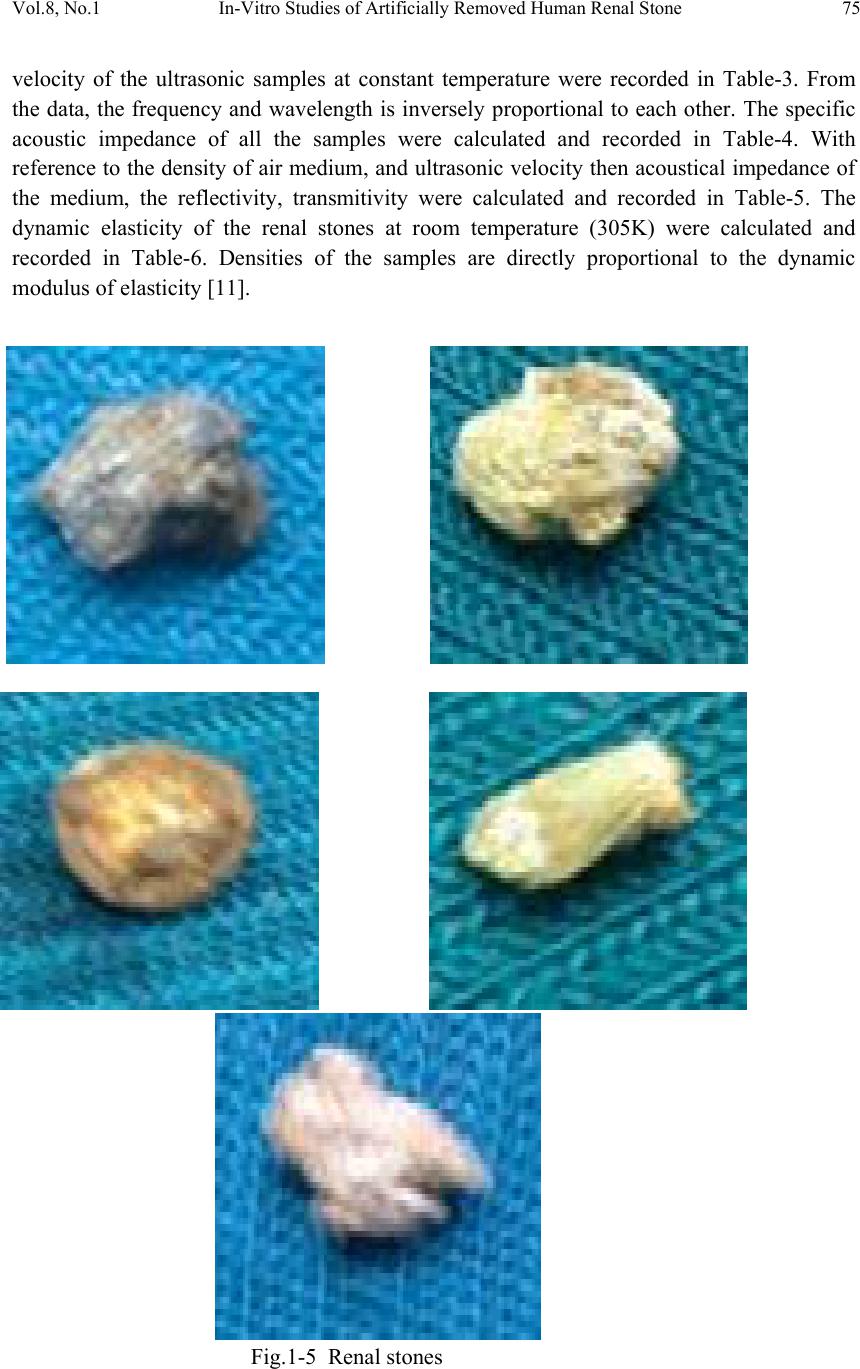 Vol.8, No.1 In-Vitro Studies of Artificially Removed Human Renal Stone 75 velocity of the ultrasonic samples at constant temperature were recorded in Table-3. From the data, the frequency and wavelength is inversely proportional to each other. The specific acoustic impedance of all the samples were calculated and recorded in Table-4. With reference to the density of air medium, and ultrasonic velocity then acoustical impedance of the medium, the reflectivity, transmitivity were calculated and recorded in Table-5. The dynamic elasticity of the renal stones at room temperature (305K) were calculated and recorded in Table-6. Densities of the samples are directly proportional to the dynamic modulus of elasticity [11]. Fig.1-5 Renal stones 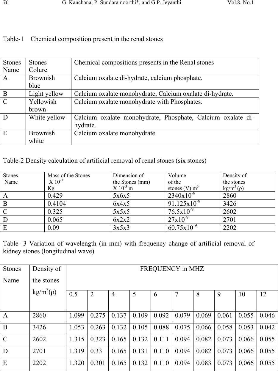 76 G. Kanchana, P. Sundaramoorthi*, and G.P. Jeyanthi Vol.8, No.1 Table-1 Chemical composition present in the renal stones Stones Name Stones Colure Chemical compositions presents in the Renal stones A Brownish blue Calcium oxalate di-hydrate, calcium phosphate. B Light yellow Calcium oxalate monohydrate, Calcium oxalate di-hydrate. C Yellowish brown Calcium oxalate monohydrate with Phosphates. D White yellow Calcium oxalate monohydrate, Phosphate, Calcium oxalate di- hydrate. E Brownish white Calcium oxalate monohydrate Table-2 Density calculation of artificial removal of renal stones (six stones) Stones Name Mass of the Stones X 10-3 Kg Dimension of the Stones (mm) X 10-3 m Volume of the stones (V) m3 Density of the stones kg/m3 (ρ) A 0.429 5x6x5 2340x10-9 2860 B 0.4104 6x4x5 91.125x10⁻⁹ 3426 C 0.325 5x5x5 76.5x10⁻⁹ 2602 D 0.065 6x2x2 27x10-9 2701 E 0.09 3x5x3 60.75x10⁻⁹ 2202 Table- 3 Variation of wavelength (in mm) with frequency change of artificial removal of kidney stones (longitudinal wave) Stones Name Density of the stones kg/m3(ρ) FREQUENCY in MHZ 0.5 2 4 5 6 7 8 9 10 12 A 2860 1.099 0.2750.1370.1090.0920.0790.069 0.061 0.0550.046 B 3426 1.053 0.263 0.132 0.105 0.088 0.075 0.066 0.058 0.053 0.042 C 2602 1.315 0.323 0.165 0.132 0.111 0.094 0.082 0.073 0.066 0.055 D 2701 1.319 0.33 0.165 0.131 0.110 0.094 0.082 0.073 0.066 0.055 E 2202 1.320 0.301 0.165 0.132 0.110 0.094 0.083 0.073 0.066 0.055 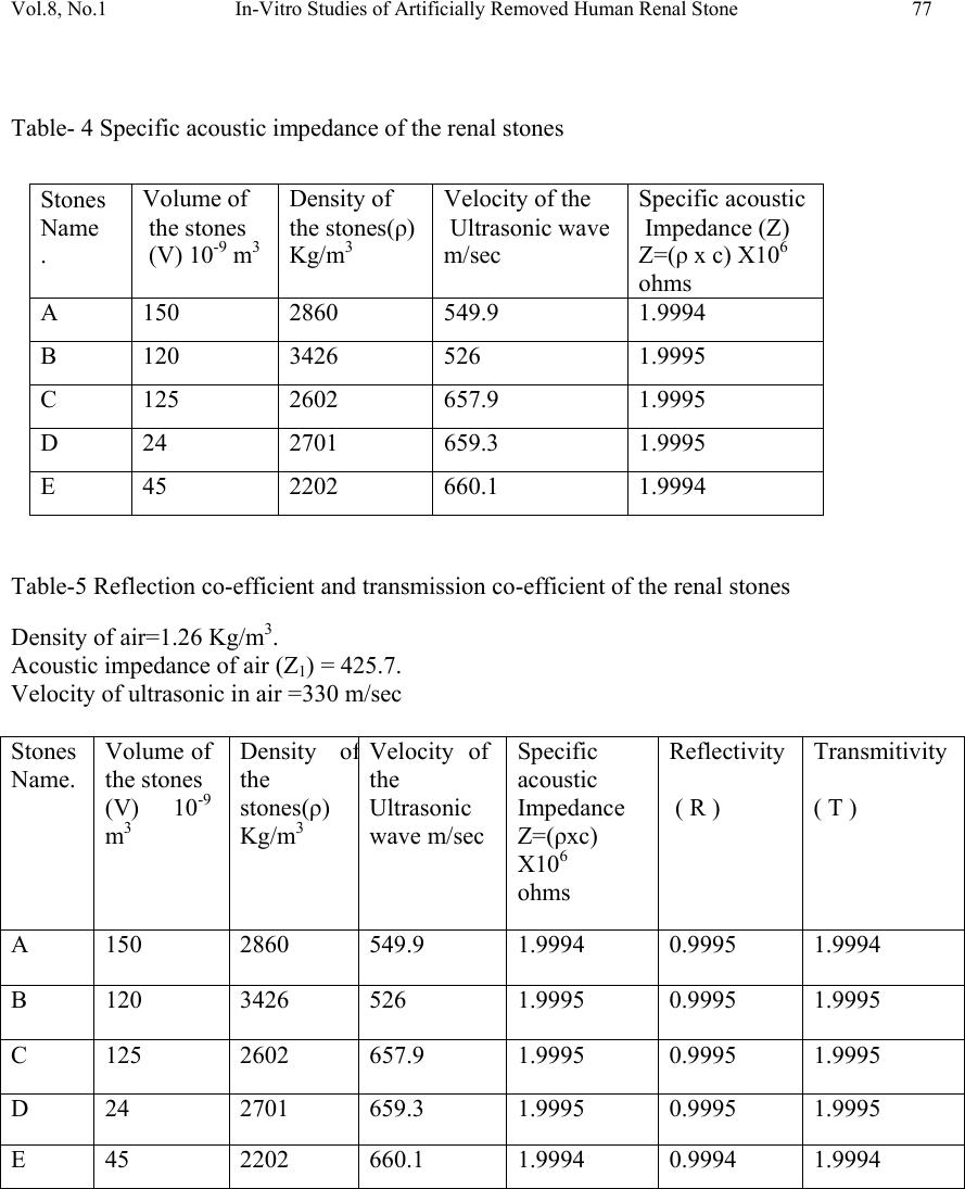 Vol.8, No.1 In-Vitro Studies of Artificially Removed Human Renal Stone 77 Table- 4 Specific acoustic impedance of the renal stones Stones Name . Volume of the stones (V) 10-9 m3 Density of the stones(ρ) Kg/m3 Velocity of the Ultrasonic wave m/sec Specific acoustic Impedance (Z) Z=(ρ x c) X106 ohms A 150 2860 549.9 1.9994 B 120 3426 526 1.9995 C 125 2602 657.9 1.9995 D 24 2701 659.3 1.9995 E 45 2202 660.1 1.9994 Table-5 Reflection co-efficient and transmission co-efficient of the renal stones Density of air=1.26 Kg/m3. Acoustic impedance of air (Z1) = 425.7. Velocity of ultrasonic in air =330 m/sec Stones Name. Volume of the stones (V) 10-9 m3 Density o f the stones(ρ) Kg/m3 Velocity of the Ultrasonic wave m/sec Specific acoustic Impedance Z=(ρxc) X106 ohms Reflectivity ( R ) Transmitivity ( T ) A 150 2860 549.9 1.9994 0.9995 1.9994 B 120 3426 526 1.9995 0.9995 1.9995 C 125 2602 657.9 1.9995 0.9995 1.9995 D 24 2701 659.3 1.9995 0.9995 1.9995 E 45 2202 660.1 1.9994 0.9994 1.9994 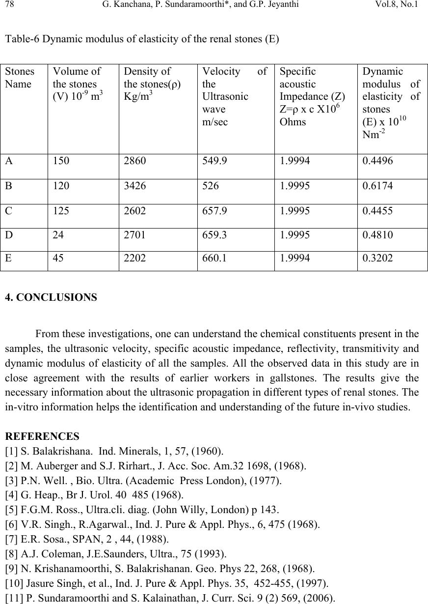 78 G. Kanchana, P. Sundaramoorthi*, and G.P. Jeyanthi Vol.8, No.1 Table-6 Dynamic modulus of elasticity of the renal stones (E) Stones Name Volume of the stones (V) 10-9 m3 Density of the stones(ρ) Kg/m3 Velocity of the Ultrasonic wave m/sec Specific acoustic Impedance (Z) Z=ρ x c X106 Ohms Dynamic modulus of elasticity of stones (E) x 1010 Nm-2 A 150 2860 549.9 1.9994 0.4496 B 120 3426 526 1.9995 0.6174 C 125 2602 657.9 1.9995 0.4455 D 24 2701 659.3 1.9995 0.4810 E 45 2202 660.1 1.9994 0.3202 4. CONCLUSIONS From these investigations, one can understand the chemical constituents present in the samples, the ultrasonic velocity, specific acoustic impedance, reflectivity, transmitivity and dynamic modulus of elasticity of all the samples. All the observed data in this study are in close agreement with the results of earlier workers in gallstones. The results give the necessary information about the ultrasonic propagation in different types of renal stones. The in-vitro information helps the identification and understanding of the future in-vivo studies. REFERENCES [1] S. Balakrishana. Ind. Minerals, 1, 57, (1960). [2] M. Auberger and S.J. Rirhart., J. Acc. Soc. Am.32 1698, (1968). [3] P.N. Well. , Bio. Ultra. (Academic Press London), (1977). [4] G. Heap., Br J. Urol. 40 485 (1968). [5] F.G.M. Ross., Ultra.cli. diag. (John Willy, London) p 143. [6] V.R. Singh., R.Agarwal., Ind. J. Pure & Appl. Phys., 6, 475 (1968). [7] E.R. Sosa., SPAN, 2 , 44, (1988). [8] A.J. Coleman, J.E.Saunders, Ultra., 75 (1993). [9] N. Krishanamoorthi, S. Balakrishanan. Geo. Phys 22, 268, (1968). [10] Jasure Singh, et al., Ind. J. Pure & Appl. Phys. 35, 452-455, (1997). [11] P. Sundaramoorthi and S. Kalainathan, J. Curr. Sci. 9 (2) 569, (2006). |

