Open Journal of Urology
Vol.07 No.12(2017), Article ID:80870,8 pages
10.4236/oju.2017.712028
External Genitalia Gangrene: Clinical, Therapeutic Aspects and Prognosis at University Hospital Souro Sanou of Bobo Dioulasso (Burkina Faso)
Timothée Kambou1, Adama Ouattara1, Abdoulkarim Paré1, Brahima Kirakoya2, Fasnéwindé Aristide Kaboré2, Hassan Dogo3, Hamidou Bako1
1Department of Urology, Sanon Souro Teaching Hospital, Bobo Dioulasso, Burkina Faso
2Department of Urology, Yalgado Ouedraogo Teaching Hospital, Ouagadougou, Burkina Faso
3Department of Surgery, Division of Urology, University of Maiduguri Teaching Hospital, Maiduguri, Nigeria

Copyright © 2017 by authors and Scientific Research Publishing Inc.
This work is licensed under the Creative Commons Attribution International License (CC BY 4.0).
http://creativecommons.org/licenses/by/4.0/


Received: October 8, 2017; Accepted: December 2, 2017; Published: December 5, 2017
ABSTRACT
Objective: To describe the clinical aspects and the management of external genitalia gangrene in the Urology department of the Bobo Dioulasso CHUSS. Patients and methods: This was a retrospective and descriptive study over a period of 6 years from January 2011 to December 2016, which covered 54 patients hospitalized in the urology department of the University Hospital Souro Sanou for external genitalia gangrene. Results: The mean age of our patients was 53.8 years. The large ulcero-necrosis of scrotum was the main reason for reference. Our patients consulted on average 8.23 days after the onset of symptomatology. A risk factor was found in 44.2% of cases and a cause was identified in 65.3% of patients. A urogenital cause was present in 32.7%. Probabilistic antibiotic therapy and medical reanimation were systematic at admission. Debridement was performed on average 18 hours after admission and 10 patients had more than one debridement. We noted a mortality rate of 5.5%. Conclusion: Fournier’s gangrene is a serious infectious disease whose diagnosis is often delayed in our context. Early surgical debridement remains the essential therapeutic gesture to reduce mortality.
Keywords:
Gangrene, External Genitalia, Debridement

1. Introduction
Gangrene of the external genital organs is a form of genital, perineal and perianal necrotizing fasciitis resulting from a polymicrobial infection. It mainly affects superficial and deep fascias in humans. The incidence is not well known. Treatment is mainly based on debridement after appropriate medical treatment. However, mortality is still high (20% to 80%), due to delayed diagnosis and management [1] . This work aims to describe the clinical aspects, the management and the prognosis of external genitals gangrene in the Department of Urology and Andrology of the University Hospital of Bobo Dioulasso.
2. Patients and Methods
This was a retrospective descriptive study over a period of 6 years from January 2011 to December 2016. The study was carried out in the Urology department of Bobo Dioulasso’s Urology Center. The sample consisted of patients received with large painful ulcero-necrosis of the scrotum with or without perineal involvement. Patients with large scrotum painfully or not only were excluded.
The variables studied were age, clinical history of the patient, reason for consultation, mode of entry, risk factors and etiology, clinical and para-clinical examination results, treatment administered and prognosis.
A preformed questionnaire has been prepared for the collection of data. Our data sources consisted of patient medical records, the operating record book and the hospitalization record. Data analysis was carried out using Epi info 3.5.1 French version 2008. The study has been performed with the approval of a medical ethics committee.
3. Results
During the study period, we received 54 cases of the external genitalia gangrene out of a total of 2375 hospitalized patients, with a hospital frequency of 2.3%.
The annual distribution of patients is presented in Figure 1. A peak of cases was noted in the year 2016.
Figure 1. The distribution of the patients with external genitalia gangrene according to the year of hospitalization.
The average age of patients was 53.8 years with extremes of 7 years and 95 years.
The distribution of patients according to age group is presented in Table 1.
Risk factors were identified in the various cases listed. Figure 2 shows the distribution of the various factors involved.
The average consultation time was 8.2 days with extremes of 2 days and 2 months. More than 2/3 of the cases (69.2%) were referred by the peripheral health centres. The main reason for consultation was painful swelling of the scrotum without necrotic ulcer and this constitutes 76.4% of cases.
Clinical examination had noted in almost all patients an infectious syndrome. In addition, 4 cases of skin cracking were observed (7.4% of cases). Figure 3
Figure 2. Distribution of identified risk factors.
Figure 3. Distribution of the different lesions reported during the clinical examination.
Table 1. Distribution of patients according to age group.
shows the distribution of the peno-scrotal lesions.
The following picture (Photo 1 and Photo 2) give an overview of lesions of the external genitalia.
The aetiology was identified in 65.3% of cases; it was uro-genital or rectal (perianal abscess and stenosis of the urethra). Table 2 gives distribution of the implicated entry gates in the occurrence of gangrene.
Hyperglycemia was noted in 37.5% of the patients, 57.2% had leukocytosis and 34.2% had kidney failure.
Case management was multidisciplinary in the majority of cases. This was a systematic medical resuscitation associated with systematic bi-antibiotic treatment at admission and anti-tetanus serotherapy. The combination of β lactams
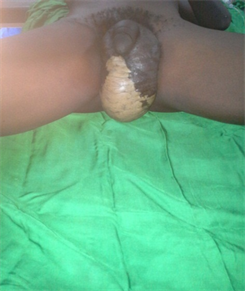
Photo 1. Ulcero-necrosis lesions of the scrotum.
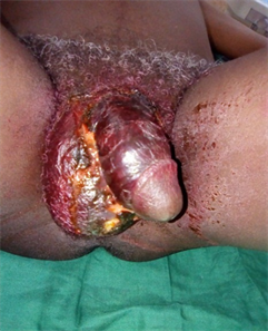
Photo 2. Ulcero-necrosis lesions peno-scrotal.
Table 2. Distribution of the implicated entry gates in the occurrence of gangrene.
and metronidazole was the rule. In some cases, an aminoglycoside (injectable gentamicin very often) was added in the absence of renal failure.
Wound debridement in the form of scrotal bipartition with necrosectomy, was performed in all patients. On average, debridement was performed 18 hours after admission and 10 patients had more than one debridement. A protective colostomy was performed in 4 patients (7.4%) and cystostomy in 3 patients (5, 5%). The following pictures (Photo 3 and Photo 4) present debridement external genitalia gangrene.
The average hospital stay was 23 days with extremes of 1 day and 192 days. Almost ¾ of the patients (86.5%) were cured and discharged with acceptable cutaneous healing. Photo 5 presents an external genitalia gangrene.
However, 3 patients (5.5%) left against medical advice. The mortality rate was 5.5%.
4. Discussion
External genitalia gangrene is a necrotizing fasciitis; a severe infection of the soft tissues affecting the superficial and deep fascias. Its incidence is poorly understood and varies from one country to another with a high incidence in developing countries. An average of 97 cases per year was reported from 1989 to 1998 by Marynowski [2] . In our series, 54 cases were managed in 6 years (frequency of 2.3%) and Rimtebaye in N’djamena in Chad [3] in its study had reported 51 cases in 4 years.
The gangrene of external genitalia occurs at any age; it ranges from a few days to 89 years [4] . In our series, the mean age of the patients was 53.8 years. Fall B et al. [5] in Dakar in Senegal and Fakhfakh M [6] in Tunis, Tunisia, had found in their study mean age of 50 years and 54 years respectively. Factors favoring in particular are advanced age, anorectal and uro-genital diseases would explain this state of affairs. In fact, diabetes was present in 29.6% of patients and chronic ingestion of alcohol in 9.25% of patients. Retroviral HIV serology was positive in 7 patients (12.9%).
Extreme ages, poor hygiene, HIV infection were blamed by several authors [7] [8] .
The average consultation time was 8.2 days with extremes of 2 days and 2 months. Borki K found an average of 14 days [9] . The gangrene of external genitalia touching the intimate organs is still a taboo in our African societies and
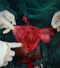
Photo 3. Scrotal necrosis done.
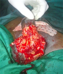
Photo 4. Penile-scrotal necrosis done.
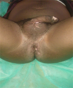
Photo 5. External genitalia gangrene healing.
the discomfort of life linked to the foul odor emitted by the ulcerated skin often make the patients to consult late in the health centres. The large necrotic ulcerative pouch was the main reason for consultation in our series (76.4%).
Systematic medical resuscitation + systematic bi-antibiotherapy at admission + anti-tetanus serotherapy must be the rule.
Debridement should be done as soon as possible after stabilization of the hemodynamic state of the patient, as the infection progresses very rapidly within hours [10] .
Mortality rates reported in the literature range from 0 to 80% [2] [11] [12] . The mortality rate in our series was 5.5%. The late diagnosis and the favorable factors are factors aggravating the prognosis of this pathology. The low rate in our series would be linked to better care, especially during the course of case management. Early debridement and better follow-up result in better survival rates.
5. Conclusion
The gangrene of the genitals constitutes a surgical emergency. It is a serious infectious disease that is often diagnosed late in our context. Early medical resuscitation and early surgical debridement are the essential therapeutic gestures to reduce mortality.
Cite this paper
Kambou, T., Ouattara, A., Paré, A., Kirakoya, B., Kaboré, F.A., Dogo, H. and Bako, H. (2017) External Genitalia Gangrene: Clinical, Therapeutic Aspects and Prognosis at University Hospital Souro Sanou of Bobo Dioulasso (Burkina Faso). Open Journal of Urology, 7, 235-242. https://doi.org/10.4236/oju.2017.712028
References
- 1. Sarkis, P., Farran, F., Khoury, R., Kamel, G., Nemr, E., Bianjini, J. and Merheje, S. (2009) Gangrène de Fournier: revue de la littérature récente. Progrès en Urologie, 19, 75-84. https://doi.org/10.1016/j.purol.2008.09.050
- 2. Marynowski, M.T. and Aronson, A.A. (2008) Fournier’s Gangrene Medicine. http://emedicine.medscape.com/article/2028899-overview
- 3. Rimtebaye, K., Niang L., Ndoye, M., Traore, I., Vadandi, V., Gueye, S.M. and Noar, T. (2014) Gangrène de Fournier: aspects épidémiologique, clinique, diagnostique et thérapeutique au service d’Urologie de N’djaména. Uro’andro, 1, 91-98.
- 4. Clayton, M.D., Fowler, J.E., Sharifi, R. and Pearl, R.K. (1990) Causes, Presentation and Survival of Fifty-Seven Patients with Necrotizing Fascitis of the Male Genitalia. Surgery, Gynecology & Obstetrics, 170, 49-55.
- 5. Fall, B., Fall, A.A., Diao, B., Kpatcha, M.T., Sow, Y., Kaboré, F.A., Ali, M., Ndoye, A.K., Ba, M. and Diagne, B.A. (2009) Les gangrènes des organes génitaux externes dans le service d’urologie-andrologie, CHU Aristide-Le-Dantec: à propos de 102 cas. Andrologie, 19, 45-49.
- 6. Fakhfakh, H., Chabchoub, K., Slimen, M.H., Bahloul, A. and Mhiri, M.N. (2006) La gangrène des organes génitaux externes de l’homme: Facteurs de risque et pronostic. Andrologie, 16, 229-234.
- 7. Nambiar, P.K., Lander, S. and Midha, M. (2005) Fournier Gangrene in Spinal Cord Injury: A Case Report. Journal of Spinal Cord Medicine, 28, 121-124. https://doi.org/10.1080/10790268.2005.11753809
- 8. Hubert, J., Fournier, G., Mangin, P. and Punga-Maole, M. (1995). Gangrène des organes génitaux externes. Progrès en Urologie, 5, 911-924.
- 9. Borki, K., Ait Ali, A., Choho, A., Daali, M., Alkandry, S. and André, J.L. (2002) La gangrène périnéo-scrotale: à propos de 60 cas. E-mémoires de l’Académie Nationale de Chirurgie, 1, 49-54.
- 10. Mejean, A., Codet, Y.P., Vogt, B., Cazalaa, J.B., Chrétien, Y. and Dufour, B. (1999) Gangrène de Fournier étendue à la totalité du scrotum: Traitement par excisions chirurgicales itératives multiples, colostomie de dérivation, triple antibiothérapie et réanimation post-opératoire. Progrès en Urologie, 9, 721-726.
- 11. Smith, G.L., Bunker, C.B. and Dinneen, M.D. (1998) Fournier’s Gangrene. British Journal of Urology, 81, 347-355. https://doi.org/10.1046/j.1464-410x.1998.00532.x
- 12. Schaeffer, E.M. and Schaeffer, A.J. (2007) Infections of the Urinary Tract. In: Wein, A., Ed., Campbell-Walsh Urology, Saunders Elsevier, Philadelphia, PA, 301-303.


