Open Journal of Veterinary Medicine
Vol.06 No.07(2016), Article ID:68296,7 pages
10.4236/ojvm.2016.67015
Gastro-Intestinal Parasites of Camels (Camelus dromedarius) from Mogadishu, Somalia
Abdalla M. Ibrahim1, Ahmed A. H. Kadle2*, Abdulkarim A. Yusuf1
1Abrar Research and Training Centre, Abrar University, Mogadishu, Somalia
2International Committee of the Red Cross, Somalia Delegation, Mogadishu, Somalia

Copyright © 2016 by authors and Scientific Research Publishing Inc.
This work is licensed under the Creative Commons Attribution International License (CC BY).
http://creativecommons.org/licenses/by/4.0/


Received 21 June 2016; accepted 10 July 2016; published 13 July 2016
ABSTRACT
Somalia has the largest dromedary (Camelus dromedarius) population in the world. However, scientific research and camel diseases investigation in the country were lacking since 1980s. A total of 167 camels (131 semi-intensive dairy camels system and 36 free grazing systems) were sampled from three districts in Mogadishu citybetween December 2015 and March 2016 to investigate the prevalence rate of gastro-intestinal parasites in camels from Mogadishu city using different coprological techniques. The overall prevalence of camel gastro-intestinal parasites was 50.3%. The prevalence rate were significantly varies between the extensive and semi-intensive production systems (p = 0.000) and between the different districts (p = 0.000). Nematodes revealed higher prevalence rate (47.9%) than Cestodes (6.0%), Trematodes (4.2%) and Coccidia (0.65%). Eggs of eight genera of nematodes, two genera of trematodes and two genera of cestodes as well as Eimeria sp. Oocysts were identified in this study. Dictyocaulus sp. and Trichostrongylus sp. were the most prevalent followed by Parascaris equorum and Strongyloides sp. About 56.0% of the infected camels revealed mix-infection of up to five different parasite species. Moreover, 35.7% of these animals revealed heavy infection. The important role of the camel has inspired investigators and researchers to look for diseases that may threaten camel health and production. However, in Somalia in spite of having the largest counts of camels but in depth scientific data are not available. Therefore, the present paper was to be the first detailed data on camel gastro-intestinal parasites in Mogadishu area and may be in whole Somalia. We came to conclude that gastrointes- tinal parasites were highly prevalent in camels from Mogadishu particularly in nomadic herds of camels. Studies on the effect of gastro-intestinal parasites on camel production and productivity are recommended. Moreover, the intensive system of camel production should be encouraged.
Keywords:
Gastro-Intestinal Parasites, Camels, Abrar, Mogadishu, Somalia

1. Introduction
The world camel population is estimated at 18.26 million. Somalia has the largest (38.34%) dromedary population in the world which is estimated at 7 million [1] [2] . The highest density (30%) of that population (1.87 million) is found in Central Somalia [3] .
The economic importance of the dromedary for Somalia is due to milk and meat Production as well as the social issues. The dromedary is also used for transport of milk, water and nomadic household migration. It is the unique to survive and produce under extreme arid and semi-arid conditions of Somalia. The dromedary is used in recent years to supply milk to towns. Therefore, it is kept in rural and around urban areas such as Mogadishu. Moreover, export of live dromedaries and hides provide for a great deal of the country’s hard currency earnings.
Gastro-intestinal parasites of one humped camels (Camelus dromedarius) are one of the major causes of reduction in production and productivity.
Despite this valuable economic importance, scientific data about Somali camel were missed since 1980s [4] - [6] . These authors reported some general information about gastro-intestinal parasites affecting Somali camels such as Eimeria cameli, Trichostrongylidae, Trichuridae, Moniezia and Stilezia.
Due to the scarcity of camel researches in the region, this study aims to investigate in details, the prevalence rate of camel gastro-intestinal parasites in two different grazing systems so as to establish a baseline data on gastro-intestinal parasites affecting camels in the country.
2. Materials and Methods
2.1. Study Area
Mogadishu is the largest and capital city of Somalia. It is located in Benadir region, south-eastern Somalia. The region itself is coextensive with the city. It consists of seventeen districts. Three of them were purposively included in this study namely Deynile, Kahda and Yaqshid. These districts are the main camel raring areas available in Benadir region.
2.2. Study Population
Adult (≥2 years old) one humped camels (Camelus dromedaries) in the three districts in Mogadishu city of Somalia were examined for presence of egg, larvae or oocyst of gastro-intestinal parasites between December 2015 and March 2016. Both nomadic (extensive camel production system) and camel dairy farms (semi-intensive camel production system) were visited for faecal sample collection. Most of the investigated camels were adult lactating females.
2.3. Faecal Sample Collection
A total of 167 faecal samples were collected from camels irrespective of their clinical status between December 2015 and March 2016. These samples were collected directly from the rectum by disposable gloves and placed into plastic containers and labeled. Then the samples were transferred to Abrar Research and Training Centre laboratory which belongs to Abrar University, Mogadishu, Somalia for parasitological examination freshly without preservation.
2.4. Coprological Examination
Each faecal sample were examined by simple wet smear, flotation method using saturated sodium chloride and sedimentation techniques described by Taylor et al., 2007 [7] . Fecal egg or oocysts counts (EPG) were determined by modified McMaster egg counting technique as stated by Taylor et al., 2007 [7] . We also use their key for identification of gastro-intestinal parasite affecting different animals as well as the level of infection (Heavy, moderate or mild infection).
2.5. Statistical Analysis
Collected data were entered, coded and stored in a Microsoft® Excel spread sheet for Windows® 2007 data base before transfered to SPSS sheet. The Statistical Package for Social Sciences (SPSS) for Windows® version 20 was used for all appropriate statistical analysis. The differences were considered statistically significant when p ≤ 0.05.
Photos of the detected eggs of parasites were captured directly from microscope eye piece using digital camera (Sony, 16.1 MP) and stored in computer.
3. Results and Discussion
3.1. General Prevalence Rate
The overall prevalence of camel gastro-intestinal parasites in the investigated area was 50.3% (Table 1). Higher prevalence rate (62.7%) was reported in Tanzania [8] and lesser prevalence rate (41.1%) was reported in Egypt [9] . Gastro-intestinal parasites were found to be more prevalent significantly (p = 0.000) in free grazingcamel production system (88.9%) than the semi-intensive dairy farm system (39.7%). It is worth mentioning that camel dairy farms are increasing in Mogadishu area and most of the farm managers are aware of the importance of using anthelmintics to reduce the effect of gastro-intestinal parasites on milk production. Some of the owners also consult veterinarians. The prevalence varies significantly (p = 0.000) among the different districts. Higher prevalence rate was recorded in camels from Yaqshid district (83.3%) followed by Deynile (69.4%). Lower prevalence rate was recorded in camels from Kahda district (21.4). That is just because all camels examined from this district were under semi-intensive production system with more effective control strategy. The variation (p = 0.317) of the gastro-intestinal parasites infection between male and female in the present study could not be considered because only four (2.4%) adult male camels were examined.
As presented in Plate 1 and Plate 2 , four different classes of gastro-intestinal parasites were recorded in the present study, including Nematodes (47.9%), Cestodes (6.0%), Trematodes (4.2%) and Coccidia (0.65%). The significant values of the effect of production system, districts and sex were presented in Table 2. Nematodes and Cestodes were previously reported in Somali camels [4] - [6] . However, Trematodes were not recorded by the later authors. Eimeria cameli and other Eimeria sp. were also reported in Somali camels [4] .
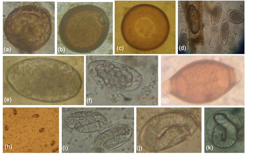
Plate 1. Eggs of different nematodes species affecting Somali camels in Mogadishu area. (a) Parascaris equorum (b), (c) Toxocara sp., (d) Trichostrongylus sp., (e) Chabertia ovina (f) Haemonchus sp., (g) Trichuris sp., (h) (i) Dictyocaulus sp., (j), (k) Strongyloides sp.
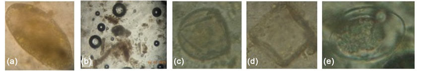
Plate 2. Eggs of some flat worms and Coccidia affecting Somali camels in Mogadishu area. (a) Paramphistomum cervi, (b) Fasciola sp., (c) Moniezia sp., (d) Anoplcephala sp., (e) Eimeria sp.

Table 1. Prevalence rate of gastro-intestinal parasites in camels from Mogadishu, Somalia.
Table 2. Prevalence rate of different classes of gastro-intestinal parasites in camels from Mogadishu, Somalia.
3.2. Prevalence Rate of Nematodes
In this study, Nematodes were found to be more prevalent (Table 2). Eggs of different eight genera of nematodes were identified including Dictyocaulus (26.9%), Trichostrongylus (23.4%), Parascaris (12.6%), Strongyloides (4.8%), Trichuris (4.2%), Toxocara (3.0%), Chabertia (3.0%) and Haemonchus (1.8%). The detailed prevalence rate in the different districts and different grazing system was presented in Table 3. Trichostrongylidae, Trichridae, Moniezia and Stilezia were previously reported [4] - [6] , but without information about their
Table 3. Prevalence rate of different Nematodes in camels from Mogadishu, Somalia.
prevalence rate. The identified eggs of nematodes in the present study were presented in Plate 1 . Most of these nematodes species were also reported in other camel zones such as Sudan, Egypt, Tanzania and Iran [8] - [11] .
Prevalence of Haemonchus sp. In our study was found to be very few compare to that reported in Iran [11] and Sudan [10] . Moreover, it was only detected from camels of dairy farms, and this may be attributed to the change in grazing behaviour of dairy camels.
3.3. Prevalence Rate of Flat Worms
As presented in Table 4 and Plate 2 , four genera of flat worms were identified in the present study including Paramphistomum (4.2%), Fasciola (0.6%), Moniezia (4.2%) and Anoplcephala (1.8%).
3.4. Mix-Infection and Level of Infection of Camel Gastro-Intestinal Parasites
Nematodes represented 95.24% (80/84) of the total infection of gastro-intestinal parasites in the present investigated Somali camels. About half (48.75%) of these infected camels showed co-existence ( Plate 3 ) of more than one nematode (2 - 4 different nematode species). When co-existence of other gastro-intestinal parasites (Flat worms and Coccidia) with Nematodes considered, most of the investigated camels (56.0%) were found to be mix infected with up to five different parasite species (Table 5). According to egg counts estimation 35.7% of the infected camels showed heavy infection followed by 29.8% moderate and 34.5% mild infection (Figure 1). The significance of the effect of co-existence and the severity of these parasites on camel production will be published elsewhere (Ibrahim et al. under preparation). Sand ingestion was also observed in 4 (2.4%) of the investigated camels. Abdurahman and Bornstein (1991) reported 5 (6%) cases of sand ingestion [6] . That is may be due to some nutritional (Vitamins and minerals) deficiency.
4. Conclusion and Recommendations
The study concludes that gastrointestinal parasites is a major problem of indigenous camels with traditional husbandry, so parasite control programs must be established to increase the production and productivity of this useful animal and the industrial system for breeding camels should be encouraged. More studies on the economical impact of gastro-intestinal parasites in camel dairy farms are also recommended to improve the lively hood of camel herders in Somalia.

Figure 1. Severity of Gastro-intestinal parasites in camels from Mogadishu, Somalia.
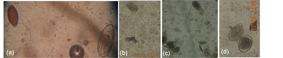
Plate 3. Co-existence of eggs of different gastro-intestinal parasites camels from Mogadishu, Somalia. (a) Trichuris sp. with Dictyocaulus sp., (b) Dictyocaulus sp. with Strongyloides sp., (c) Dictyocaulus sp. with Strongyloides sp. and Trichostrongylus sp., (d) Trichostrongylus sp. with Dictyocaulus sp.
Table 4. Prevalence rate of different flat worms in camels from Mogadishu, Somalia.
Table 5. Co-existence of gastro-intestinal parasites in camels from Mogadishu, Somalia.
Conflict of Interest
The authors declare that they have no conflict of interest regarding the publication of this article.
Cite this paper
Abdalla M. Ibrahim,Ahmed A. H. Kadle,Abdulkarim A. Yusuf, (2016) Gastro-Intestinal Parasites of Camels (Camelus dromedarius) from Mogadishu, Somalia. Open Journal of Veterinary Medicine,06,112-118. doi: 10.4236/ojvm.2016.67015
References
- 1. Wilson, R.T., Araya, A. and Melaku, A. (1990) The One-Humped Camel. An Analytical and Annotated Bibliography. Technical Paper Series 3, The United Nations Sudano-Sahelian Office (UNSO), Nairobi.
- 2. Anonymous (1988) Somali Livestock Statistics 1987/1988. Department of Planning and Statistics. Ministry of Livestock, Forestry and Range. Mogadishu, Somalia.
- 3. Anonymous (1981) Annual report of the Veterinary Laboratory, Kismayo. Department of Veterinary Services, Ministry of Livestock, Forestry and Range, Mogadisho.
- 4. Cankovik, M. (1984) Technical Report on Parasitology. Field Document, UNDP/FAO, Rome. SOM/87/006.
- 5. Abdurahman, O.Sh. (1987) Pulmonary Lesions among Slaughtered Camels in Mogadishu, Somalia. Camel Forum, Working Paper No 20. Somlia Academy of Sciences and Arts (SOMAC)/Scondinavian Institute of African Studies (SIAS), Mogadishu and Uppsala.
- 6. Abdurahman O.Sh. and Bornstein, S. (1991) Diseases of Camels in Somalia and Prospect for Better Health. Nomadic Peoples, No. 29, 104-112.
- 7. Taylor, M.A., Coop, R.L. and Wall, R.L. (2007) Veterinary Parasitology. 3rd Edition, Blackwell Publishing, Hoboken, 121-258.
- 8. Swai, E.S., Moshy, W., Mshanga, D., Lutatina, J. and Bwanga, S. (2011) Intestinal Parasitic Infections of Camels in the Agro and Pastoral Areas of Northern Tanzania. Veterinary Research, 4, 34-38
- 9. Mahmoud, M.A., Amin, M.M., Youssef, R.R., El-Kattan, A., Goda, A.S.A. and Abou El-Naga, T.R. (2008) Studies on Some Endoparasites of Camels in the Southeastern Area of Egypt. SCVMJ, XIII, 81-92.
- 10. Abdel Rahman, M.B., Osman, A.Y. and Hunter, A.G. (2001) Parasites of the One-Humped Camel (Camelus dromedarius) in the Sudan: A Review. The Sudan Journal of Veterinary Research, 17, 13 p.
- 11. Radfar, M.H. and Gowhari, M.A. (2013) Common Gastrointestinal Parasites of Indigenous Camels (Camelus dromedarius) with Traditional Husbandry Management (Free-Ranging System) in Central Deserts of Iran. Journal of Parasitic Diseases, 37, 225-230.
http://dx.doi.org/10.1007/s12639-012-0170-8
NOTES

*Corresponding author.