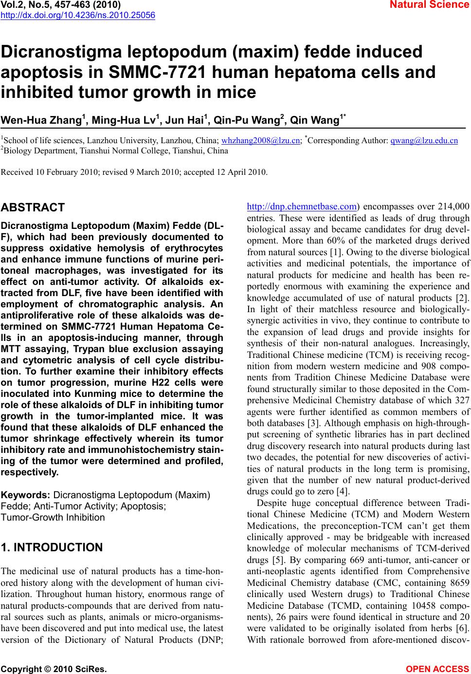
 Vol.2, No.5, 457-463 (2010) Natural Science http://dx.doi.org/10.4236/ns.2010.25056 Copyright © 2010 SciRes. OPEN ACCESS Dicranostigma leptopodum (maxim) fedde induced apoptosis in SMMC-7721 human hepatoma cells and inhibited tumor growth in mice Wen-Hua Zhang1, Ming-Hua Lv1, Jun Hai1, Qin-Pu Wang2, Qin Wang1* 1School of life sciences, Lanzhou University, Lanzhou, China; whzhang2008@lzu.cn; *Corresponding Author: qwang@lzu.edu.cn 2Biology Department, Tianshui Normal College, Tianshui, China Received 10 February 2010; revised 9 March 2010; accepted 12 April 2010. ABSTRACT Dicranostigma Leptopodum (Maxim) Fedde (DL- F), which had been previously documented to suppress oxidative hemolysis of erythrocytes and enhance immune functions of murine peri- toneal macrophages, was investigated for its effect on anti-tumor activity. Of alkaloids ex- tracted from DLF, five have been identified with employment of chromatographic analysis. An antiproliferative role of these alkaloids was de- termined on SMMC-7721 Human Hepatoma Ce- lls in an apoptosis-inducing manner, through MTT assaying, Trypan blue exclusion assaying and cytometric analysis of cell cycle distribu- tion. To further examine their inhibitory effects on tumor progression, murine H22 cells were inoculated into Kunming mice to determine the role of these alkaloids of DLF in inhibiting tumor growth in the tumor-implanted mice. It was found that these alkaloids of DLF enhanced the tumor shrinkage effectively wherein its tumor inhibitory rate and immunohistochemistry stain- ing of the tumor were determined and profiled, respectively. Keywords: Dicranostigma Leptopodum (Maxim) Fedde; Anti-Tumor Activity; Apoptosis; Tumor-Growth Inhibition 1. INTRODUCTION The medicinal use of natural products has a time-hon- ored history along with the development of human civi- lization. Throughout human history, enormous range of natural products-compounds that are derived from natu- ral sources such as plants, animals or micro-organisms- have been discovered and put into medical use, the latest version of the Dictionary of Natural Products (DNP; http://dnp.chemnetbase.co m) encompasses over 214,000 entries. These were identified as leads of drug through biological assay and became candidates for drug devel- opment. More than 60% of the marketed drugs derived from natural sources [1]. Owing to the diverse biological activities and medicinal potentials, the importance of natural products for medicine and health has been re- portedly enormous with examining the experience and knowledge accumulated of use of natural products [2]. In light of their matchless resource and biologically- synergic activities in vivo, they continue to contribute to the expansion of lead drugs and provide insights for synthesis of their non-natural analogues. Increasingly, Traditional Chinese medicine (TCM) is receiving recog- nition from modern western medicine and 908 compo- nents from Tradition Chinese Medicine Database were found structurally similar to those deposited in the Com- prehensive Medicinal Chemistry database of which 327 agents were further identified as common members of both databases [3]. Although emphasis on high-through- put screening of synthetic libraries has in part declined drug discovery research into natural products during last two decades, the potential for new discoveries of activi- ties of natural products in the long term is promising, given that the number of new natural product-derived drugs could go to zero [4]. Despite huge conceptual difference between Tradi- tional Chinese Medicine (TCM) and Modern Western Medications, the preconception-TCM can’t get them clinically approved - may be bridgeable with increased knowledge of molecular mechanisms of TCM-derived drugs [5]. By comparing 669 anti-tumor, anti-cancer or anti-neoplastic agents identified from Comprehensive Medicinal Chemistry database (CMC, containing 8659 clinically used Western drugs) to Traditional Chinese Medicine Database (TCMD, containing 10458 compo- nents), 26 pairs were found identical in structure and 20 were validated to be originally isolated from herbs [6]. With rationale borrowed from afore-mentioned discov-  W.-H. Zhang et al. / Natural Science 2 (2010) 457-463 Copyright © 2010 SciRes. OPEN ACCESS 458 eries and previous findings that Dicranostigma leptopo- dum (maxim.) fedde (DLF) possessed physiological re- levance of antipyretic and analgesic, detumescence, etc. (Dictionary of Traditional Chinese Medicine 1986), we further investigated its activities implicated in the induc- tion of apoptosis of cancerous cells. The scientific name of DLF was for the first time coined and collectively classified by Harvard University Herbaria in 1987 (http://www.gbif.net/o ccurrences/86270582/). Enhanced effects of DLF on immune-suppression were determined in vivo [7] and its effect on suppressing oxidative hemo- lysis of erythrocytes was also validated [8]. Efforts made to separate and characterize the components of DLF have identified five crystals from DLF of which three were isocorydine, corydine and protopine [9] and five alkaloids isolated were dicranostigmine, isocorydine, corydine, protopine and sinoacutine [10]. In this study, we treated SMMC-7721Human Hepatoma Cells with extracted alkaloids of DLF, aiming to examine effect of alkaloids of DLF on inducing apoptosis of cancerous cells. Moreover, we treated H1-implanted Kunming Mice with alkaloids of DLF to have determined tumor growth-inhibiting effects of DLF. 2. EXPERIMENTAL MATERIALS AND METHOD 2.1. Materials 2.1.1. Extraction of Alakoids from DLF The powdered material of roots, stems and leaves of DLF (12.5 g) was mixed with 75% alcohol (400 mL) for 1 h in a hermetic glass container and then disrupted con- tinuously with an ultrasonic purge. The whole material was filtered in vacuum followed by a distillation process. Add alcohol to the mixture of filtrate to adjust its alcohol concentration to 85%. After 24 h, adjust the filtrate to pH 8.0 using NaOH and then filter and distill the filtrate until the alcohol is deprived. Adjust the filtrate to pH 7.0 using HCL [11]. 2.1.2. Determination of Alkaloids of DLF Extracts 20 mg/mL standard DLF extracts were diluted to con- centrations of 0.1 mg/mL, 0.2 mg/mL, 0.3 mg/mL, 0.4 mg/mL, 0.5 mg/mL, 0.6 mg/mL and 0.7 mg/mL, respec- tively. The standard curve was made with these gradient concentrations and de-ionized water as control [12]. The separation of the alkaloids from DLF extracts was per- formed using Sephadex G-50 chromatograph analysis during which samples (50 ul) flowed through the Sepha- dex G-50 columns (0.1 mL) steadily at rate of 1 mL/min. The eluted proteins were determined at 254 nm wave- length by WD-9430D UV spectrophotometer. 2.1.3. Animals and Cells Kunming mice were purchased from the Experimental Animal Center at Lanzhou University. The use and treat- ment of mice were in accordance with institutional guideline for Laboratory Animal Care. Muring H22 and SMMC-7721 Human Hepatoma cells were obtained from cell library of Institute of Cancer Biology and Drug Discovery, Lanzhou University. Cells were grown in RPMI 1640 medium (Gibco) supplemented with 10% fetal bovine serum (Lanzhou Minhai Biotechnology), 2 mM l-glutamine, 100 units/mL penicillin and 100 mg/ mL streptomycin and incubated at 37℃ in a humidified atmosphere of 5% CO2 and 95% air. 2.2. Methods 2.2.1. MTT Assay SMMC-7721 were plated in 96-well microtiter plates at a density of 4,000/well for culture and incubated in a humidified 5% CO2-95% air atmosphere at 37℃ for 24 h. Then cells were treated with different concentrations of alkaloids of DLF (0-24 mg/mL) and incubated for additional 24 h, 48 h, and 72 h respectively. Cell viabil- ity was determined by MTT assay [13] whereby 20 μL of 5 mg/mL MTT was added to each well and incubated for another 4 hours at 37℃. The supernatant was subse- quently removed and 100 μL/well DMSO was added to dissolve the formazan crystal. After shaking plates for 1 min, the absorbance of each well was read at 570 nm wavelength with microplate reader (Bio-Rad). The vi- ability of SMMC-7721 cells was calculated employing the formula below: Viability = (A57 0 of treated cells/A570 of untreated cells) × 100% 2.2.2. Trypan Blue Exclusion Assay Trypan blue dye, which would be excluded by normal cells but could diffused into cells with disrupted mem- brane integrity, was employed to determine the number of the viable cells after treatment with alkaloids of DLF [14]. 24 hours after the SMMC-7721cells were treated with PMS-1077, measurements were conducted after trypan blue (Sigma) was incubated with SMMC-7721 cells for 5 min at room temperature. At least 500 cells were counted per sample. The cell viability was calcu- lated with the following equation: %100 cells ofnumber Total cells blue ofNumber - cells ofnumber Total Viability 2.2.3. Morphological Analysis To determine the morphological changes of SMMC- 7721 cells upon treatment with alkaloids of DLF, SMMC-7721 cells which were treated with alkaloids of DLF for 24 h were observed under inverted dark field microscope (Nikon, Japan) and photographed after- wards. 2.2.4. Single Cell Gel Electrophoresis Assay The single cell gel electrophoresis (SCGE) assay [15]  W.-H. Zhang et al. / Natural Science 2 (2010) 457-463 Copyright © 2010 SciRes. OPEN ACCESS 459 459 commonly referred to as “comet assay” allowed the very sensitive detection of DNA breakage induced by genoto- xic agents at single cell level. Thus, this method was adopted to determine the DNA damaging effects of al- kaloids of DLF on SMMC-7721 cells. Treated with dif- ferent concentrations of alkaloids of DLF for 24 h, SMMC-7721 cells were lysed by alkaline lysis solution (2.5 M NaCl, 100 mM EDTA•Na2, 10 mM Tris pH 10), 10% DMSO and 1% triton X-100 (Sigma) at 4℃ for 1 h. Another 20 min was allowed for the DNA to unwind in electrophoresis running buffer solution (300 mM NaOH, and 1 mM EDTA•Na2, pH 13). Electrophoresis was per- formed for 20 minutes at 50 V and 300 mA. After elec- trophoresis, the slides were gently removed and alkaline pH neutralized with 0.4 M Tris (pH 7.5). Then Ethidium bromide (75 mL of a 20 mg/mL solution, Sigma) was added to each slide and a cover glass was placed on the gel. DNA migration was analyzed on a fluorescence mi- croscope (Olympus, Japan) (Filter G-2A) and photo- graphed afterwards. 2.2.5. Flow Cytometric Analysis SMMC-7721 cells that were treated with alkaloids of DLF for 24 h were washed twice with PBS and then fixed with 70% ethanol at -20℃ for about 12 h. Then cells were washed again twice with PBS and suspended with 1 mL 100 μg/mL RNase (Sigma) containing 0.1% Triton-100 and 50 μg/mL propidium iodide (Sigma). Cells were stained with DNA dye for 30 min and ana- lyzed by flow cytometer (EPICS-XL, Beckman). 2.2.6. In Vivo Inhibition of Tumor Growth Six-week old inbred female Kunming mice were inocu- lated with murine Hepatoma 2.0 × 10⁷ mg/mL H22 cells [16]. From the second day after the implantation of H22 cells, 40 tumor-bearing mice were grouped randomly into five groups as the following: 1) Blank control, 2) Model control, 3) 5-Fu positive control, 4) High dose control, 5) Low dose control. 48h after tumor implanta- tion, these mice were I.P. injected with a daily dose of 0.2 mL of 3.0 mg/mL, 0.2 mL 6.0 mg/mL, 0.2 mL, 2 mg/mL, 0.2 mL physiological saline (0.9%) and 0.2 mL physiological saline (0.9%) of alkaloids of DLF for High dose group, Low dose group, 5-Fu positive group, Blank group and Model group, respectively. Mice were exe- cuted within 24 h after the last dose of alkaloids of DLF and their tumor was obtained and analyzed. And Spleen and thymus indexes were examined. Tumor inhibitory rate (Ri) and organ index were expressed as the follow- ing formula, respectively [17]: % m m tumor tumor 100) untreated treated 1(Ri mouse organ m m IndexOrgan 2.2.7. Immunohistochemistry Analysis The tumors of mice were excised and fixed in 4% para- formaldehyde for 24 h. Paraffin sections were prepared for immunohistochemical staining and hematoxylin and cosin (H & E) staining [18,19]. Sections for immuno- histochemical staining were deparaffinized and then hy- drated by transferring them through the following solu- tions xylene bath twice for 5 min, 100% ethanol for 5 min twice, and then 90% ethanol, 80% ethanol, 70% ethanol, and PBS, for 3 min each. Subsequently, sections were placed in a microwave oven for 15 min at 100℃ in sodiumcitrate buffer (0.01 M, pH 5.7) to expose epitopes. After that, sections were incubated at 37℃ with PCNA antibody for about 1.5 h followed by the visualization using immunosystem kit (Santa Cruz, CA). 2.2.8. Statistical Analysis All experimental data were expressed as mean ± SD, and statistical analysis was performed using Student’s t-test to compare the results from the untreated group. 3. RESULTS AND DISCUSSION 3.1. Extraction and Determination of Alkaloids from DLF The alkaloids extracted from DLF in roots, leaves and whole part were 5.31%, 5.91% and 5.57%, respectively. The average content of alkaloids in whole DLF part was 5.59%. Five main alkaloids were separated using chro- matography and their contents were determined to be unevenly distributed, as indicated by the peaks of chro- matographic profile (Figure 1). (a) Growing Time (weeks) (b) Figure 1. Extraction and determination alkaloids from DLF. (a) The comparison of alkaloids content in different organs of DLF during different growing periods; (b) Separation of alkaloids from DLF extracts and the determination of their proportion.  W.-H. Zhang et al. / Natural Science 2 (2010) 457-463 Copyright © 2010 SciRes. OPEN ACCESS 460 3.2. Anti-Proliferative Effect of Alkaloids of DLF on SMMC-7721 Cells Treated with assigned concentrations of alkaloids of DLF for indicated time, SMMC-7721 cells exhibited compromised cell viability. A concentration-dependent anti-proliferative effect of alkaloids of DLF on SMMC- 7721cells was determined by MTT assay and Trypan Blue Exclusion assay (Figure 2). 3.3. Induction of Apoptosis of SMMC-7721 Cells by Alkaloids of DLF Upon treatment with 8.5 mg/mL alkaloids of DLF for 24 h, SMMC-7721 cells exhibited the characteristic features of apoptosis including cell shrinkage, cell detachment and vesicle formation (Figure 3). Flow cytometric ana- lysis demonstrated that cell cycles were arrested (Figure 4) and cell cycle distribution analysis manifested that cells were arrested predominately at G1 phase (Figure 5). 3.4. Alkaloids of DLF-Induced DNA Damage of SMMC-7721 Cells Upon treatment with various concentrations of alkaloids of DLF, SMMC-7721 cells exhibited characteristic fea- 0 10 20 30 40 50 60 70 80 90 100 0 1.5 36912 18 24 Viability Alkaloids of DLF(mg/ml ) 24h 48h 72h (a) 0 10 20 30 40 50 60 70 80 90 100 01.5369 1218 Viability(%) Alkaloids of DLF(mg/ml) SMMC-7721 (b) Figure 2. Determination of anti-proliferative effect of DLF on SMMC-7721 cells. (a) MTT Assay: Treated with DLF for 24 h, 48 h and 72 h, respectively, cells viability was inhibited in a concentration-dependent manner; (b) Trypan Blue Ex- clusion Assay: Effect of alkaloids of DLF on inhibiting the cell viability was concentration-dependent. tures of DNA damage. Examination of DNA damage by Single cell gel electrophoresis revealed appreciable DNA damage in terms of its length of migrating tails (Figure 6). It’s also observed that the damage was concentration- dependent. 3.5. Inhibition of Tumor Growth by Alkaloids of DLF was Determined in Vivo Murine Hepatoma H22 cells were inoculated to six-week old Kunming mice and, from 24 h after the inoculation, a Figure 3. Morphological analysis of Alkaloids-treated SMMC- 7721 by inverted dark field microscopy (X200). Cells were treated with different concentrations of alkaloids of DLF as indicated: (a) control; (b) 3.0 mg/ml; (c) 6.0 mg/ml and (d) 12.0 mg/ml. Figure 4. Flow cytometric analysis of cell cycle distribution upon treatment with alkaloids of DLF. (a) 0 mg/ml; (b) 3.0 mg/ml; (c) 6.0 mg/ml; (d) 12.0 mg/ml.  W.-H. Zhang et al. / Natural Science 2 (2010) 457-463 Copyright © 2010 SciRes. OPEN ACCESS 461 461 Figure 5. Cell cycle distribution analysis by flow cytometry. Figure 6. Determination of DNA damage of AlkaloidsDLF- treated SMCC-7721 cells by Single Cell Gel Electrophoresis. (a) control; (b) 3.0 mg/ml; (c) 6.0 mg/ml and (d) 12.0 mg/ml. daily dosage of alkaloids of DLF was I.P. injected into these tumor-bearing mice. After 12 days, mice were ex- ecuted and their tumors, spleens and thymus were ex- cised for analysis. Alkaloids of DLF exerted a role as potent as 5-Fu in enhancing tumor shrinkage. 5-Fu in- hibited the tumor growth by 55.75% while 6.0 mg/mL alkaloids of DLF and 12.0 mg/mL alkaloids of DLF in- hibited the growth of tumor by 42.50% and 51.00%, respectively (Table 1). By examining effects of alkaloids of DLF on the growth of spleen and thymus, it revealed that DLF exerted significant effect on inhibiting the ab- errant progression thymus of tumor-bearing mice while the effects of alkaloids of DLF on the growth of spleen of tumor-bearing mice were not discernible (Table 2). 3.6. Alkaloids of DLF-Mediated Inhibition of Tumor Progression Analyzed by HE Staining and Immunohistochemistry Histological section of tumors excised from tumor- bearing mice which were treated with 0.9% physiologi- cal saline, 5-FU, 6.0 mg/mL DLF and 12.0 mg/mL alka- loids of DLF were examined with employment of HE staining and Immunohistochemistry (Figure 7). Both demonstrated that alkaloids of DLF exerted pronounced effects on inhibiting of the growth and progression of tumor. 3.7. Discussion In response to the recognition that fewer side effects have been documented in phytotherapy and natural prod- uct-based therapy and therapeutic potential of natural products [5,20,21], we further explored to study the anti- tumor activities of alkaloids of DLF. In this study, approx. 5.7% of alkaloids were extracted from the whole part of DLF and five alkaloids were identified using chromatographic analysis which was in consistence with the separation of alkaloids from DLF [10]. Anti-prolif- erative effect was determined for alkaloids of DLF on SMCC-7721 cells and IC50 of alkaloids of DLF on SMCC-7721 cells was 8.5 mg/ml. Upon treatment with DLF, SMCC-7721 cells exhibited apoptotic features as a rule [11]. Flow cytometric analysis of cell cycle distribu- tion upon treatment with alkaloids of DLF revealed that cells were arrested in the G1 phase (Figure 4) during which DNA damage was determined (Figure 6). With in vitro anti-tumor activity of alkaloids of DLF validated, in vivo effect of anti-tumor of alkaloids of DLF was further explored. Tumor implantation was completed with inoculation of Muring H22 cells in Kunming mice. Inhibitory rates of 51% and 42% of tumor growth inhibi- tion were determined for 12.0 mg/ml alkaloids of DLF and 6.0 mg/ml, respectively, through examining the tu- mor weight upon treating tumor-bearing mice with alka- loids of DLF. In addition, effects of alkaloids of DLF on inhibiting the progression tumor were determined by HE staining of histological section of tumors (Figure 7). Table 1. The inhibition effect of the drug to transplanted tumor H22. X ± s n = 8. Group Tumor weight Inhibitory rate Tumor control 0.40 ± 0.23 0 5-FU control 0.18 ± 0.10* 55.75% AlkaloidsDLF 6.0 mg/ml 0.20 ± 0.09* 42.00% AlkaloidsDLF12.0 mg/ml 0.23 ± 0.12* 51.50% Table 2. The effects of DLF on growth of spleen and thymus of tumor-bearing miceX ± s n = 8. Group Spleen index (%) Thymus index (%) Healthy group 3.48 ± 1.31* 4.26 ± 0.80 Tumor group 6.28 ± 1.08 4.88 ± 0.88 5-FU group 2.48 ± 1.08 0.98 ± 0.40* AlkaloidsDLF 6.0 mg/ml 11 ± 1.28* 2.84 ± 0.90* AlkaloidsDLF 12.0 mg/ml6.66 ± 1.37 2.84 ± 0.60*  W.-H. Zhang et al. / Natural Science 2 (2010) 457-463 Copyright © 2010 SciRes. OPEN ACCESS 462 Figure 7. Histological Profile of tumor upon treatment. HE staining of histological section of tumors: (a) 6.0 mg/ml Alka- loidsDLF; (b) 12.0 mg/ml AlkaloidsDLF; (c) 5-FU; (d) 0.9% physiological saline. PCNA assaying of histological section of tumors; (e) 6.0 mg/ml AlkaloidsDLF; (f) 12.0 mg/ml Alka- loidsDLF; (g) 5-FU; (h) 0.9% physiological saline. Taken together, an anti-tumor effect of alkaloids of DLF was determined by means of both in vitro and in vivo examinations. However, the elaborate molecular mechanism underlying its anti-tumor activity remains elusive for these five alkaloids. Since determining the biological properties of plants used in traditional medi- cine is helpful to drug screening, thus, further research is deserved to isolate the very compounds responsible for the observed biological activity of alkaloids of DLF. 4. CONCLUSIONS With promising effects of alkaloids of DLF determined both in vitro and in vivo on anti-proliferating of SMCC- 7721 cells and enhancing shrinkage of tumor, DLF de- serves further characterization of its very components entailing its anti-tumor activities and mechanisms un- derlying its anti-tumor effect. Moreover, its anti-prolif- erating effect and anti-tumor activities remains to be extended to other cancerous cell lines and tumors as well. A comprehensive understanding of its activities on a spectrum of cancerous cells and tumors will shed light on its mechanistic elucidation of anti-tumor effects. 5. ACKNOWLEDGEMENTS This work was jointly supported by International Cooperation Pro- grams in Gansu Province (NO. 0708WCGA149) and International Science and Technology Cooperation Project (NO. 2009DFA30990). REFERENCES [1] Molinari, G. (2009) Natural products in drug discovery: Present status and perspectives. Advances in Experimen- tal Medicine and Biology, 655(1), 13-27. [2] Ji, H.F., Li, X.J. and Zhang, H.Y. (2009) Natural products and drug discovery: Can thousands of years of ancient medical knowledge lead us to new and powerful drug combinations in the fight against cancer and dementia? EMBO Report, 10(3), 194-200. [3] Kong, D.X., Li, X.J., Tang, G.Y. and Zhang, H.Y. (2008) How many traditional Chinese medicine components have been recognized by modern Western medicine? A chemoinformatic analysis and implications for finding multicomponent drugs. ChemMedChem, 3(2), 233-236. [4] Li, J.W. and Vederas, J.C. (2009) Drug discovery and natural products: end of an era or an endless frontier? Science, 325(5937), 161-165. [5] Efferth, T., Li, P.C., Konkimalla, V.S. and Kaina, B. (2007) From traditional Chinese medicine to rational cancer therapy. Trends in Molecular Medicine, 13(8), 353-361 [6] Li, X.J. and Zhang, H.Y. (2008) Western-medicine-vali- dated anti-tumor agents and traditional Chinese medicine. Trends in Molecular Medicine, 14(1), 1-2. [7] Wang, X., Zhang, L.H. and Wang, Q. (2006) Enhanced effect of Dicranostigma Leptopodum (Maxim.) Fedde on experimental immunosuppression in mice. Journal of Lanzhou University (Natural Sciences), 36(4), 6-12. [8] Zhao, Q., Han, Y., Du, Y.P., Wang, T.P. and Wang, Q. (2006) The effect of Dicranostigma Leptopodum (Maxim) Fedde (DLF) extraction on suppressing oxidative hemo- lysis of erythrocytes and its mechanism. Journal of Lan- zhou University Medical Sciences, 3, 3-11. [9] Ruo, X., Chang, H.W. and Ma, G.G. (1981) An analysis of the chemical components of Dicranostigma Leptopo- dum (Maxim.) Fedde. Chinese Pharmaceutical Bulletin, 16(2), 118-123. [10] Yan Dang HG, Junxi Liu, Sijiu Yu. 2009. Alkaloid from Dicranostigma Leptopodum (Maxim) Fedde. Chinese Chemical Letters, 20(10), 1218-1220. [11] Copper, C.L., Newman, C.I. and Collins, G.E. (2008) Simple and rapid extraction, separation, and detection of  W.-H. Zhang et al. / Natural Science 2 (2010) 457-463 Copyright © 2010 SciRes. OPEN ACCESS 463 463 alkaloids in beverages. Journal of Separation Science, 31(21), 3727-3731. [12] Chen, J., Wang, F., Liu, J., Lee, F.S., Wang, X. and Yang, H. (2008) Analysis of alkaloids in Coptis chinensis Franch by accelerated solvent extraction combined with ultra performance liquid chromatographic analysis with photodiode array and tandem mass spectrometry detec- tions. Analytical Chimica Acta, 613(2), 184-195. [13] Marks, D.C., Belov, L., Davey, M.W., Davey, R.A. and Kidman, A.D. (1992) The MTT cell viability assay for cytotoxicity testing in multidrug-resistant human leuke- mic cells. Leukemia Research, 16(12), 1165-1173. [14] Darlington, G.J. (2007) Viability Staining of Mammalian Cell Cultures. Cold Spring Harbor Protocols, pdb. prot4769, New York. [15] Winter, H.K., Ehrlich, V.A., Grusch, M., Lackner, A., Schulte-Hermann, R., et al. (2008) Use of four new human-derived liver-cell lines for the detection of geno- toxic compounds in the single-cell gel electrophoresis (SCGE) assay. Mutation Research, 657(2), 133-139. [16] Cai, J., Lei, L.S., Chi, D.B. (2009) Antineoplastic effect of koumine in mice bearing H22 solid tumor. Journal of Southern Medical University, 29(9), 1851-1852. [17] Zhu, B.D., Yuan, S.J., Zhao, Q.C., Li, X., Li, Y., Lu, Q.Y. (2005) Antitumor effect of Gefitinib, an epidermal growth factor receptor tyrosine kinase inhibitor, com- bined with cytotoxic agent on murine hepatocellular car- cinoma. World Journal of Gastroenterology, 11(9), 1382-1386. [18] Herrmann, H.J. and Schlosser, G. (1974) Detection of osteoid tissues and degenerated intercellular bone sub- stance using a modification of the hematoxylin-cosin staining technique (author’s transl). Zentralbl Allgemeine Pathology, 118, 342-347. [19] Abe, Y., Yonemura, K., Nishida, K. and Takagi, K. (1994) Giant cell tumor of bone: analysis of proliferative cells by double-labeling immunohistochemistry with anti-pro- liferating cell nuclear antigen antibody and culture pro- cedure. Nippon Seikeigeka Gakkai Zasshi, 68(5), 407- 414. [20] Baker, J.T., Borris, R.P., Carte, B., Cordell, G.A., Soe- jarto, D.D., et al. (1995) Natural product drug discovery and development: new perspectives on international col- laboration. Journal of Natural Products, 58(9), 1325- 1357. [21] Itokawa, H., Morris-Natschke, S.L., Akiyama, T. and Lee, K.H. (2008) Plant-derived natural product research aim- ed at new drug discovery. Journal of Nature Medicine, 62(3), 263-280. [22] Belicza, M. (2009) Evaluation of morphologically deter- mined apoptotic index. Acta Medica Croatica, 63(Suppl 2), 3-12.
|