J. A. Rubano et al. / Open Journal of Internal Medicine 2 (2012) 27-30
28
meral bony lesion, that was biopsied several years earlier
at another hospital. Once ACS was ruled out, the patient
began to focus her complaints more on her right shoulder
pain. The patient denies any history of trau ma to the right
upper extremity. Review of her hospital records revealed
some evidence of bony destruction of the right humerus
from ten years earlier.
On examination, the patient was noted to have an ob-
vious deformity of her right upper extremity, with an
apparent area of swelling extending from approximately
the right shoulder to the distal asp ect of the right deltoid.
Range of motion was grossly limited, especially involv-
ing abduction, and extension. She also had grossly di-
minished strength to 3/5 for both flexion and extension.
Sensation was grossly diminished when compared to the
left.
Chest x-ray showed destruction of the right humeral
head and anterior glenoid. CT revealed an expansile right
axillary soft tissue mass with adjacent bony destruction,
most consistent with a neoplasm. The prior biopsy report
was obtained, and demonstrated a low grade spindle cell
neoplasm most consistent with a low grade neoplasm of
the bone. Given this history, her physical exam and ra-
diographic findings, it was arranged for an open bone
biopsy. Destruction of the osseous structures and joint
was obvious. Multiple samples were sent to pathology,
with no evidence of malignancy.
Based upon the pathologic results, as well as the
clinical picture of the patient, it was determined she was
likely suffering from neuropathic arthropathy. An MRI of
the cervical spine was obtained, which revealed a syrinx
extending from approximately C1 throu gh T2, with glio-
sis at the C2-3 level.
4. DISCUSSION
4.1. Background/History
Neuropathic arthropathy (NA), also known as Charcot
Joint, is associated with d ecreased sensory innervation of
the involved joints. Although Mitchell was the first to
describe this entity in 1 831, Charcot brought attention to
the disorder in 1868 [2].
4.2. Theories of Pathogenesis
Both Charcot and Mitch ell speculated th at the changes in
the involved joint were secondary to damage in the tro-
phic centers of the central nervous system (CNS), later
known as the French Theory. Shortly thereafter, the
German theory, proposed by both Volkmann and Vir-
chow contend ed that NA was the result of multiple years
of repeated, insensible trauma results in total joint de-
struction [2]. This theory has limitations , as NA is known
to develop in bedridden patients, with no history of
trauma. In addition, the neurotraumatic theory proposes
that when the CNS is damaged, the joint exceeds the safe
limits of normal range of motion due to decreased pro-
prioception, resulting in the aforementioned repeated
microtrauma, similar to the German theory, and ulti-
mately total joint destruction [2]. The neurovascular the-
ory holds that CNS damage results in a loss of vascular
reflex, which produces locally increased blood flow, with
bone resorption due to increased osteoclast activity. Al-
though not perfor med in the two cases above, this can be
correlated with increased uptake in nuclear bone scans
and angiography demonstrating hypervascularity of the
joint [2].
4.3. Clinical Features
NA has been reported to occur with a variety of diseases
including: diabetes, tabes dorsalis, leprosy, syringomye-
lia, poliomyelitis, rheumatoid arthritis, multiple sclero sis,
congenital neuropathy, traumatic injury, iatrogenic
causes, and tertiary syphilis [3]. Both upper motor neu-
ron (UMN) and lower motor neuron (LMN) lesions can
potentiate sensory impairment an d lead to NA [1].
The majority of cases of NA are seen in patients with
underlying DM, usually accompanying peripheral neu-
ropathy [4], and a painless monoarthritis. Currently,
prevalence of NA is estimated to range from 0.08% in
the general diabetic population to 13% in high-risk dia-
betic patients [4]. The development of arthropathy in this
subset of patients with diabetes is likely multifactorial
with a complex interaction between mechanical and vas-
cular factors, vasomotor changes, and cytokine related
changes to osteoclastic activity [5].
As demonstrated by both of our patients, NA can also
be associated with syringomyelia [1,6], of which 20% to
25% of patients with syringomyelia developing NA, with
a predilection for upper extremity joints [3]. An uncom-
mon process, syringomyelia is characterized by a longi-
tudinal cavitatio n of the spinal cord, leading to the loss of
pain and sensory innervation of the involved joint, pre-
disposing the patient to the development of NA. The
involvement of a single join t helps categorize this arthri-
tis in the group of monoarthropathies. The clinician
should keep in mind that the differential diagnosis for a
monoarthopathy is diverse, and appropriate history and
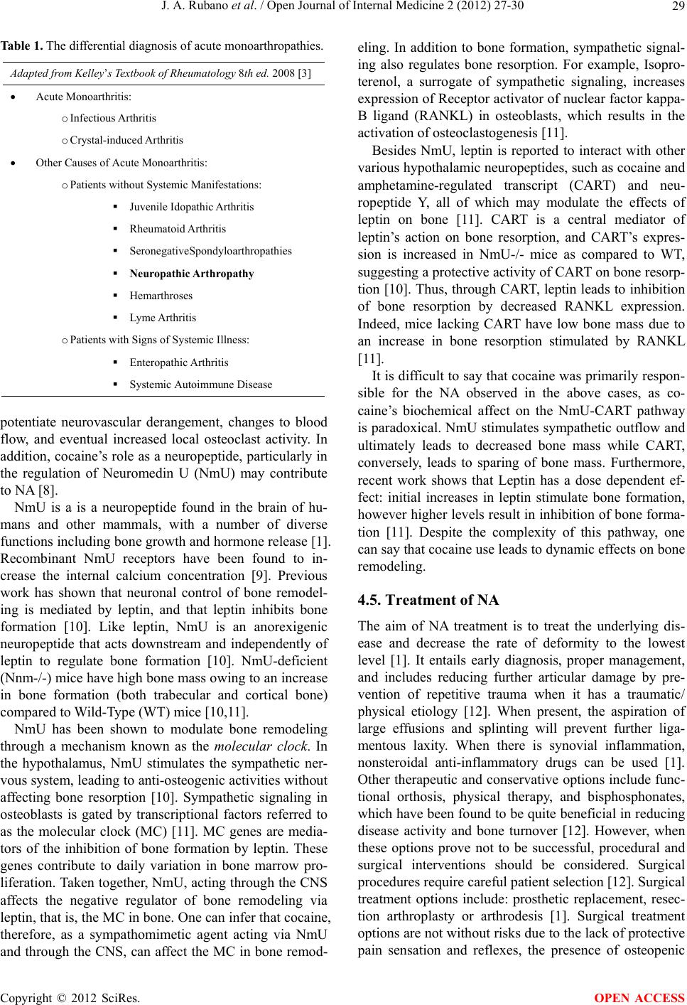
examination is needed to distinguish (see Table 1).
4.4. Role of Cocaine
Of interest both patients reported in this series had a his-
tory of cocaine use. Whether cocaine use has contributed
to the unusual findings in these two cases is unclear. It
has been well documented that cocaine use can cause
clinically significant vasospasm and ischemia [7]. It may
be possible that a similar vaso-occulusive process can
Copyright © 2012 SciRes. OPEN ACCESS