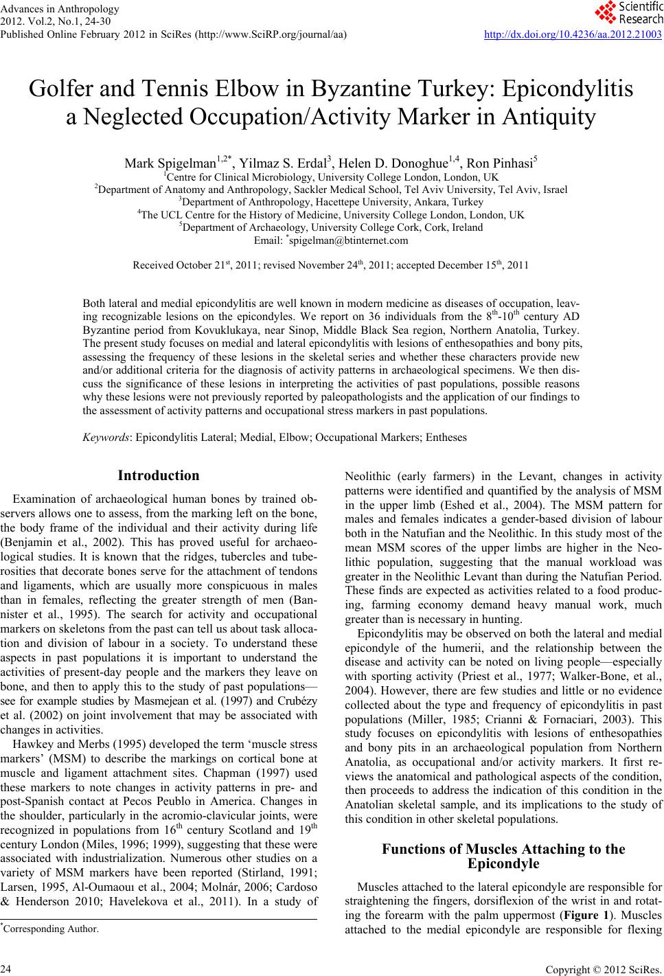
 Advances in Anthropology 2012. Vol.2, No.1, 24-30 Published Online February 2012 in SciRes (http://www.SciRP.org/journal/aa) http://dx.doi.org/10.4236/aa.2012.21003 Copyright © 2012 SciRes. 24 Golfer and Tennis Elbow in Byzantine Turkey: Epicondylitis a Neglected Occupation/Activity Marker in Antiquity Mark Spigelman1,2*, Yilmaz S. Erdal3, Helen D. Donoghue1,4, Ron Pinhasi5 1Centre for Clinical Microbiology, University College London, London, UK 2Department of Anatomy and Anthropology, Sackler Medical School, Tel Aviv University, Tel Aviv, Israel 3Department of Anthropology, Hacettepe University, Ankara, Turkey 4The UCL Centre for the History of Medicine, University College London, London, UK 5Department of Archaeology, University College Cork, Cork, Ireland Email: *spigelman@btinternet.com Received October 21st, 2011; revised November 24th, 2011; accepted December 15th, 2011 Both lateral and medial epicondylitis are well known in modern medicine as diseases of occupation, leav- ing recognizable lesions on the epicondyles. We report on 36 individuals from the 8th-10th century AD Byzantine period from Kovuklukaya, near Sinop, Middle Black Sea region, Northern Anatolia, Turkey. The present study focuses on medial and lateral epicondylitis with lesions of enthesopathies and bony pits, assessing the frequency of these lesions in the skeletal series and whether these characters provide new and/or additional criteria for the diagnosis of activity patterns in archaeological specimens. We then dis- cuss the significance of these lesions in interpreting the activities of past populations, possible reasons why these lesions were not previously reported by paleopathologists and the application of our findings to the assessment of activity patterns and occupational stress markers in past populations. Keywords: Epicondylitis Lateral; Medial, Elbow; Occupational Markers; Entheses Introduction Examination of archaeological human bones by trained ob- servers allows one to assess, from the marking left on the bone, the body frame of the individual and their activity during life (Benjamin et al., 2002). This has proved useful for archaeo- logical studies. It is known that the ridges, tubercles and tube- rosities that decorate bones serve for the attachment of tendons and ligaments, which are usually more conspicuous in males than in females, reflecting the greater strength of men (Ban- nister et al., 1995). The search for activity and occupational markers on skeletons from the past can tell us about task alloca- tion and division of labour in a society. To understand these aspects in past populations it is important to understand the activities of present-day people and the markers they leave on bone, and then to apply this to the study of past populations— see for example studies by Masmejean et al. (1997) and Crubézy et al. (2002) on joint involvement that may be associated with changes in activities. Hawkey and Merbs (1995) developed the term ‘muscle stress markers’ (MSM) to describe the markings on cortical bone at muscle and ligament attachment sites. Chapman (1997) used these markers to note changes in activity patterns in pre- and post-Spanish contact at Pecos Peublo in America. Changes in the shoulder, particularly in the acromio-clavicular joints, were recognized in populations from 16th century Scotland and 19th century London (Miles, 1996; 1999), suggesting that these were associated with industrialization. Numerous other studies on a variety of MSM markers have been reported (Stirland, 1991; Larsen, 1995, Al-Oumaouı et al., 2004; Molnár, 2006; Cardoso & Henderson 2010; Havelekova et al., 2011). In a study of Neolithic (early farmers) in the Levant, changes in activity patterns were identified and quantified by the analysis of MSM in the upper limb (Eshed et al., 2004). The MSM pattern for males and females indicates a gender-based division of labour both in the Natufian and the Neolithic. In this study most of the mean MSM scores of the upper limbs are higher in the Neo- lithic population, suggesting that the manual workload was greater in the Neolithic Levant than during the Natufian Period. These finds are expected as activities related to a food produc- ing, farming economy demand heavy manual work, much greater than is necessary in hunting. Epicondylitis may be observed on both the lateral and medial epicondyle of the humerii, and the relationship between the disease and activity can be noted on living people—especially with sporting activity (Priest et al., 1977; Walker-Bone, et al., 2004). However, there are few studies and little or no evidence collected about the type and frequency of epicondylitis in past populations (Miller, 1985; Crianni & Fornaciari, 2003). This study focuses on epicondylitis with lesions of enthesopathies and bony pits in an archaeological population from Northern Anatolia, as occupational and/or activity markers. It first re- views the anatomical and pathological aspects of the condition, then proceeds to address the indication of this condition in the Anatolian skeletal sample, and its implications to the study of this condition in other skeletal populations. Functions of Muscles Attaching to the Epicondyle Muscles attached to the lateral epicondyle are responsible for straightening the fingers, dorsiflexion of the wrist in and rotat- ing the forearm with the palm uppermost (Figure 1). Muscles attached to the medial epicondyle are responsible for flexing *Corresponding Author. 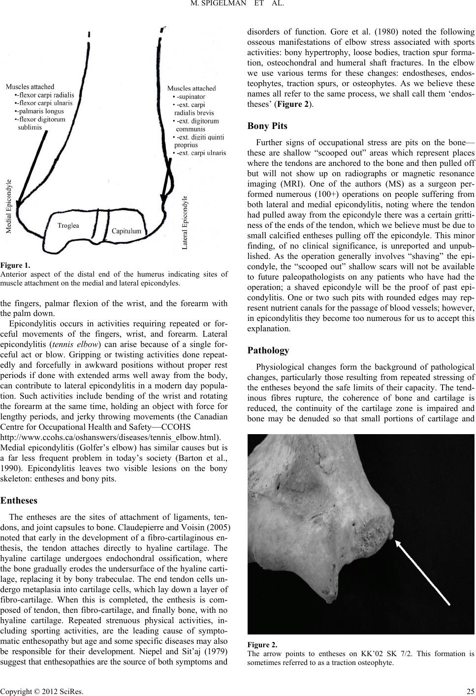 M. SPIGELMAN ET AL. Figure 1. Anterior aspect of the distal end of the humerus indicating sites of muscle attachment on the medial and lateral epicondyles. the fingers, palmar flexion of the wrist, and the forearm with the palm down. Epicondylitis occurs in activities requiring repeated or for- ceful movements of the fingers, wrist, and forearm. Lateral epicondylitis (tennis elbow) can arise because of a single for- ceful act or blow. Gripping or twisting activities done repeat- edly and forcefully in awkward positions without proper rest periods if done with extended arms well away from the body, can contribute to lateral epicondylitis in a modern day popula- tion. Such activities include bending of the wrist and rotating the forearm at the same time, holding an object with force for lengthy periods, and jerky throwing movements (the Canadian Centre for Occupational Health and Safety—CCOHS http://www.ccohs.ca/oshanswers/diseases/tennis_elbow.html). Medial epicondylitis (Golfer’s elbow) has similar causes but is a far less frequent problem in today’s society (Barton et al., 1990). Epicondylitis leaves two visible lesions on the bony skeleton: entheses and bony pits. Entheses The entheses are the sites of attachment of ligaments, ten- dons, and joint capsules to bone. Claudepierre and Voisin (2005) noted that early in the development of a fibro-cartilaginous en- thesis, the tendon attaches directly to hyaline cartilage. The hyaline cartilage undergoes endochondral ossification, where the bone gradually erodes the undersurface of the hyaline carti- lage, replacing it by bony trabeculae. The end tendon cells un- dergo metaplasia into cartilage cells, which lay down a layer of fibro-cartilage. When this is completed, the enthesis is com- posed of tendon, then fibro-cartilage, and finally bone, with no hyaline cartilage. Repeated strenuous physical activities, in- cluding sporting activities, are the leading cause of sympto- matic enthesopathy but age and some specific diseases may also be responsible for their development. Niepel and Sit’aj (1979) suggest that enthesopathies are the source of both symptoms and disorders of function. Gore et al. (1980) noted the following osseous manifestations of elbow stress associated with sports activities: bony hypertrophy, loose bodies, traction spur forma- tion, osteochondral and humeral shaft fractures. In the elbow we use various terms for these changes: endostheses, endos- teophytes, traction spurs, or osteophytes. As we believe these names all refer to the same process, we shall call them ‘endos- theses’ (Figure 2). Bony Pits Further signs of occupational stress are pits on the bone— these are shallow “scooped out” areas which represent places where the tendons are anchored to the bone and then pulled off but will not show up on radiographs or magnetic resonance imaging (MRI). One of the authors (MS) as a surgeon per- formed numerous (100+) operations on people suffering from both lateral and medial epicondylitis, noting where the tendon had pulled away from the epicondyle there was a certain gritti- ness of the ends of the tendon, which we believe must be due to small calcified entheses pulling off the epicondyle. This minor finding, of no clinical significance, is unreported and unpub- lished. As the operation generally involves “shaving” the epi- condyle, the “scooped out” shallow scars will not be available to future paleopathologists on any patients who have had the operation; a shaved epicondyle will be the proof of past epi- condylitis. One or two such pits with rounded edges may rep- resent nutrient canals for the passage of blood vessels; however, in epicondylitis they become too numerous for us to accept this explanation. Pathology Physiological changes form the background of pathological changes, particularly those resulting from repeated stressing of the entheses beyond the safe limits of their capacity. The tend- inous fibres rupture, the coherence of bone and cartilage is reduced, the continuity of the cartilage zone is impaired and bone may be denuded so that small portions of cartilage and Figure 2. The arrow points to entheses on KK’02 SK 7/2. This formation is sometimes referred to as a traction osteophyte. Copyright © 2012 SciRes. 25 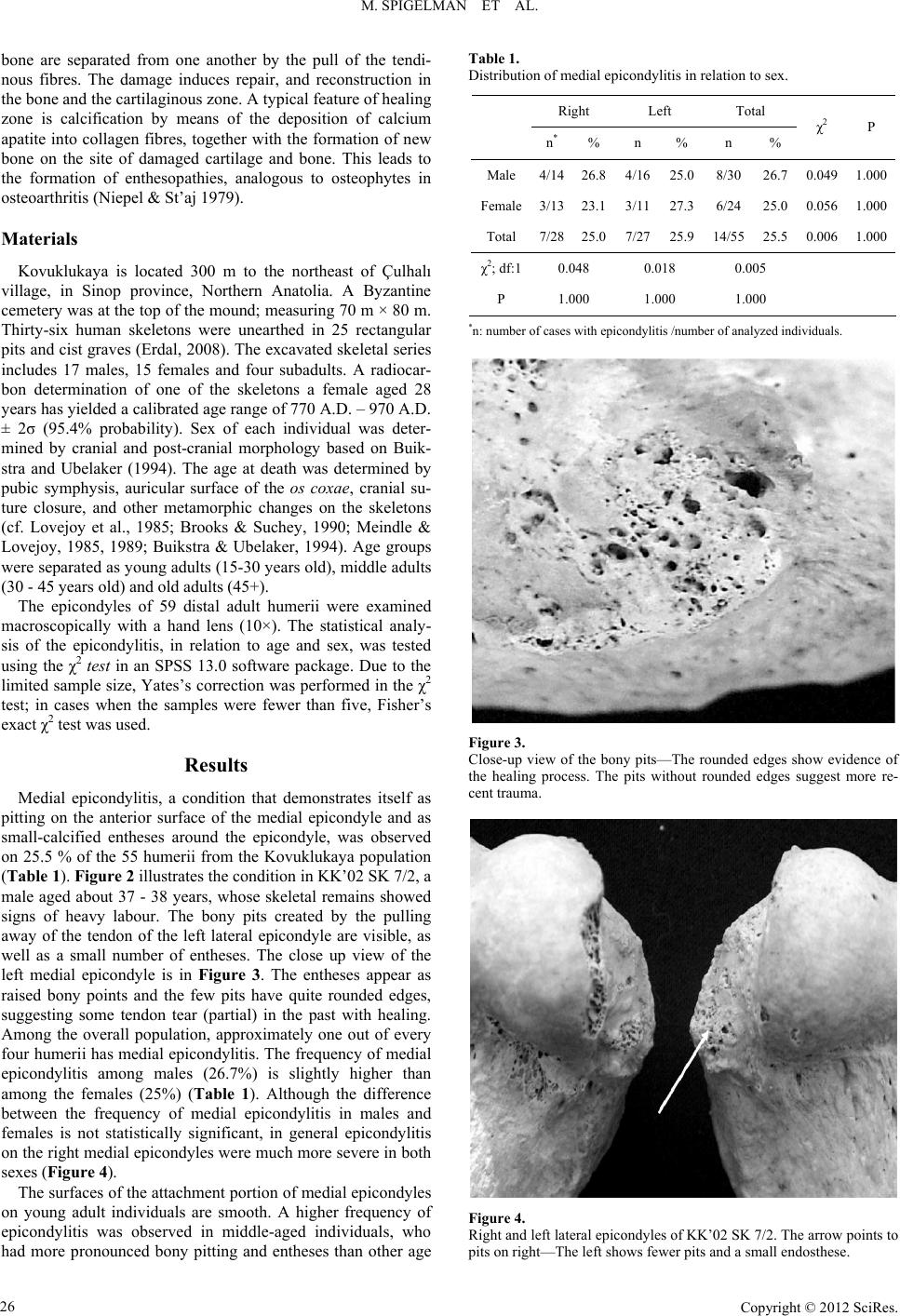 M. SPIGELMAN ET AL. bone are separated from one another by the pull of the tendi- nous fibres. The damage induces repair, and reconstruction in the bone and the cartilaginous zone. A typical feature of healing zone is calcification by means of the deposition of calcium apatite into collagen fibres, together with the formation of new bone on the site of damaged cartilage and bone. This leads to the formation of enthesopathies, analogous to osteophytes in osteoarthritis (Niepel & St’aj 1979). Materials Kovuklukaya is located 300 m to the northeast of Çulhalı village, in Sinop province, Northern Anatolia. A Byzantine cemetery was at the top of the mound; measuring 70 m × 80 m. Thirty-six human skeletons were unearthed in 25 rectangular pits and cist graves (Erdal, 2008). The excavated skeletal series includes 17 males, 15 females and four subadults. A radiocar- bon determination of one of the skeletons a female aged 28 years has yielded a calibrated age range of 770 A.D. – 970 A.D. ± 2σ (95.4% probability). Sex of each individual was deter- mined by cranial and post-cranial morphology based on Buik- stra and Ubelaker (1994). The age at death was determined by pubic symphysis, auricular surface of the os coxae, cranial su- ture closure, and other metamorphic changes on the skeletons (cf. Lovejoy et al., 1985; Brooks & Suchey, 1990; Meindle & Lovejoy, 1985, 1989; Buikstra & Ubelaker, 1994). Age groups were separated as young adults (15-30 years old), middle adults (30 - 45 years old) and old adults (45+). The epicondyles of 59 distal adult humerii were examined macroscopically with a hand lens (10×). The statistical analy- sis of the epicondylitis, in relation to age and sex, was tested using the χ2 test in an SPSS 13.0 software package. Due to the limited sample size, Yates’s correction was performed in the χ2 test; in cases when the samples were fewer than five, Fisher’s exact χ2 test was used. Results Medial epicondylitis, a condition that demonstrates itself as pitting on the anterior surface of the medial epicondyle and as small-calcified entheses around the epicondyle, was observed on 25.5 % of the 55 humerii from the Kovuklukaya population (Table 1). Figure 2 illustrates the condition in KK’02 SK 7/2, a male aged about 37 - 38 years, whose skeletal remains showed signs of heavy labour. The bony pits created by the pulling away of the tendon of the left lateral epicondyle are visible, as well as a small number of entheses. The close up view of the left medial epicondyle is in Figure 3. The entheses appear as raised bony points and the few pits have quite rounded edges, suggesting some tendon tear (partial) in the past with healing. Among the overall population, approximately one out of every four humerii has medial epicondylitis. The frequency of medial epicondylitis among males (26.7%) is slightly higher than among the females (25%) (Table 1). Although the difference between the frequency of medial epicondylitis in males and females is not statistically significant, in general epicondylitis on the right medial epicondyles were much more severe in both sexes (Figure 4). The surfaces of the attachment portion of medial epicondyles on young adult individuals are smooth. A higher frequency of epicondylitis was observed in middle-aged individuals, who had more pronounced bony pitting and entheses than other age Table 1. Distribution of medial epicondylitis in relation to sex. Right Left Total n * % n % n % χ2 P Male 4/1426.84/1625.0 8/30 26.7 0.0491.000 Female3/1323.13/1127.3 6/24 25.0 0.0561.000 Total 7/2825.07/2725.9 14/55 25.5 0.0061.000 χ2; df:10.048 0.018 0.005 P 1.000 1.000 1.000 *n: number of cases with epicondylitis /number of analyzed individuals. Figure 3. Close-up view of the bony pits—The rounded edges show evidence of the healing process. The pits without rounded edges suggest more re- cent trauma. Figure 4. Right and left lateral epicondyles of KK’02 SK 7/2. The arrow points to pits on right—The left shows fewer pits and a small endosthese. Copyright © 2012 SciRes. 26 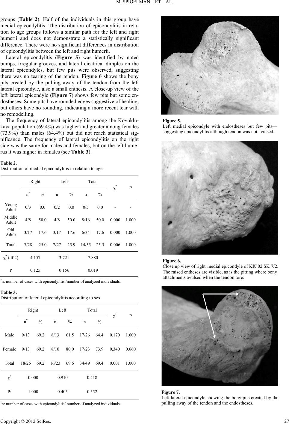 M. SPIGELMAN ET AL. groups (Table 2). Half of the individuals in this group have medial epicondylitis. The distribution of epicondylitis in rela- tion to age groups follows a similar path for the left and right humerii and does not demonstrate a statistically significant difference. There were no significant differences in distribution of epicondylitis between the left and right humerii. Lateral epicondylitis (Figure 5) was identified by noted bumps, irregular grooves, and lateral cicatrical dimples on the lateral epicondyles, but few pits were observed, suggesting there was no tearing of the tendon. Figure 6 shows the bony pits created by the pulling away of the tendon from the left lateral epicondyle, also a small enthesis. A close-up view of the left lateral epicondyle (Figure 7) shows few pits but some en- dostheses. Some pits have rounded edges suggestive of healing, but others have no rounding, indicating a more recent tear with no remodelling. The frequency of lateral epicondylitis among the Kovuklu- kaya population (69.4%) was higher and greater among females (73.9%) than males (64.4%) but did not reach statistical sig- nificance. The frequency of lateral epicondylitis on the right side was the same for males and females, but on the left hume- rus it was higher in females (see Table 3). Table 2. Distribution of medial epicondylitis in relation to age. Right Left Total n * % n % n % χ2 P Young Adult 0/3 0.0 0/2 0.0 0/5 0.0 - - Middle Adult 4/8 50,0 4/8 50.08/16 50.0 0.0001.000 Old Adult 3/17 17.6 3/17 17.6 6/34 17.6 0.0001.000 Total 7/28 25.0 7/27 25.914/55 25.5 0.0061.000 χ2 (df:2) 4.157 3.721 7.880 P 0.125 0.156 0.019 *n: number of cases with epicondylitis /number of analyzed individuals. Table 3. Distribution of lateral epicondylitis according to sex. Right Left Total n * % n % n % χ2 P Male 9/13 69.2 8/13 61.517/26 64.4 0.1701.000 Female 9/13 69.2 8/10 80.017/23 73.9 0,3400.660 Total 18/26 69.2 16/23 69.6 34/49 69.4 0.001 1.000 χ2 0.000 0.910 0.418 P: 1.000 0.405 0.552 *n: number of cases with epicondylitis/ number of analyzed individuals. Figure 5. Left medial epicondyle with endostheses but few pits— suggesting epicondylitis although tendon was not avulsed. Figure 6. Close up view of right medial epicondyle of KK’02 SK 7/2. The raised entheses are visible, as is the pitting where bony attachments avulsed when the tendon tore. Figure 7. Left lateral epicondyle showing the bony pits created by the pulling away of the tendon and the endostheses. Copyright © 2012 SciRes. 27 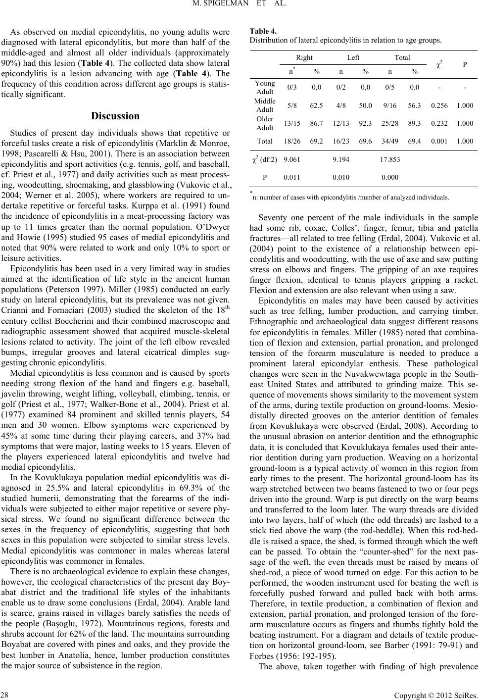 M. SPIGELMAN ET AL. As observed on medial epicondylitis, no young adults were diagnosed with lateral epicondylitis, but more than half of the middle-aged and almost all older individuals (approximately 90%) had this lesion (Table 4). The collected data show lateral epicondylitis is a lesion advancing with age (Table 4). The frequency of this condition across different age groups is statis- tically significant. Discussion Studies of present day individuals shows that repetitive or forceful tasks create a risk of epicondylitis (Marklin & Monroe, 1998; Pascarelli & Hsu, 2001). There is an association between epicondylitis and sport activities (e.g. tennis, golf, and baseball, cf. Priest et al., 1977) and daily activities such as meat process- ing, woodcutting, shoemaking, and glassblowing (Vukovic et al., 2004; Werner et al. 2005), where workers are required to un- dertake repetitive or forceful tasks. Kurppa et al. (1991) found the incidence of epicondylitis in a meat-processing factory was up to 11 times greater than the normal population. O’Dwyer and Howie (1995) studied 95 cases of medial epicondylitis and noted that 90% were related to work and only 10% to sport or leisure activities. Epicondylitis has been used in a very limited way in studies aimed at the identification of life style in the ancient human populations (Peterson 1997). Miller (1985) conducted an early study on lateral epicondylitis, but its prevalence was not given. Crianni and Fornaciari (2003) studied the skeleton of the 18th century cellist Boccherini and their combined macroscopic and radiographic assessment showed that acquired muscle-skeletal lesions related to activity. The joint of the left elbow revealed bumps, irregular grooves and lateral cicatrical dimples sug- gesting chronic epicondylitis. Medial epicondylitis is less common and is caused by sports needing strong flexion of the hand and fingers e.g. baseball, javelin throwing, weight lifting, volleyball, climbing, tennis, or golf (Priest et al., 1977; Walker-Bone et al., 2004). Priest et al. (1977) examined 84 prominent and skilled tennis players, 54 men and 30 women. Elbow symptoms were experienced by 45% at some time during their playing careers, and 37% had symptoms that were major, lasting weeks to 15 years. Eleven of the players experienced lateral epicondylitis and twelve had medial epicondylitis. In the Kovuklukaya population medial epicondylitis was di- agnosed in 25.5% and lateral epicondylitis in 69.3% of the studied humerii, demonstrating that the forearms of the indi- viduals were subjected to either major repetitive or severe phy- sical stress. We found no significant difference between the sexes in the frequency of epicondylitis, suggesting that both sexes in this population were subjected to similar stress levels. Medial epicondylitis was commoner in males whereas lateral epicondylitis was commoner in females. There is no archaeological evidence to explain these changes, however, the ecological characteristics of the present day Boy- abat district and the traditional life styles of the inhabitants enable us to draw some conclusions (Erdal, 2004). Arable land is scarce, grains raised in villages barely satisfies the needs of the people (Başoglu, 1972). Mountainous regions, forests and shrubs account for 62% of the land. The mountains surrounding Boyabat are covered with pines and oaks, and they provide the best lumber in Anatolia, hence, lumber production constitutes the major source of subsistence in the region. Table 4. Distribution of lateral epicondylitis in relation to age groups. Right Left Total n * % n % n % χ2 P Young Adult 0/3 0,0 0/2 0,0 0/5 0.0 - - Middle Adult 5/8 62.5 4/8 50.0 9/16 56.3 0.2561.000 Older Adult 13/1586.7 12/13 92.3 25/28 89.3 0.2321.000 Total18/26 69.2 16/23 69.6 34/49 69.4 0.0011.000 χ2 (df:2)9.061 9.194 17.853 P 0.011 0.010 0.000 *n: number of cases with epicondylitis /number of analyzed individuals. Seventy one percent of the male individuals in the sample had some rib, coxae, Colles’, finger, femur, tibia and patella fractures—all related to tree felling (Erdal, 2004). Vukovic et al. (2004) point to the existence of a relationship between epi- condylitis and woodcutting, with the use of axe and saw putting stress on elbows and fingers. The gripping of an axe requires finger flexion, identical to tennis players gripping a racket. Flexion and extension are also relevant when using a saw. Epicondylitis on males may have been caused by activities such as tree felling, lumber production, and carrying timber. Ethnographic and archaeological data suggest different reasons for epicondylitis in females. Miller (1985) noted that combina- tion of flexion and extension, partial pronation, and prolonged tension of the forearm musculature is needed to produce a prominent lateral epicondylar enthesis. These pathological changes were seen in the Nuvakwewtaga people in the South- east United States and attributed to grinding maize. This se- quence of movements shows similarity to the movement system of the arms, during textile production on ground-looms. Mesio- distally directed grooves on the anterior dentition of females from Kovuklukaya were observed (Erdal, 2008). According to the unusual abrasion on anterior dentition and the ethnographic data, it is concluded that Kovuklukaya females used their ante- rior dentition during yarn production. Weaving on a horizontal ground-loom is a typical activity of women in this region from early times to the present. The horizontal ground-loom has its warp stretched between two beams fastened to two or four pegs driven into the ground. Warp is put directly on the warp beams and transferred to the loom later. The warp threads are divided into two layers, half of which (the odd threads) are lashed to a stick tied above the warp (the rod-heddle). When this rod-hed- dle is raised a space, the shed, is formed through which the weft can be passed. To obtain the “counter-shed” for the next pas- sage of the weft, the even threads must be raised by means of shed-rod, a piece of wood turned on edge. For this action to be performed, the wooden instrument used for beating the weft is forcefully pushed forward and pulled back with both arms. Therefore, in textile production, a combination of flexion and extension, partial pronation, and prolonged tension of the fore- arm musculature occurs as fingers and thumbs tightly hold the beating instrument. For a diagram and details of textile produc- tion on horizontal ground-loom, see Barber (1991: 79-91) and Forbes (1956: 192-195). The above, taken together with finding of high prevalence Copyright © 2012 SciRes. 28  M. SPIGELMAN ET AL. rates of both lateral and medial epicondylitis in the Kovukkaya sample, imply that these pathological changes were caused in males by the use of an axe, and in females by yarn and textile production on ground-looms as well as grinding of cereals— activities that require the continuous use of both hands. There- fore, the lack of statistically significant differences between the right and left epicondylitis in the Kovuklukaya population can be explained. However the pitting and enthesopathies on the right side were more severe indicating more stress on the do- minant right arm. The risk of developing medial epicondylitis is much higher in the presence of lateral epicondylitis (Walker-Bone et al., 2004), which may contribute to the high incidence of medial epicondylitis among the Kovuklukaya population. Lateral epi- condylitis is most frequently observed among individuals ap- proximately aged 45 - 54 years (Walker-Bone et al., 2004). The increasing prevalence of lateral epicondylitis in Kovuklukaya with age resembles this modern pattern. The greater proportion of older females in Kovuklukaya (females lived on average 6 years more than males and demonstrated an average of 49.5 years of age at death) may explain the higher frequency of lat- eral epicondylitis among females. Conclusion The frequency of medial and lateral epicondylitis was high in the 8th-10th century Byzantine Kovuklukaya population and indicates a similarity of the movements of the forearm and fin- gers of these individuals to modern-day golf and tennis players. Repetitive activities such as using an axe, producing yarn, and weaving textile on a ground-loom are consistent with the flex- ion, extension and pronation of the arms, which today are char- acteristic injuries of golf and tennis, amongst a range of other activities. In studies oriented towards identifying the life style in an- cient human societies, the presence of epicondylitis is a useful indication of activity patterns in archaeological populations. Analyzing these occupational markers on a population-based level may in certain instances help us understand division of labour in relation to (1) sex, (2) social complexity and (3) age. This study therefore highlights the potential of accurate patho- logical diagnosis of these conditions in skeletal series in con- junction with available archaeological and osteological data regarding sex, age, status, other pathologies and various addi- tional indicators of the activities of past populations. Acknowledgements We acknowledge the help of Professor Israel Hershkovitz of Tel Aviv University and for the C14 dating, Dr. Elisabetta Boa- retto, Radiocarbon Dating Laboratory, Weizmann Institute of Science, Rehovot, Israel. We thank the Director of Sinop Mu- seum, Musa Ozcan, and the Museum assistant Fuat Dereli for permission to work on the Kovuklukaya skeletal material. REFERENCES Al-Oumaoui, I., Jiménez-Brobeil, S., & du Souich, P. (2004). Markers of activity patterns in some populations of the Iberian Peninsula. In- ternational Journal of O s t eo a r c h a e o logy, 14, 343-359. Bannister, L. H., Berry, M. M., Collins, P., Dyson, M., Dussek, J. E., & Ferguson, M. W. J. (1995). In: Gray’s Anatomy, 38th edition (pp. 512-515). Edinburgh: Churchill-Livingstone. Barber, E. J. W. (1991). Prehistoric textile: The development of cloth in the Neolithic and Bronze Ages with special reference to the Aegean. Princeton: Princeton University Press. Barton, N. J., Hooper, G., Noble, J., & Steel, W. M. (1990). Report of BOA Working Party on occupational causes of upper limb disorders. London: British Orthopaedic Association. Basoglu, B. (1972). Boyabat ve Çevresi Tarihi. Ankara: British Institute at Ankara. Benjamin, M., Kumai, T., Milz, S., Boszczyk, B. M., Boszczyk, A. A., & Ralphs J R. 2002. The skeletal attachment of tendons—Tendon “enthuses”. Comparative Biochemistry and Physiology Part A: Mo- lecular & Integrative Physiology, 133, 931-945. doi:10.1016/S1095-6433(02)00138-1 Brooks, S., & Suchey, J. M. (1990). Skeletal age determination based on the os pubis: A comparison of the Ascadi-Nemeskeri and Suchey- Brooks methods. Human Evolution, 5, 227-238. doi:10.1007/BF02437238 Buikstra, J., & Uberlaker, D. H. (1994). Standards for data collection from human skeletal remains. Proceedings of a seminar at the Field Museum of Natural History, Fayetteville: Arkansas Archaeological Survey. Cardoso, F. C., & Henderson, C. Y. (2010). Enthesopathy formation in the humerus: Data from known age-at-death and known occupation skeletal collections. America n J ou rn al of Physical Anthropology, 141, 550-560. Chapman, N. E. M. (1997). Evidence for Spanish influence on activity induced musculoskeletal stress markers at Pecos Pueblo. Interna- tional Journal of Osteoarcheology, 7, 497-506. doi:10.1002/(SICI)1099-1212(199709/10)7:5<497::AID-OA394>3.0 .CO;2-H Claudepierre, P., & Voisin, M.-C. (2005). The entheses: Histology, pathology, and pathophysiology. Joint Bone Spine, 72, 32-37. doi:10.1016/j.jbspin.2004.02.010 Crianni, R., & Fornaciari, G. (2003). Luigi Boccheriniand the Barocco Cello: An 18th Century striking case of occupational disease. Inter- national Journal of Os t e o a rc h a e o l o gy, 13, 294-302. doi:10.1002/oa.701 Crubézy, E., Goulet, J., Bruzek, J., Jelinek, J., Rouge, D., & Ludes, B. (2002). Epidemiology of osteoarthritis and enthesopathies in a Euro- pean population dating back 7700 years. Joint Bone Spine, 69, 580-588. doi:10.1016/S1297-319X(02)00455-4 Erdal, Y. S. (2004). Kovuklukaya (Boyabat/Sinop) İnsanlarının Sağlık Yapısı ve Yaşam Biçimleriyle İlişkisi. Anadolu Arastirmalari, 17, 169-176. Erdal, Y. S. (2008). Occlusal grooves in anterior dentition among Ko- vuklukaya inhabitants (Sinop, Northern Anatolia, 10th century AD). International Jour n a l o f O st e o a r c h aology, 18, 152-166. doi:10.1002/oa.925 Eshed, V., Gopher, A., Galili. E., & Hershkovitz, I. (2004). Muscu- loskeletal stress markers in Natufian hunter-gatherers and Neolithic farmers in the Levant: The upper limb. American Journal of Physical Anthropology, 123, 303-315. doi:10.1002/ajpa.10312 Forbes, R. J. (1956). Studies ancient technology, Volume IV. Ledien: E.J. Brill. Havelokova, P., Villotte, S., Velemínský, P., Poláček, L. & Dobisíková, M. (2011). Enthesopathies and activity patterns in the Early Medie- val Great Moravian population: Evidence of division of labour. In- ternational Journal of O s t eo a r c h a e o logy, 21, 487-504. Gore, R. M., Rogers, L. F., Bowerman, J., Suker, J., & Compere, C. L. (1980). Osseous manifestations of elbow stress associated with sports activities. American Journal of Roent genol ogy, 134, 971-977. Hawkey, D. E., & Merbs, C. F. (1995). Activity-induced musculoskeletal stress markers (MSM) and subsistence strategy changes among an- cient Hudson Bay Eskimos. International Journal of Osteoarchae- ology, 5, 324-338. doi:10.1002/oa.1390050403 Kurppa, K., Viikari-Juntura, E., Kuosma, E., Huuskonen, M., & Kivi, P. (1991). Incidence of tenosynovitis or peritendinitis and epicondylitis in a meat-processing factory. Scandinavian Journal of Work, Envi- ronment and Health, 17, 32-37. doi:10.5271/sjweh.1737 Larsen, C. S. (1995). Biological changes in human populations with Copyright © 2012 SciRes. 29  M. SPIGELMAN ET AL. Copyright © 2012 SciRes. 30 agriculture. Annual Review of Anthropology, 24, 185-213. doi:10.1146/annurev.an.24.100195.001153 Lovejoy, C. O., Meindl, R. S., Pryzbeck, T. R., & Mensforth, R. P. (1985). Chronological metamorphosis of the auricular surface of the ilium: A new method for the determination of adult skeletal age at death. American Journal of P hys ica l Anthropology, 68, 15-28. doi:10.1002/ajpa.1330680103 Marklin, R. W., & Monroe, J. F. (1998). Quantitative biomechanical analysis of wrist motion in bonetrimming jobs in the meat packing industry. Ergonomics , 41, 227-237. doi:10.1080/001401398187279 Masmejean, E., Dutour, O., Touam, C., & Oberlin, C. (1997). SLAC wrist bilatéral: Une entité à part: À propos d’un cas préhistorique de 7000 ans. Annales de Chirurgie de la Main et du Membre Supérieur, 16, 207-214. doi:10.1016/S0753-9053(97)80003-8 Meindl, R. S., & Lovejoy, C. O. (1985). Ectocranial suture closure: A revised method for the determination of skeletal age at death based on the lateral-anterior sutures. American Journal of Physical An- thropology, 68, 57-66. doi:10.1002/ajpa.1330680106 Meindl, R. S., & Lovejoy, C. O. (1989). Age changes in the pelvis: Implication for paleodemography. In M. Y. Işcan (Ed.), Age markers in the human skeleton (pp. 137-168). Springfield, Illinois: Charles C Thomas. Miles, A. E. W. (1996). Humeral impingement on the acromion in a Scottish island population of c.1600 AD. International Journal of Osteoarchaeology, 6, 259-288. doi:10.1002/(SICI)1099-1212(199606)6:3<259::AID-OA270>3.0.C O;2-5 Miles, A. E. W. (1999). A five-grade categorization of age-related change in the acromioclavicular joint derived from the skeletal remains of early 19th century Londoners of known sex and age. International Journal of Osteoarchaeology, 9 , 83-101. doi:10.1002/(SICI)1099-1212(199903/04)9:2<83::AID-OA461>3.0. CO;2-V Miller, R. J. (1985). Lateral epicondylitis in the prehistoric indian population from Nuvakwewtaqa (Chavez Pass), Arizona). In C. F. Merbs and R. J. Miller (Eds.), Health and diseases in the prehistoric southwest (pp. 391-400). Phoenix metropolitan area, Arizona: Ari- zona State University. Molnar, P. (2006). Tracing prehistoric activities: Muskuloskeletal stress marker analysis of a Stone-Age population on the island of Gotland in the Baltic Sea. American Journal of Physical Anthropology, 129, 12-23. doi:10.1002/ajpa.20234 Niepel, G. A., & Sit’aj, S. (1979). Enthesopathy. Clinics in Rheumatic Diseases, 5, 857-872. O’Dwyer, K. J, & Howie, C. R. (1995). Medial epicondylitis of the elbow. International Orthopaedics, 19, 69-71. Pascarelli, E. F., & Hsu, Y. P. (2001). Understanding work-related upper extremity disorders: Clinical findings in 485 computer users, musicians, and others. Journal of Occupational Rehabilitation, 11, 1- 21. doi:10.1023/A:1016647923501 Peterson, J. (1997). Tracking activity patterns through skeletal remains: A case study from Jordan and Palestine. In H. G. K. Gebel, Z. Kafafi, & G. O. Rollefson (Eds.), The prehistory of Jordan II. Perspectives from 1997. Studies in Early Near Eastern production, subsistence, and environment 4 (pp. 475-492). Berlin: Ex Oriente. Priest, J. D., Jones, H. H., Tichenor, C. J. C., & Nagel, D. A. (1977). Arm and Elbow Changes in Expert Tennis Players. Minnesota Medi- cine, 60, 399-404. Stirland A. (1991). Diagnosis of occupationally related paleopathology. Current syntheses and future options. Washington: Smithsonian. Vuković, S., Krstev, S., & Maksimović, M. (2004). Diseases of the locomotor system in forestry workers. Srpski Arhiv za Celokupno Lekarstvo, 132, 246-249 (in Serbian). Walker-Bone, K., Palmer, K. T., Reading, I., Coggon, D., & Copper, C. (2004). Prevalence and impact of musculoskeletal disorders of the upper limb in the general population. Arthritis and Rheumatism, 51, 642-651. doi:10.1002/art.20535 Werner, R. A., Franzblau, A., Gell, N., Hartigan, A., Ebersole, M., & Armstrong, T. J. (2005). Predictors of persistent elbow tendonitis among auto assembly workers. Journal of Occupational Rehabilita- tion, 15, 393-400. doi:10.1007/s10926-005-5945-6
|