Synthesis and Characterization of CaPd3O4 Crystals
Copyright © 2012 SciRes. JCPT
20
4. Conclusion [7] T. Taniguchi, Y. Nagata, T. C. Ozawa, M. Sato, Y. Noro,
T. Uchida and H. Samata, “Insulator-Metal Transition In-
duced in Sr1–xNaxPd3O4 for Small Na-Substitutions,”
Jour- nal of Alloys and Compounds, Vol. 373, No. 1-2,
2004, pp. 67-72. doi:10.1016/j.jallcom.2003.11.004
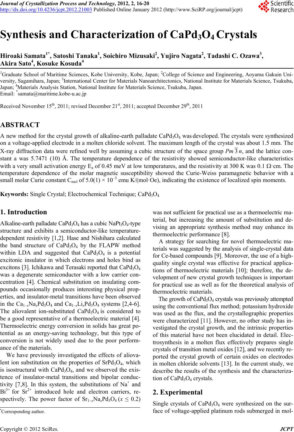
Single crystals of alkaline-earth palladate CaPd3O4 were
grown by an electrochemical technique that used voltage-
applied platinum electrodes inserted into a molten chlo-
ride solvent. Crystals with natural surfaces were obtained
on the anode, reflecting that the growth proceeded in a
liquid phase. The maximum length of the crystal was about
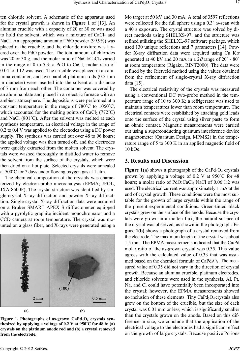
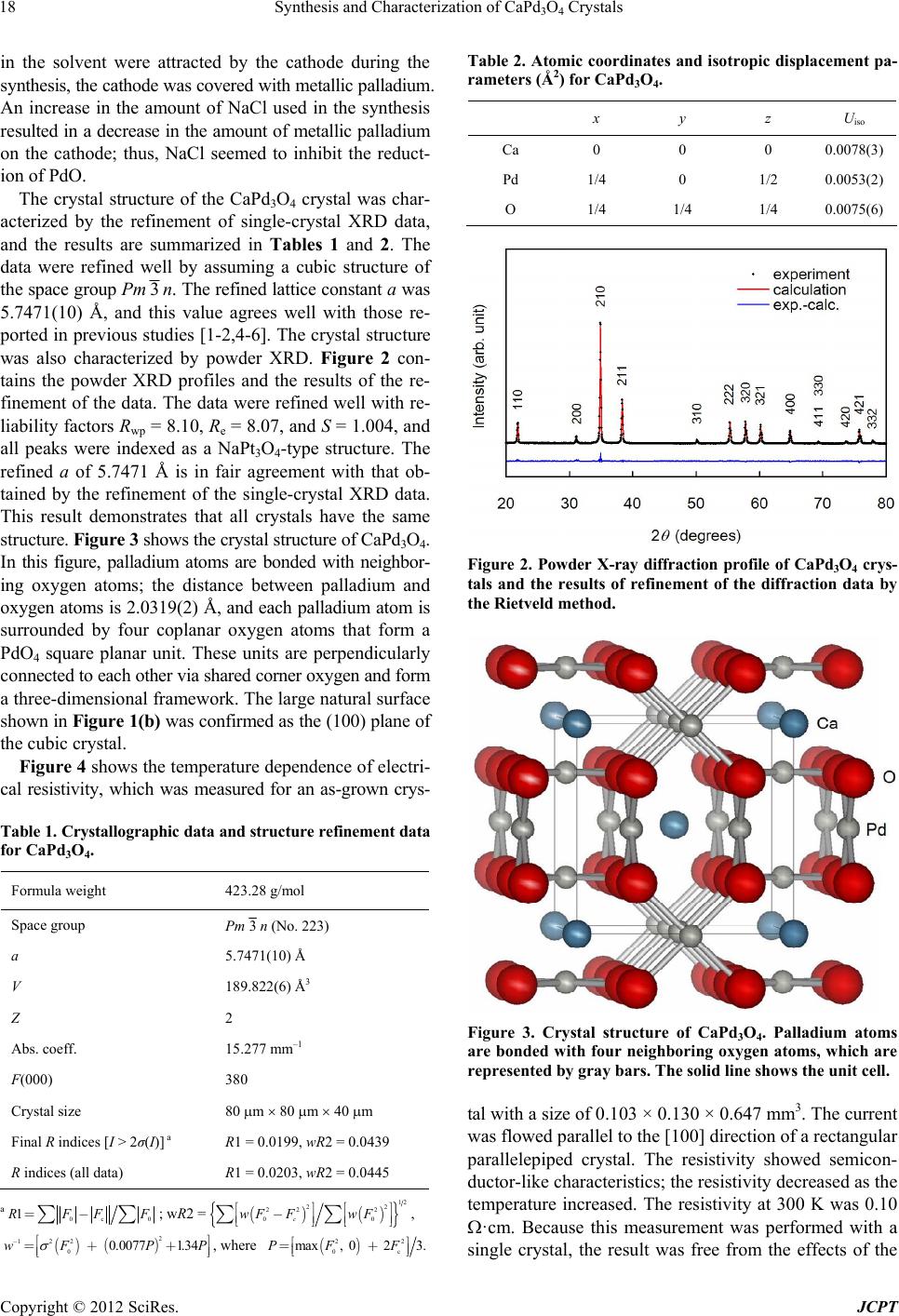
1.5 mm. The crystal structure parameters were refined well
by assuming a cubic structure of the space group Pm 3n,
and the lattice constant a was 5.7471(10) Å. The resistiv-
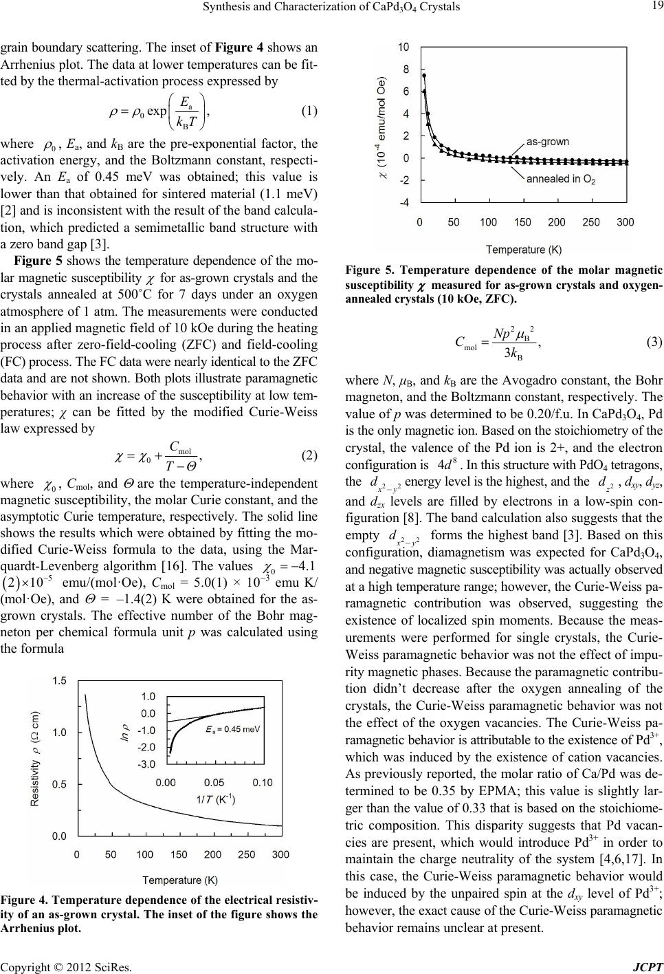
ity showed semiconductor-like characteristics with a very
small activation energy Ea of 0.45 meV at lower tempe-
ratures. The temperature dependence of the molar mag-
netic susceptibility showed the Curie-Weiss paramagne-
tic behavior, indicating the existence of localized spin mo-
ments.
[8] T. C. Ozawa, A. Matsushita, Y. Hidaka, T. Taniguchi, S.
Mizusaki, Y. Nagata, Y. Noro and H. Samata, “Synthesis
and Characterization of Electron and Hole Doped Ternary
Palladium Oxide: Sr1-xAxPd3O4 (A = Na, Bi),” Journal of
Alloys and Compounds, Vol. 448, No. 1-2, 2008, pp. 77-
83. doi:10.1016/j.jallcom.2007.03.137
[9] J. Kitagawa, T. Sasakawa, T. Suemitsu, Y. Echizen and T.
Takabatake, “Effects of Valence Fluctuation and Pseu-
dogap Formation on Phonon Thermal Conductivity of
Ce-Based Compounds with ε-TiNiSi-Type Structure,” Phy-
sical Review B, Vol. 66, No. 22, 2002, p. 224304.
doi:10.1103/PhysRevB.66.224304
[10] A. Saramat, G. Svensson, A. E. C. Palmqvist, C. Stiewe,
E. Mueller, D. Platzek, S. G. K. Williams and D. M.
Rowe, “Large Thermoelectric Figure of Merit at High Tem-
perature in Czochralski-Grown Clathrate Ba8Ga16Ge30,”
Journal of Applied Physics, Vol. 99, No. 2, 2006, p.
023708. doi:10.1063/1.2163979
5. Acknowledgements
The work performed at Kobe University and Aoyama Ga-
kuin University was supported by Grants-in-Aid for Sci-
entific Research from the Ministry of Education, Culture,
Sports, Science and Technology of Japan.
[11] P. L. Smallwood, M. D. Smith and H.-C. zur Loye, “Flux
Synthesis of Alkaline Earth Palladates,” Journal of Crystal
Growth, Vol. 216, No. 1-4, 2000, pp. 299-303.
doi:10.1016/S0022-0248(00)00432-2
REFERENCES [12] T. N. Nguyen and Hans-Conrad zur Loye, “Electrosyn-
thesis in Hydroxide Melts,” Journal of Crystal Growth,
Vo. 172, No. 1-2, 1997, pp. 183-189.
doi:10.1016/S0022-0248(96)00726-9
[1] R. C. Wnuk, T. R. Touw and B. Post, “The Crystal Struc-
ture of CaPd3O4,” IBM Journal of Research and Devel-
opment, Vol. 8, No. 2, 1964, pp. 185-186.
doi:10.1147/rd.82.0185 [13] H. Samata, Y. Saeki, S. Mizusaki, Y. Nagata, T. C. Oza-
wa and A. Sato, “Electrochemical Crystal Growth of Pe-
rovskite Ruthenates,” Journal of Crystal Growth, Vol.
311, No. 3, 2009, pp. 623-626.
doi:10.1016/j.jcrysgro.2008.09.042
[2] K. Itoh and N. Tsuda, “Metal to Semiconductor Like
Transition for Sintered Ca1–xNaxPd3O4,” Solid State
Com- munications, Vol. 109, No. 11, 1999, pp. 715-719.
doi:10.1016/S0038-1098(98)00549-3
[14] G. M. Sheldrick, SHELXTL, Version 6.10, Bruker AXS
Inc., Madison, 1997.
[3] I. Hase and Y. Nishihara, “CaPd3O4, as an Excitonic In-
sulator,” Physical Review B, Vol. 62, No. 20, 2000, pp.
13426-13429. doi:10.1103/PhysRevB.62.13426 [15] F. Izumi and T. Ikeda, “A Rietveld-Analysis Programm
RIETAN-98 and Its Applications to Zeolites,” Materials
Science Forum, Vol. 321-324, 2000, pp. 198-205.
doi:10.4028/www.scientific.net/MSF.321-324.198
[4] S. Ichikawa and I. Terasaki, “Metal-Insulator Transition
in Ca1–xLixPd3O4,” Physical Review B, Vol. 68, 2003, p.
233101. doi:10.1103/PhysRevB.68.233101
[16] W. H. Press, B. P. Flannery, S. A. Teukolsky and W. T. Vet-
terling, “Numerical Recipes,” Cambridge University Press,
Cambridge, 1986.
[5] K. Itoh, Y. Yano and N. Tsuda, “Metal to Insulator Tran-
sition for Ca1–xNaxPd3O4,” Journal of the Physical Soci-
ety of Japan, Vol. 68, No. 9, 1999, pp. 3022-3026.
doi:10.1143/JPSJ.68.3022 [17] S. J. Kim, S. Lemaux, G. Demazeau, J. Y. Kim and J. H.
Choy, “LaPdO3: The First PdIII Oxide with the Perovskite
Structure,” Journal of the American Chemical Society,
Vol. 123, No. 42, 2001, pp. 10413-10414.
doi:10.1021/ja016522b
[6] Y. Yano, M. Kanazawa, T. Fujii and N. Tsuda, “Magnetic
Susceptibility of Ca1-xNaxPd3O4,” Journal of the Physi-
cal Society of Japan, Vol. 70, No. 6, 2001, pp. 1772-1176.
doi:10.1143/JPSJ.70.1772