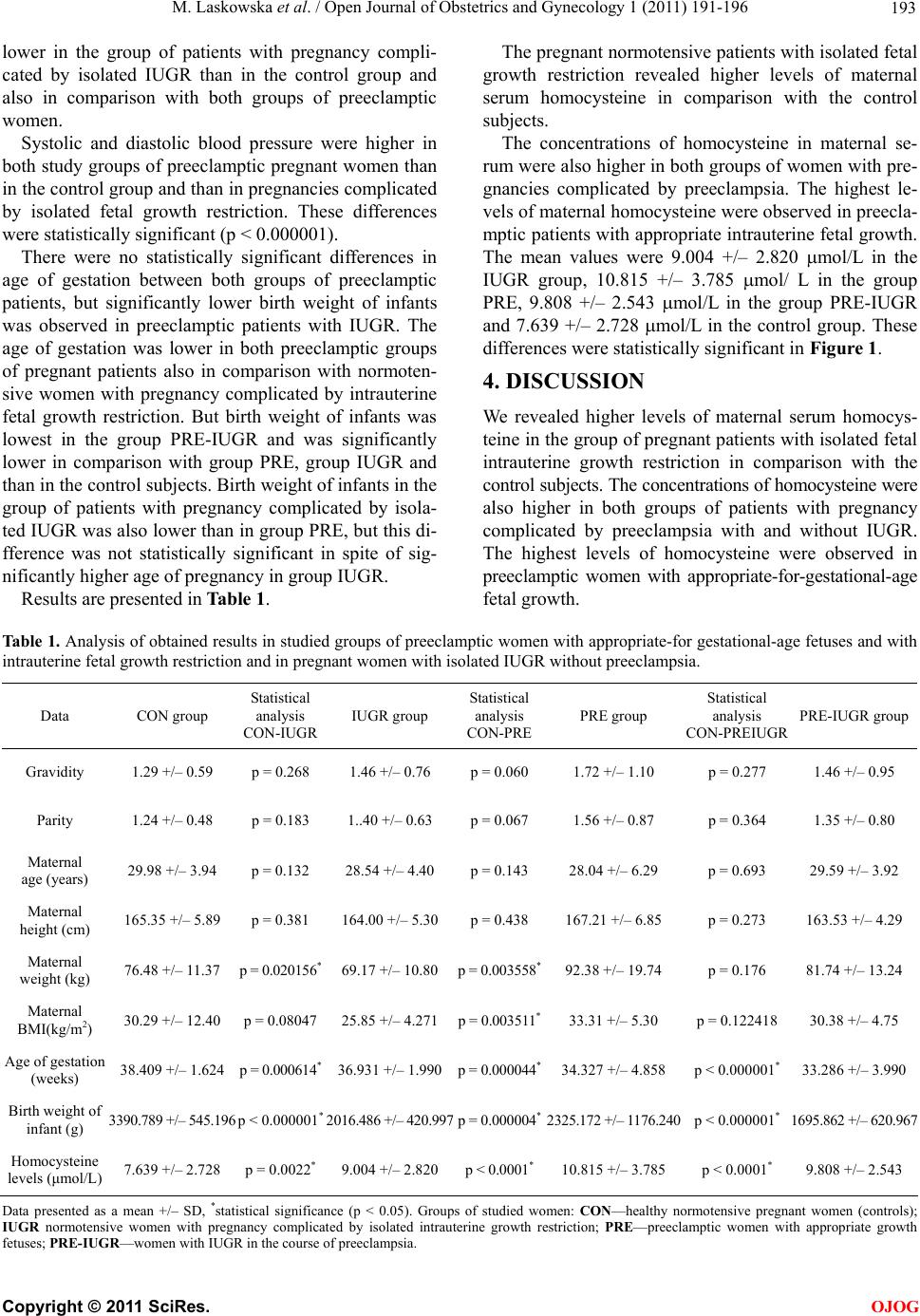
M. Laskowska et al. / Open Journal of Obstetrics and Gynecology 1 (2011) 191-196 195
However Hogg et al. [21] did not find such differen-
ces in pregnancies with IUGR. They concluded that pla-
sma homocysteine concentrations in the second trimester
do not predict the subsequent complications of preg-
nancy-induced hypertension, preeclampsia and intraute-
rine growth restriction.
Contrary to our findings also D’Anna et al. [22] didn’t
find any association between homocysteine levels in pre-
gnancy and IUGR.
Our findings about higher levels of maternal serum
homocysteine in pregnancies complicated by IUGR are
inconsistent with some earlier reports about the rela-
tionship between higher levels of homocysteine in preg-
nancy with intrauterine fetal growth restriction. This may
be due to the fact that we included into a group of wo-
men with pregnancy complicated by IUGR only patients
with fetal weight estimated below the 5th percentile for
gestational age. Furthermore we divided all pregnant
patients with fetal growth restriction into two groups,
one with IUGR in the course of preeclampsia and the se-
cond group with isolated IUGR and normotensive preg-
nancy, without any maternal or fetal condition response-
ble for this pregnancy complication. The aim of this was
to select the patients with the IUGR due to placental
dysfunction.
5. CONCLUSIONS
In conclusion, our findings seem to suggest that homo-
cysteine is a mediator of endothelial activation or dys-
function observed in pregnancies complicated by pree-
clampsia, but also in normotensive pregnancies compli-
cated by isolated IUGR.
It seems that the pathomechanism underlying the de-
velopment of preeclampsia and/or intrauterine fetal grow-
th restriction is similar, but highest levels of homocys-
teine observed in preeclamptic pregnancies may suggest
that its beginning and intensity may be more enhanced in
pregnancy complicated by hypertension and preeclamp-
sia.
Thanks to our better understanding of its physiology
and pathophysiology, homocysteine may develop towa-
rds a novel target for pharmacological intervention for
patients with these pregnancy complications.
REFERENCES
[1] Mandruzzato, G., Antsaklis, A., Botet, F., Chervenak,
F.A., Figueras, F., Grunebaum, A., Puerto, B., Skupski, D.
and Stanojevic, M. (2008) Intrauterine restriction (IUGR).
Journal of Perinatal Medicine, 36, 277-281.
doi:10.1515/JPM.2008.050
[2] Maynard, S.E., Min, J.Y., Merchan, J., Lim, K.H., Li, J.,
Mondal, S., Libermann, T.A., Morgan, J.P., Selke, F.W.,
Stillman, I.E., Epstein, F.H., Sukhatme, V.P.M. and Ka-
rumanchi, S.A. (2003) Excess placental soluble fms-like
tyrosine kinase 1 (sFlt1) may contribute to endothelial
dysfunction, hypertension, and proteinuria in preeclamp-
sia. Journal of Clinical Investigation, 111 , 600-602.
[3] Maršal, K. (2002) Intrauterine growth restriction. Current
Opinion in Obstetrics and Gynecology, 14,127-135.
doi:10.1097/00001703-200204000-00005
[4] Robinson, J.S. and Owens, J.A. (1996) Pathophysiology
of intrauterine growth failure. In: Gluckman, P.D. and
Heymann, M.A., Eds., Pediatrics and Perinatology: The
Scientific Basis, Arnold, London, 290-297.
[5] Bretelle, F., Sabatier, F., Blann, A., D’Ercole, Boutiere,
B., Mutin, M., Boubli, L., Sampol, J. and Dignat-George,
F. (2001) Maternal endothelial soluble cell adhesion
molecules with isolated small for gestational age fetuses,
comparison with preeclampsia. British Journal of Ob-
stetrics Gynaecology, 108, 1277-1282.
doi:10.1016/S0306-5456(01)00259-5
[6] Bamberger, A.M., Schulte, H.M., Thuneke, I., Erdmann,
I., Bamberger, ChM. and Asa, S.L. (1997) Expression of
the apoptosis-inducing fas ligand (FasL) in human first
and third trimester placenta and choriocarcinoma cells.
Journal of Clinical Endocrinology & Metabolism, 82,
3173-3175. doi:10.1210/jc.82.9.3173
[7] Hsu, ChD., hariah, H., Basherra, H. and Mor, A.G. (2001)
Serum soluble fas levels in preeclampsia. Obstetrics and
Gynecology, 97, 530-532.
doi:10.1016/S0029-7844(00)01227-8
[8] Roberts, J.M. (1999) Objective evidence of endothelial
dysfunction in preeclampsia. American Journal of Kid-
ney Diseases, 33, 992-997.
doi:10.1016/S0272-6386(99)70439-7
[9] Granger, J.P., Alexander, B.T., Llinas, M.T., Bennett,
W.A. and Khalil, R.A. (2001) Pathophysiology of hyper-
tension during preeclampsia linking placental ischemia
with endothelial dysfunction. Hypertension, 38, 718-722.
[10] Kassab, S., Abu-Hijleh, M.F., Al-Shaikh, H.B. and Na-
galla, D.S. (2005) Hyperhomocysteinemia in pregnant
rats: Effects on arterial pressure, kidneys and fetal
growth. European Journal of Obstetrics & Gynecology
and Reproductive, 122, 177-181.
doi:10.1016/j.ejogrb.2005.02.008
[11] Aubard, Y., Darodes, N. and Cantaloube, M. (2000) Hy-
perhomocysteinemia and pregnancy—review of our pre-
sent understanding and therapeutic implications. Euro-
pean Journal of Obstetrics & Gynecology and Reproduc-
tive, 93, 57-165.
[12] Onalan, R., Onalan, G., Gunenc, Z. and Karabulut, E.
(2006) Combining 2nd trimester maternal serum homo-
cysteine levels and uterine artery doppler for prediction
of preeclampsia and isolated intrauterine growth restric-
tion. Gynecology and Obstetrics Investigaton, 61, 142-
148. doi:10.1159/000090432
[13] Weir, D.G. and Scott, J.M. (1998) Homocysteine as a risk
factor for cardiovascular and related disease: Nutritional
implications. Nutr ition Research Reviews, 11, 311-338.
doi:10.1079/NRR19980020
[14] López-Quesada, E., Also-Razo, E. and Vilaseca, M.A.
(2003) Hyperhomocysteinemia during pregnancy as a
risk factor of preeclampsia. Clinical Medicine, 121,
350-355.
[15] Wang, J., Trudinger, B.J., Duarte, N., Wilcken, D.E. and
C
opyright © 2011 SciRes. OJOG