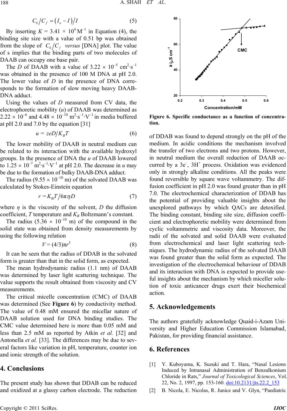
International Journal of Organic Chemistry, 2011, 1, 183-190
doi:10.4236/ijoc.2011.14027 Published Online December 2011 (http://www.SciRP.org/journal/ijoc)
Copyright © 2011 SciRes. IJOC
183
Electrochemical Characterization, Detoxification and
Anticancer Activity of
Didodecyldimethylammonium Bromide
Afzal Shah1, Erum Nosheen1, Rumana Qureshi1, Muhammad Masoom Yasinzai1,
Suzanne K. Lunsford2, Dionysios D. Dionysiou3, Zia ur Rehman1, Muhammad Siddiq1,
Amin Badshah1, Saqib Ali1*
1Department of Chemistry, Quaid-i-Azam University, Islamabad, Pakistan
2Department of Chemistry, Wright State University, Dayton, USA
3School of Energy, Environmental, Biological, and Medical Engineering (SEEBME),
705 Engineering Research Center, University of Cincinnati, Cincinnati, USA
E-mail: *drsa54@yahoo.com
Received October 20, 2011; revised November 27, 2011; accepted December 6, 2011
Abstract
Quaternaryammonium compounds (QACs) are cationic surfactants with broad range applications. They enter
into aquatic environment through the discharge of sewage effluents and adversely affect the ecosystem due
to toxicity. Modern electrochemical methods have been used for characterization and detoxification of an
extensively used QAC, didodecyldimethylammonium bromide (DDAB) in a wide pH range. The diffusion
coefficient of DAAB was determined by Randles-Sevcik equation. In acidic conditions the electrochemical
reduction of DAAB occurred by two electrons and two protons process. However, in neutral medium the
overall reduction of DAAB followed electronation-protonation mechanism involving 3e and 3H+. Although
DDAB is toxic yet it is bestowed with cancer preventing ability. Hence, for getting insights into the under-
standing of DAAB anticancer effect, its DNA binding parameters has been evaluated.
Keywords: Didodecyldimethylammonium Bromide, Detoxification, Anticancer Effect,
DNA Binding Parameters
1. Introduction
Surfactants are widely used as household and industrial
cleaning agents. They are classified as: cationic, anionic,
non-ionic and zwitterionic. Quaternary ammonium com-
pounds (QAC) are the most extensively studied surface ac-
tive agents. They are mainly used as slimicides in swim-
ming pools, antistatic agents in hair conditioners and wet-
ting agents in nasal sprays [1,2]. Moreover, they also find
use in preservatives, shampoos and dish washing materials
[3].
The used surfactants find their way to various environ-
mental segments and thus pose serious health hazards.
They have the ability to adsorb strongly onto negatively
charged suspended particulates and sludge. Intensive in-
vestigations have been carried out on their behavior, fate
and biological effects [4,5]. Long exposure to QACs can
cause sensitization, haemolysis and toxic effects by all
routes of exposure including inhalation, ingestion, der-
mal application and irrigation of body cavities [6]. Their
toxicity in the mg·l–1 range with a fatal dose of 1 - 3 g
has been reported [7]. The concentrated solutions of
QACs result in allergic reactions, hypersalivation, vomit-
ing, haematemesis and diarrhoea [8]. In severe cases they
may cause respiratory paralysis, convulsions, coma and
cardiorespiratory arrest [9]. A part of the current article is
devoted to voltammetric characterization and detoxifica-
tion of a quaternary ammonium compound, didodecyldi-
methylammonium bromide (DDAB) by modern electro-
chemical techniques.
DDAB (Scheme 1) is toxic as well as cancer prevent-
ing agent [10]. Toxicity is a major nuisance of anticancer
drugs. Most of the clinically used anticancer drugs have
a narrow therapeutic index, with a small difference in
their dose for antitumor effect and toxicity. Therefore, the
doses recommended for such compounds are determined
A. SHAH ET AL.
184
Scheme 1. Chemical structure of DAAB.
according to their toxicity endpoints. Considerable re-
search work has been carried out on the cancer preven-
tion ability of DAAB but the mechanism of action has
not been explored. The present work is an effort to estab-
lish a critical role of DDAB in exercising its anticancer
effect.
The main cause of several diseases such as diabetes,
hemophilia and cancer is related to the alteration in the
replication machinery of DNA. Therefore, drug interact-
tions with DNA which can affect the replication proc-
esses, are potential treatments for such ailments [11-13].
Normally, cancers are difficult to be diagnosed at a stage
of small hidden tumors, so the surgical eradication can
not be practiced. At such an early stage, chemotherapy is
the only weapon to combat with cancer. The research fo-
cused on the action mechanism of anticancer drugs is ex-
pected to accelerate the discovery of effective chemo-
therapeutic agents.
A variety of techniques has been employed [14-18] for
the study of toxicity alleviation and drug-DNA interact-
ion with relative advantages and disadvantages. How-
ever, most of these suffer from high cost, low sensitivity
and procedural complication. Electrochemical techniques
were used for the characterization, detoxification and
DNA binding studies of DAAB due to their cost effect-
tivity, high sensitivity and ease of operation.
2. Experimental
2.1. Materials and Reagents
DDAB (98% purity) was purchased from Fluka and used
without further purification. Its 5.0 mM stock solution
was prepared and stored at 4˚C. The working solutions
were prepared in 1:1 mixed solvent of ethanol and buffer.
All supporting electrolyte solutions (Table 1) were pre-
pared using analytical grade reagents. Calf thymus DNA
was purchased from Sigma and used as received. 2.0 mM
stock solution of DNA was prepared and its concentra-
tion was determined by UV absorbance at 260 nm using
a molar absorptivity (ε) of 6600 M–1cm–1 [19,20]. As the
absorbance ratio of pure DNA at 260 and 280 nm
(A260/A280) should not be less than 1.8, so a ratio of 1.85
indicated protein free DNA [21]. Doubly distilled water
was used throughout.
Table 1. List of 0.1 M supporting electrolytes.
pHComposition pH Composition
1.0HCl + KCl 7.0 NaH2PO4 + Na2HPO4
2.0HCl + KCl 8.0 NaH2PO4 + Na2HPO4
3.0HAcO + NaAcO 9.0 NaHCO3 + NaOH
4.0HAcO + NaAcO 10.0 NH3 + NH4Cl
5.0HAcO + NaAcO 12.0 NaOH + KCl
6.0HAcO + NaAcO 13.0 NaOH + KCl
2.2. Equipments and Measurements
Voltammetric experiments were performed using µAuto-
lab running with GPES 4.9 software, Eco-Chemie, The
Netherlands. A glassy carbon (GC) (A = 0.07 cm2) was
used as working electrode, a Pt wire served as counter
electrode and a saturated calomel electrode (SCE) was
employed as the reference. The differential pulse volt-
ammetry (DPV) and square wave voltammetry (SWV)
were carried out at a scan rate of 10 and 100 mV·s–1. Cy-
clic voltammetry (CV) was performed at different scan
rates. Before each experiment the surface of GCE was
polished with alumina powder followed by thorough rin-
sing with distilled water. All the voltammetric experiments
were conducted in a high purity nitrogen (99.995%) at-
mosphere at room temperature (25˚C ± 1˚C).
Viscosity and density measurements were made on an
Anton Paar Stabinger Viscometer SVM 3000. The dyna-
mic laser light scattering experiment was carried out by a
commercial LLS spectrometer BI-200SM motor-driven
goniometer equipped with BI-9000AT digital autocorre-
lator and a cylindrical 22 mW uniphase He-Ne laser (wa-
velength = 637 nm) and BI-ISTW software.
3. Results and Discussion
3.1. Cyclic Voltammetry
Cyclic voltammetric (CV) behavior of DDAB has been
examined in pH range 1 - 13. In acidic and neutral media,
DDAB was found to give two cathodic peaks correspon-
ding to two step reduction while a single anodic peak re-
lated to oxidation was registered in alkaline media. Fig-
ures 1(a)-(c) show typical cyclic voltammograms of 1.0
mM DDAB in different pH media. In strongly acidic
conditions (pH 2.0) two close reduction peaks were re-
corded in the forward scan with no corresponding oxida-
tion peaks in the reverse scan. In a medium buffered at
pH 7.0, a broad reduction peak (1c) was observed which
may be due to the overlapping two close cathodic peaks
appeared in harsh acidic environment. The 2nd cathodic
peak appeared at high negative value. The CV of DAAB
at pH 13.0 showed only oxidation. The variation in CV
response indicates that the redox mechanism of DDAB is
pH dependent.
Copyright © 2011 SciRes. IJOC
185
A. SHAH ET AL.
(a) (b)
(c)
Figure 1. CVs 1.0 mM DDAB in pH (a) 2.0, (b) 7.0 and (c)
3.0 at = 100 mV·s–1. 1
The dependence of peak current Ip, on the scan rate, ν,
is an important diagnostic criterion for establishing the
type of mechanism by cyclic voltammetry. Therefore,
CVs of DDAB were recorded at different scan rates (100 -
500 mV·s–1). The shift in peak potentials to more nega-
tive values with the increase in scan rate indicated the
irreversibility of the reduction processes. In strongly
acidic conditions (pH 2.0), the current of peak 1c in-
creased linearly with the square root of , indicating dif-
fusion-limited reduction of analyte. The peak current in
amperes for a diffusion-controlled irreversible cathodic
process is given by equation [22]
512*12
=2.9910 ()
pcco o
InnACD
12
 (1)
where Ipc is the cathodic peak current in amperes, n the
number of electrons transferred during the reduction, A
the geometric area of the electrode in cm2,
c the catho-
dic charge transfer coefficient, Do the diffusion coeffi-
cient in cm2·s–1, n the number of electrons involved in the
electrochemical process, Co
* the bulk concentration of
the ox-3 n–1
idant in mol cm and the scan rate i V·s.
The difference between peak potential Epc and the po-
tential at half peak height Epc/2 of ~50 mV implied an
irreversible 1e reduction. The cn with a value of 0.95
was calculated from the relation [23]

2 47.7
pc pcc
EE n
 (2)
The diffusion coefficient of DDAB with a value of 5.71
10–4 cm2 s
–1 was evaluated from the measured slope
(–2.5 10–3 A/(V·s–1)1/2) of Ipc vs 1/2. The D (1.15 10–7
cm2·s–1) of DDAB was also determined at pH 7.0. The
more current carrying ability as evidenced from high
peak current and concomitant D value in strongly acidic
medium can be attributed to the greater charge density of
cationic DDAB due to non-availability of hydroxide ions.
The slightly smaller diffusion coefficient of DAAB at pH
7.0 than the D (3.70 10–7 cm2·s–1) of a closely related
surfactant, cetyltrimethylammonium bromide under simi-
lar conditions [24] may be due to its comparatively larger
molecular mass, thus confirming the idea that heavy mo-
lecule diffuses slowly to the electrode surface.
3.2. Differential Pulse Voltammetry
The pH dependence of DDAB reduction was also invest-
tigated by DPV in acidic, basic and neutral media. Figure
2 shows typical DP voltammograms of 1.0 mM DDAB.
Like CV, DDAB showed two reduction peaks in neutral
medium but unlike CV, peak 1c did not split into two sub
peaks at pH 2.0. The reason could be the low scan rate
(10 mV·s–1) at which the DPV was recorded. Thus the 2nd
component of peak 1c corresponds to the formation of an
electroactive reduction product of DDAB (PDDAB) that
can only be observed at higher scan rates. Hence, it is
necessary to scan the potential faster than the rate of the
homogenous reaction (hydrolysis) that PDDAB undergoes
in order to observe its redox signals. The absence of peak
2c in the DPV of DDAB at pH 2.0 and its presence in
neutral buffer support the results obtained from CV. As
the broad peak in DPV was due to two overlapping sub
peaks (as described above) so the half peak width (W1/2)
was divided by 2. A value of 91 mV obtained in this way
indicated the transfer of one electron [25].
With the increase in pH, the potential of peak 1c was
displaced to more negative values (Figure 3). Epc varied
linearly (see Inset) with increase in pH (Epc = 0.8443 -
0.0636 pH). The slope of 63.6 mV per pH unit close to
-1.5 -1-0.500.51
E / V vs. SCE
4 A
1
c
2
c
1
c
Figure 2. Base line corrected DPVs of 1.0 mM DDAB in pH
2 () and 7 () at a scan rate of 10 mV·s–1.
Copyright © 2011 SciRes. IJOC
A. SHAH ET AL.
186
Figure 3. Base line corrected DPVs of 1.0 mM DDAB in pH
1.0 (), 2.0 (), 5.0 () and 7.0 () at = 10 mV·s–1. Inset
shows Epc as a function of pH.
the theoretical value of 59.5, suggested the involvement
of the same number of electrons and protons in the elec-
trochemical reduction of the analyte. Hence, the reduc-
tion of DDAB followed 1e, 1H+ process in a pH de-
pendent manner.
In strongly alkaline media the analyte registered only a
single anodic peak corresponding to oxidation at Epa =
1.21 V. The DPV under these conditions showed similar
features to CV.
3.3. Square Wave Voltammetry
SW voltammetry is preferred over other electroanalytical
techniques as it is associated with the added advantage of
recording forward, backward and net current in only one
scan which manifests the reversibility or irreversibility of
the electron transfer reaction. Since the current is sam-
pled in both positive and negative-going pulses, peaks
corresponding to oxidation and reduction of the electro-
active species at the electrode surface can be obtained in
the same experiment. Like CV and DPV, both the reduc-
tion peaks of 1.0 mM DDAB in pH 7.0 were observed in
the forward scan of SW voltammogram at Epc
1 = 0. 51 V
and Epc
2 = –0.59 V (Figure 4(a)). But a dramatically dif-
ferent situation from CV was encountered in the back-
ward scan which showed the reversibility of both peaks.
Moreover, the reversibility of peak 1c and 2c was also
evidenced by the equality of the components of total cur-
rent. Due to the comparatively higher sensitivity of SWV,
the reversibility of oxidation peak obtained in alkaline
media (Figure 4(b)) was also proved in the same manner
and during the same experiment.
Figure 4(c) shows the SW voltammetric behavior of
1.0 mM DDAB at pH 2.0. Like CV a couple of cathodic
(a) (b)
(c)
Figure 4. SWV of 1.0 mM DDAB in pH (a) 7.0, (b) 13.0 and
(c) 2.0 at = 100 mV·s–1. 1st scan in solution showing total
current (), forward current () and backward current
().
peaks corresponding to two close charge transfer reac-
tions were observed in the forward scan. But in contrast to
CV, the backward scan indicated the reversibility of both
the charge transfer processes. Unlike the SWV at pH 7.0,
peak 2c of DDAB was not observed at pH 2.0. The be-
havior points to the fact that the redox reactions of DA-
AB follow different mechanistic pathways in acidic and
neutral media.
3.4. Redox Mechanism
The results obtained from CV, SWV and DPV enabled to
propose the redox mechanism of DAAB (Scheme 2).
The appearance of cathodic peak 1c and 2c indicated the
reduction of DDAB at the GCE. The splitting of peak 1c
into two sub peaks at high scan rate revealed the overall
reduction of DDAB to occur in three steps. In media buf-
fered at pH < 7.0 the components of 1c are attributable to
the electrochemical reduction of DDAB (1) to quaternary
dodecyldimethylammonium ion (2) and quaternary di-
methylammonium ion (3) by a mechanism involving two
1e, 1H+ processes. The presence of corresponding oxi-
dation peaks in the reverse scan (Figure 4(c)) suggested
the reversibility of DDAB redox processes. Under neu-
tral conditions, the components of peak 1c merged into a
broad peak that can be related to the formation of reduc-
tion product (3) by a mechanism involving 2e, 2H+
transfer. The appearance of 2nd cathodic peak 2c at pH =
Copyright © 2011 SciRes. IJOC
187
A. SHAH ET AL.
N+
CH
3
H
3
C
C
12
H
25
C
12
H
25
1e
-
1H+
N+
CH
3
H
3
CH
C
12
H
25
1e
-
1H+
N+
CH
3
H
3
CH
H
pH < 7.0
(1) (2) (3)
N+
CH
3
H
3
C
C
12
H
25
C
12
H
25
2e
-
2H+
1e
-
1H+
N+
H
3
CH
H
pH = 7.0
(1) (3) (4)
N+
CH
3
H
3
CH
H
H
-1e
-
pH = 13.0
N+
CH
3
H
3
C
C
12
H
25
C
12
H
25
(1) (5)
+ 2 C
12
H
25
OHN (CH
3
)
2-
2OH
-
Scheme 2. Proposed mechanism for the electrochemical re-
duction of DDAB.
7.0 pointed to the formation of quaternary methylammo-
nium ion (4). The appearance of anodic peak at pH 13.0
can presumably be due to the oxidation of DAAB to pro-
duct (5 ). The mechanism suggested here is supported by
the work of Nishiyama et al., [26] on the microbial deal-
kylation of alkyl trimethylammonium compounds. The
presence of dealkylated intermediates of alkyltrimethy-
lammonium salts (triethylamine, dimethylamine and me-
thyllamine) in activated sludge obtained from municipal
sewage treatment plant [27] further supported our sug-
gested mechanism. The increase in number of non-me-
thyl alkyl groups in QACs cause difficulty in biodegrad-
ability which in turn results in toxicity [27]. Hence, the
results of our experiments reveal that QACs can be de-
toxified to biodegradable products by electrochemical
methods.
3.5. DNA Binding Study
The effect of increasing concentration of DNA on the cyc-
lic voltammetric response of 1.0 mM DDAB has been
shown in Figure 5. Unlike the general trend, the peak
current of DDAB increased in the presence of DNA.
This anomalous behavior may be due to the dissociation
of DDAB micelles by DNA and concomitant increase in
number of monomers. The breakdown of micellar ag-
gregates can be due to electrostatic interaction of anionic
oxygen with ammonium moiety and the affinity of hy-
drophobic interior of DNA for the lipophilic hydrocarbon
section of DAAB. The increase in peak current further
points to the fact that the number of monomers produced
by the breakdown of micelles is greater in the bulk than
those attached with DNA. The peak potential shift to
more negative and then to positive values is attributed to
-2 -1.6-1.2-0.8-0.400.4 0.81.2
E/ V v s. SCE
1 mA
Figure 5. 1st scan CVs of 1.0 mM DDAB (obtained in pH 2.0)
in the absence of DNA () and presence of 20 () and 40
M DNA ().
mixed binding mode. A part of the hydrocarbon tail of
DDAB is suggested to intercalate and the rest may oc-
cupy the grooves. The cationic ammonium moiety may
interact electrostatically with anionic phosphate back-
bone of DNA. The interaction is expected to effect the
DNA of cancerous cells in such a way that the cell can
not replicate further.
The peak current of DAAB diminished with further in-
cremental addition of DNA. The rationale behind the di-
minution in peak current may be the decrease in number
of free monomers due to the formation of macromolecu-
lar DAAB-DNA complex with lower current carrying abi-
lity.
The decay in peak current (Ip) of DAAB by the incre-
mental addition of DNA was exploited for the quantifi-
cation of binding constant according to the following
equation [28]

log 1DNAloglogo
K
II I  (3)
where K is the binding constant, Io and I are the peak
currents of the drug in the absence and presence of DNA.
The K with a value of 3.41 × 10⁴ M–1 was obtained from
the intercept of log (1/[DNA]) versus log(I/(Io I)) plot.
The value is comparable with the reported K (3.60 × 10⁴
M–1) of 1,8-dihydroxyanthraquinone interacting with
DNA by mixed binding mode [29].
For the determination of binding site size the follow-
ing equation was used [30]
DNA 2
bf
CC Ks (4)
where s is the binding site size in terms of base pairs, Cf
the concentration of the free species, Cb the concentra-
tion of DNA-bound species and K the binding constant.
The values of bf
CCwere determined from the experi-
mental peak currents by the following equation [30].
Copyright © 2011 SciRes. IJOC
A. SHAH ET AL.
188

bf o
CCIII (5)
By inserting K = 3.41 × 10⁴ M–1 in Equation (4), the
binding site size with a value of 0.51 bp was obtained
from the slope of bf
CC
versus [DNA] plot. The value
of s implies that the binding parts of two molecules of
DAAB can occupy one base pair.
The D of DAAB with a value of 3.22 10–5 cm2·s–1
was obtained in the presence of 100 M DNA at pH 2.0.
The lower value of D in the presence of DNA corre-
sponds to the formation of slow moving heavy DAAB-
DNA adduct.
Using the values of D measured from CV data, the
electrophoretic mobility (u) of DAAB was determined as
2.22 10–6 and 4.48 10–10 m2·s–1·V–1 in media buffered
at pH 2.0 and 7.0 by the equation [31]
= B
uzeDKT (6)
The lower mobility of DAAB in neutral medium can
be related to its interaction with the available hydroxyl
groups. In the presence of DNA the u of DAAB lowered
to 1.25 10–7 m2·s–1·V–1 at pH 2.0. The decrease in u may
be due to the formation of bulky DAAB-DNA adduct.
The radius (9.55 10–10 m) of the solvated DAAB was
calculated by Stokes-Einstein equation
B
=6πrKT D
(7)
where η is the viscosity of the solvent, D the diffusion
coefficient, T temperature and KB Boltzmann’s constant.
The radius (5.36 10–10 m) of the compound in the
solid state was obtained from density measurements by
using the following relation
V = (4/3)πr3 (8)
It can be seen that the radius of DDAB in the solvated
form is greater than that in the solid form, as expected.
The mean hydrodynamic radius (1.1 nm) of DAAB
was determined by laser light scattering technique. The
value supports the result obtained from viscosity and CV
measurements.
The critical micelle concentration (CMC) of DAAB
was determined (See Figure 6) by conductivity method.
The value of 0.48 mM ensured the micellar nature of
DAAB solution used for DNA binding studies. The
CMC value determined here is more than 0.05 mM and
less than 2.5 mM as reported by Atkin et al. [32] and
Antonella et al. [33]. The differences may be due to sev-
eral factors like variation in pH, temperature, counter ion
and ionic strength of the solution.
4. Conclusions
The present study has shown that DDAB can be reduced
and oxidized at a glassy carbon electrode. The reduction
20
40
60
80
0.20.30.40.5
Concentration/mM
k/S c m
-1
0.6
CMC
Figure 6. Specific conductance as a function of concentra-
tion.
of DDAB was found to depend strongly on the pH of the
medium. In acidic conditions the mechanism involved
the transfer of two electrons and two protons. However,
in neutral medium the overall reduction of DAAB oc-
curred by a 3e, 3H+ process. Oxidation was evidenced
only in strongly alkaline conditions. All the peaks were
found reversible by square wave voltammetry. The dif-
fusion coefficient in pH 2.0 was found greater than in pH
7.0. The electrochemical characterization of DDAB has
the potential of providing valuable insights about the
unexplored pathways by which QACs are detoxified.
The binding constant, binding site size, diffusion coeffi-
cient and electrophoretic mobility were determined from
cyclic voltammetric and viscosity data. Moreover, the
radii of the solvated and solid DAAB were evaluated
from electrochemical and laser light scattering tech-
niques. The hydrodynamic radius of the solvated DAAB
was found greater than the solid form as expected. The
investigation of the electrochemical behaviour of DDAB
and its interaction with DNA is expected to provide use-
ful insights about the mechanism by which miceller solu-
tion of toxic anticancer drugs exert their biochemical
action.
5. Acknowledgements
The authors gratefully acknowledge Quaid-i-Azam Uni-
versity and Higher Education Commission Islamabad,
Pakistan, for providing financial assistance.
6. References
[1] Y. Kuboyama, K. Suzuki and T. Hara, “Nasal Lesions
Induced by Intranasal Administration of Benzalkonium
Chloride in Rats,” Journal of Toxicological Sciences, Vol.
22, No. 2, 1997, pp. 153-160. doi:10.2131/jts.22.2_153
[2] B. Nicola, E. Nicolas, R. Junice and V. Glyn, “Paediatric
Copyright © 2011 SciRes. IJOC
189
A. SHAH ET AL.
Toxicology Handbook of Poisoning in Children,” Mac-
millan Reference Ltd., London, 1997.
[3] R. Beasley, D. Fishwick, J. F. Miles and L. Hendeles,
“Preservatives in Nebulizer Solutions: Risks without
Benefit,” Pharmacotherapy, Vol. 18, 1998, pp. 130-139.
[4] P. S. Boeris, A. S. Liffourrena, M. A. Salvano and G. I.
Lucchesi, “Physiological Role of Phosphatidylcholine in
the Pseudomonas putida A ATCC 12633 Response to
Tetradecyltrimethylammonium Bromide and Alumin-
ium,” Letters in Applied Microbiology, Vol. 49, No. 4,
2009, pp. 491-496.
doi:10.1111/j.1472-765X.2009.02699.x
[5] R. P. Singh, N. Gupta, S. Singh, A. Singh, R. Suman and
K. Annie, “Toxicity of Ionic and Non Ionic Surfactants to
Six Microbes Found in Agra, India,” Bulletin of Envi-
ronmental Contamination and Toxicology, Vol. 69, No. 2,
2002, pp. 265-270. doi:10.1007/s00128-002-0056-z
[6] F. Placucci, A. Benini and A. Tosti, “Occupational Aller-
gic Contact Dermatitis from Disinfectant Wipes Used in
Dentistry,” Contact Dermatitis, Vol. 35, No. 5, 1996, p.
306. doi:10.1111/j.1600-0536.1996.tb02397.x
[7] D. Chataigner, R. Garnier, S. Sans and M. L. Efthymiou,
“Intoxication Aigue Accidentelle Par un Désinfectant
Hospitalier. 45 cas dont 13 d’évolution Mortelle,” La
Presse Médicale, Vol. 20, 1991, pp. 741-743.
[8] M. Van Berkel and F. A. de Wolff, “Survival after Ben-
zalkonium Chloride Poisoning,” Human Toxicology, Vol.
7, 1988, pp. 191-193. doi:10.1177/096032718800700216
[9] R. Ren, D. Liu, K. Li, J. Sun and C. Zhang, “Adsorption
of Quaternary Ammonium Compounds onto Activated
Sludge,” Journal of Water Resource and Protection, Vol.
3, No. 2, 2011, pp. 105-113.
doi:10.4236/jwarp.2011.32012
[10] M. T. Garcia, E. Campos, J. Sanchez-Leal and I. Risoba,
“Anaerobic Degradation and Toxicity of Commercial Ca-
tionic Surfactants in Anaerobic Screening Tests,” Che-
mosphere, Vol. 41, No. 5, 2000, pp. 705-710.
doi:10.1016/S0045-6535(99)00455-5
[11] H. T. Chifotides and K. R. Dunbar, “Interactions of Met-
al-Metal-Bonded Antitumor Active Complexes with DNA
Fragments and DNA,” Accounts of Chemical Research,
Vol. 38, No. 2, 2005, pp. 146-156.
doi:10.1021/ar0302078
[12] D. D. Li, J. L. Tian, W. Gu, X. Liu, H. H. Zeng and S. P.
Yan, “DNA Binding, Oxidative DNA Cleavage, Cyto-
toxicity, and Apoptosis-Inducing Activity of Copper(II)
Complexes with 1,4-Tpbd(N,N,N’,N-tetrakis(2-yridylme-
thyl)benzene-1,4-diamine) Ligand,” Journal of Inorganic
Biochemistry, Vol. 105, No. 6, 2011, pp. 894-901.
doi:10.1016/j.jinorgbio.2011.03.012
[13] M. Egli, L. D. Williams, C. A. Frederick and A. Rich,
“DNA-Nogalamycin Interactions,” Biochemistry, Vol. 30,
No. 5, 1991, pp. 1364-1372.
[14] A. Fontana, P. D. Maria, G. Siani and B. H. Robinson,
“Kinetics of Breakdown of Vesicles from Didodecyldi-
methylammonium Bromide Induced by Single Chain
Surfactants and by Osmotic Stress in Aqueous Solution,”
Colloids and Surfaces B: Biointerfaces, Vol. 32, No. 4,
2003, pp. 365-374.
doi:10.1016/j.colsurfb.2003.08.003
[15] M. Gradzielski, “Recent Developments in the Charac-
terization of Microemulsions,” Current Opinion in Col-
loid & Interface Science, Vol. 13, No. 4, 2008, pp. 263-
269. doi:10.1016/j.cocis.2007.10.006
[16] K. Kusumoto and T. Ishikawa, “Didodecyldimethylam-
monium Bromide (DDAB) Induces Caspase-Mediated
Apoptosis in Human Leukemia HL-60 Cells,” Journal of
Controlled Release, Vol. 147, No. 2, 2010, pp. 246-252.
doi:10.1016/j.jconrel.2010.07.114
[17] T. Neumann, S. Gajria, N. Bouxsein, L. Jaeger and M.
Tirrell, “Structural Responses of DNA-DDAB Films to
Varying Hydration and Temperature,” Journal of the
American Chemical Society, Vol. 132, No. 20, 2010, pp.
7025-7037. doi:10.1021/ja909514j
[18] N. Subramanian, S. K. Ghosal, A. Acharya and S. P.
Moulik, “Formulation and Physicochemical Characteriza-
tion of Microemulsion System Using Isopropyl Myristate,
Medium-Chain Glyceride, Polysorbate 80 and Water,”
Chemical & Pharmaceutical Bulletin, Vol. 53, No. 12,
2005, pp. 1530-1535. doi:10.1248/cpb.53.1530
[19] A. Shah, A. M. Khan, R. Qureshi, F. L. Ansari, M. F.
Nazar and S. S. Shah, “Redox Behavior of Anticancer
Chalcone on a Glassy Carbon Electrode and Evaluation
of Its Interaction Parameters with DNA,” International
Journal of Molecular Sciences, Vol. 9, No. 8, 2008, pp.
1424-1434. doi:10.3390/ijms9081424
[20] M.-J. Han, Z.-M. Duan, et al., “Molecular Light Switches
for Calf Thymus DNA Based on Three Ru(II) Bipyridyl
Complexes with Variations of Heteroatoms,” The Journal
of Physical Chemistry C, Vol. 111, No. 44, 2007, pp.
16577-16585. doi:10.1021/jp075194k
[21] D. L. Guo, Y. Xin, P. C. Zeng, L. S. Guo and Q. Y. Ru,
“Interaction of Metal Complexes of Bis(salicylidene)-
ethylenediamine with DNA,” Analytical Sciences, Vol.
16, No. 12, 2000, pp. 1255-1260.
doi:10.2116/analsci.16.1255
[22] C. M. A. Brett and A. M. O. Brett, “Electrochemistry.
Principles, Methods and Applications,” Oxford Univer-
sity Press, Oxford, 1993.
[23] V. C. Diculescu, T. A. Enache, P. J. Oliveira and A. M. O.
Brett, “Electrochemical Oxidation of Berberine and of Its
Oxidation Products at a Glassy Carbon Electrode,” Elec-
troanalysis, Vol. 21, No. 9, 2009, pp. 1027-1034.
doi:10.1002/elan.200804516
[24] B. M. Asit and U. N. Balachandran, “Cyclic Voltammet-
ric Technique for the Determination of the Critical Mi-
celle Concentration of Surfactants, Self-Diffusion Coeffi-
cient of Micelles, and Partition Coefficient of an Electro-
chemical Probe,” Journal of Physical Chemistry, Vol. 95,
No. 22, 1991, pp. 9008-9013. doi:10.1021/j100175a106
[25] A. Shah, V. C. Diculescu, R. Qureshi and A. M. O. Brett,
“Electrochemical Behavior of Dimethyl-2-oxoglutarate
on Glassy Carbon Electrode,” Bioelectrochemistry, Vol.
77, No. 2, 2010, pp. 145-150.
Copyright © 2011 SciRes. IJOC
A. SHAH ET AL.
Copyright © 2011 SciRes. IJOC
190
doi:10.1016/j.bioelechem.2009.07.012
[26] N. Nishiyama, Y. Toshima and Y. Ikeda, “Biodegrada-
tion of Alkyltrimethylammonium Salts in Activated
Sludge,” Chemosphere, Vol. 30, 2009, pp. 593-603.
doi:10.1016/0045-6535(94)00406-K
[27] D. Y. Guang, “Fate, Behaviour and Effects of Surfactants
and Their Degradation Products in the Environment,”
Environmental International, Vol. 32, No. 3, 2006, pp.
417-431. doi:10.1016/j.envint.2005.07.004
[28] Q. Feng, N. Q. Li and Y. Y. Jiang, “Electrochemical Stu-
dies of Porphyrin Interacting with DNA and Determina-
tion of DNA,” Analytica Chimica Acta, Vol. 344, No. 1-2,
1997, pp. 97-104.
doi:10.1016/S0003-2670(97)00008-1
[29] M. B. Gholivand, S. Kashanian, H. Peyman and H. Ro-
shanfekr, “DNA-Binding Study of Anthraquinone De-
rivatives Using Chemometrics Methods,” European Jour-
nal of Medicinal Chemistry, Vol. 46, No. 7, 2011, pp.
2630-2638. doi:10.1016/j.ejmech.2011.03.034
[30] M. Aslanoglu and N. Oge, “Voltammetric, UV Absorp-
tion and Viscometric Studies of the Interaction of Nore-
pinephrine with DNA,” Turkish Journal of Chemistry,
Vol. 29, 2005, pp. 477-485.
[31] D. F. Evans and H. Wennerstrom, “The Colloidal Do-
main: Where Physics, Chemistry, Biology, and Technol-
ogy Meet,” VCH Publishers Inc., New York, 1994.
[32] R. Atkin, V. S. J. Craig, E. J. Wanless and S. Biggs,
“Mechanism of Cationic Surfactant Adsorption at the
Solid-Aqueous Interface,” Advances in Colloid and In-
terface Science, Vol. 103, No. 3, 2003, pp. 219-304.
doi:10.1016/S0001-8686(03)00002-2
[33] F. Antonella, D. M. Paolo, S. Gabriella and H. R. Brian,
“Kinetics of Breakdown of Vesicles from Didodecylam-
monium Bromide Induced by Single Chain Surfactants
and by Osmotic Stress in Aqueous Solution,” Colloids
and Surfaces B: Biointerfaces, Vol. 32, No. 4, 2003, pp.
365-374. doi:10.1016/j.colsurfb.2003.08.003