World Journal of Nano Science and Engineering, 2011, 1, 89-92
doi:10.4236/wjnse.2011.14014 Published Online December 2011 (http://www.SciRP.org/journal/wjnse)
Copyright © 2011 SciRes. WJNSE
Impact of Laser Energy on Synthesis of Iron Oxid e
Nanoparticles in Liquid Medium
Alemu Kebede1, Ashok V. Gholap1, Awadhesh K. Rai2*
1Department of Physics , Addis Ababa University, Addis Ababa, Ethiopia
2Laser Spectroscopy Research Laboratory, University of Allahabad, Allahabad, India
E-mail: nuuftoleeta@gmail.com, *awadheshkrai@rediffmail.com
Received May 30, 201 1; revised November 10, 2011; accepted November 20, 2011
Abstract
We present echofriendly laser ablation technique of Synthesizing iron oxide nanoparticle in pure water and
discuss the impact of laser energy on the size, shape and morphology of the nanoparticle. The synthesized
nanoparticle was characterized by UV/Visible absorption spectroscopy and morphological study was per-
formed by scanning electron microscope (SEM). Intensity and wave length of the absorption peak of the
colloidal nanoparticle prepared in water are dependent on the laser energy. Red-shift in the absorption band
was observed at increasing laser energy. The intensity of absorption peak also changed when ablating laser
energy was increased. The spherical natures of the nanoparticle is lost as the laser energy gradually increases
and finally triangular shaped structures is observed as the laser energy increases from 9.3 mJ to 75 mJ.
Keywords: Laser, Ablation, SEM, UV/Visible, Nanoparticle, Iron Oxide
1. Introduction
The electronic and optical properties and the chemical
reactivity of small clusters/nanoparticles are completely
different from the known property of each component in
the bulk or at extended surfaces. The interaction forces
crucially determine the properties of individual and col-
lective nanoparticles. This interaction between nanopar-
ticles resulting in aggregates may influence on their be-
havior [1]. Iron nanoparticles are of great interest due to
their unique physicochemical properties and have been
used for the development of imaging, purifying agents,
magnetic and electrical applications. The structure and
properties of the nanoparticle have been more investi-
gated [1-6]. Much work on the synthesis of iron oxide
nanoparticles have been done on bottom-up method in
which chemicals are required for its synthesis [6,7]. But
these methods are not echo friendly and may result in
pollution of the environment. In addition to this there is
possibility of the presence of impurities which create
hindrance in using composites of the nanoparticle for
medical purposes.
The laser ablation of a solid target immersed in a liq-
uid environment has become an increasingly important
top-down method for fabricating nano-structured materi-
als [8,9] which are free from chemicals. The physical
approach was originally used to produce noble metal
nanoparticles with bare surfaces, which cannot be achieved
with wet chemical methods. Recently, Abhimanyu K. et
al. [8] used pulsed laser ablation in liquid medium to
synthesized and characterize noble metal nanoparticles.
In this paper, we present an efficient and chemical-free
method of preparation of colloidal iron oxide nanoparti-
cle in water using laser ablation, and its characterization.
The synthesized iron oxide colloidal nanoparticle loaded
with insulin have been used for oral delivery in diabetic
management.
2. Experimental Details
Iron slug (99.95% pure), 9.5 mm dia × 9.5 mm length
and 10 g mass was purchased from Alfa Aesar, USA.
Ultra pure de-ionized water (HPLC grade) was pur-
chased from Merck, Germany. Laser ablation of iron
oxide nano-particle in pure water was performed by us-
ing Nd:YAG, pulsed laser (Continuum Surelite III-10,
USA), using its second harmonic, (532 nm) wavelength,
and having 4Hz pu lse repetition rate. The n anoparticle of
iron oxide was synthesized at three different laser ener-
gies: 25.8 m, 75 mJ and 255 mJ. A long focal length lens,
300 mm was use to focus th e laser beam on to the mate-
rial (Iron slug) emersed in 10 ml ultra pure water. Abla-
A. KEBEDE ET AL.
90
tion was made for 30 minutes. The experimental set-up
for synthesis of iron oxide nanoparticle in water is shown
in Figure 1.
3. Results and Discussion
Three different laser energies; 9.3 mJ, 25.8 mJ, 75 mJ at
4 Hz pulse repetition rate have been used for ablation
purpose of iron slug immersing into 10 ml pure water
(Figure 1). The color of the solutions has gradually
changed to dark brown (Figure 2) after ablation of the
slug for 30 minutes which clearly indicates the formation
of iron oxide colloidal nanoparticles.
UV/Visible spectroscopic technique has been used to
verify the formation of iron oxide nanoparticles. The
UV-Visible absorption spectrum of synthesized nanopar-
ticle were recorded using Perkin Elmer Lambda-35 UV/
Vis spectrometer. Our experimental results clearly dem-
onstrates that the position of absorption band and peaks
(Figure 3) depends up on the laser energy which had
been used to synthesize th e iron oxide colloidal nanopar-
ticles. The peak of the iron oxide colloidal nanoparticles
shifted from 205 nm to 215 nm as the laser energy
changed from 9.3 mJ to 75 mJ (Figure 3 and Table 1).
The change in position of absorption peak of the iron
oxide colloidal nanoparticles may be due to the change in
the size of the colloidal nanoparticles. Our experimental
results clearly reveales that smaller size nanoparticles are
synthesized using lower laser energy 9.3 mJ (Figure
4(a)), and a tendency of agglomeration/cementation was
also seen at higher laser energy 75 mJ (Figure 4(b)).
This may cause a decrease in the reactivity of the nanoparti-
cle as suggested by Paul G. et al. [10]. Further, it was ob-
served tht the shape of the nanoparticles is changing with
the laser energy, i.e., when the laser energy is increased
the shape of the nanoparticlesit changes from spherical
nature to triangular nature (Figure 4(c)). Therefore one
may conclude that the spherical nanoparticles may be
synthesized at lower laser energy. The other difficulty in
synthesizing iron oxide nanoparticles at higher laser en-
ergy is associated with mechanical difficulties because
because increasing laser energy resulted in the propaga-
tion of shock waves throughout the liquid medium and it
is found difficult to control the rod at a fixed position
during the ablation. At very high energy of the laser
beam splashing of water from the container takes place
and water from the container jumps out and collects at
the surface of the focusing lens which prevents the
transmission of the laser beam and finally stopped the
ablation and this needs frequent cleaning of the focusing
lens and there would also be loss of nanoparticles along
with water splashing out of the container. The high local
temperature of plasma during ablation of the nanoparti-
cles in liquid solution may also have an impact on de-
termining the optical properties of th e nanoparticles.
Figure 1. Experimental set up of laser ablation of iron oxide
nanoparticle in water, 1: Laser source; 2: Laser light; 3:
Right angled prism; 4: Converging lense; 5: Glass; 6: Water;
7: Iron slug; 8: Plasma.
Figure 2. Iron oxide nanoparticle synthe sized in water.
Figure 3. UV/Visible spectra of iron oxide nanoparticle in
water.
Copyright © 2011 SciRes. WJNSE
A. KEBEDE ET AL.
Copyright © 2011 SciRes. WJNSE
91
(a) (b) (c)
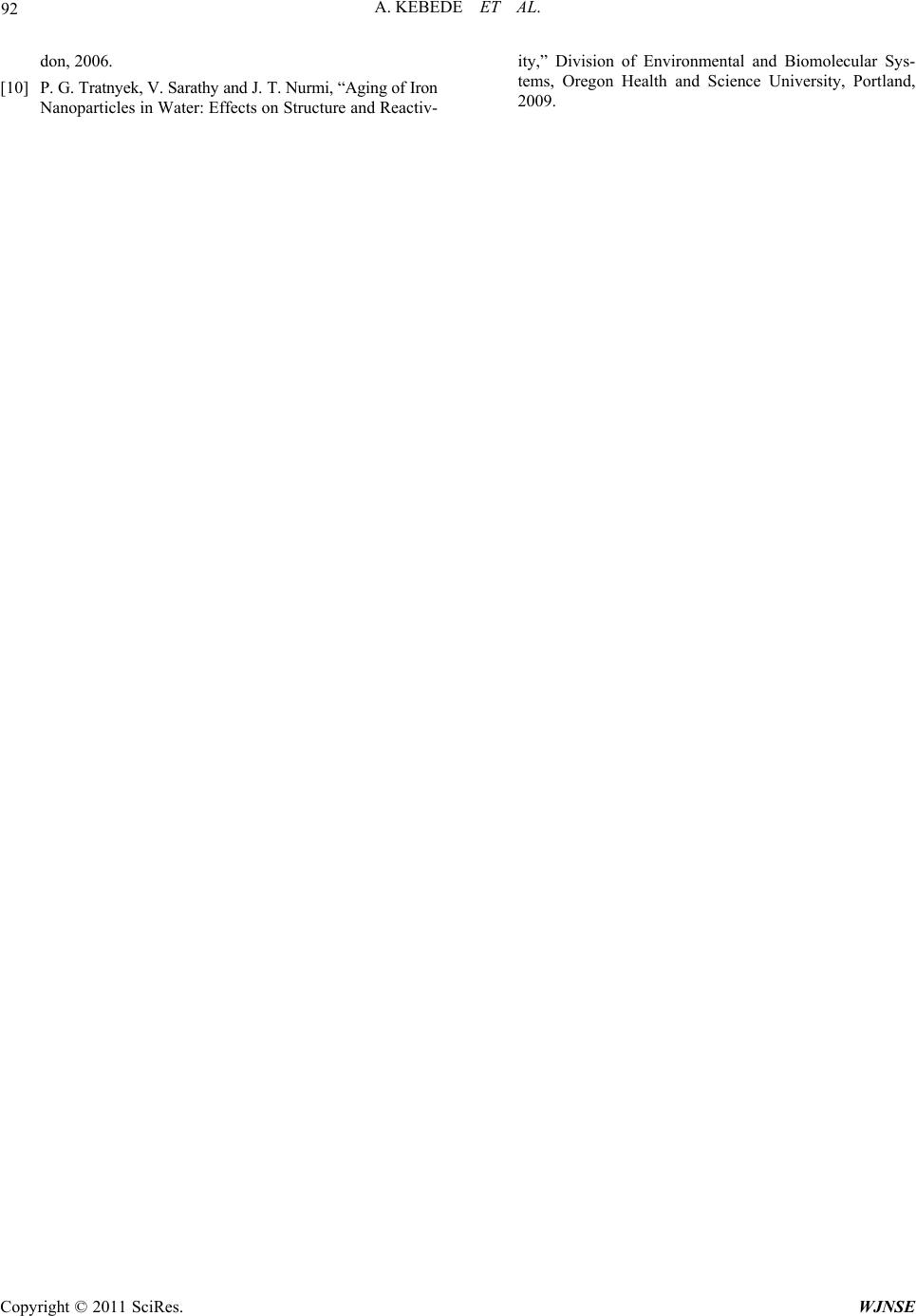
Figure 4. SEM image of colloidal iron oxide nanoparticles in water synthesized by laser ablation at (a) 25.8 mJ; (b) cementa-
tion of nanoparticles 75 mJ; (c) shape of nanoparticles (at the edge of a slide) at 75 mJ.
Table 1. Dependence wave length and absorbance on laser
energy.
Absorbance Wave length
(nm) Laser energy
(mJ)
0.425 205 9.3
1.717 210 25
1.729 215 75
[2] P. L. Apopa, Y. Qian, R. Shao, L. N. Guo, Diane Schwe-
gler-Berry, M. Pacurari, D. Porter, X. L. Shi, V. Vallya-
than, V. Castranova and C. D. Flynn, “Iron Oxide Nanopa-
rticles Induce Human Microvascular Endothelial Cell Per-
meability through Reactive Oxygen Speceous Production
and Microtubule Remodeling,” Particle and Fibre Toxico-
logy, Vol. 6, 2009, p. 1. doi:10.1186/1743-8977-6-1
[3] K.-C. Huang and K.-S. Chou, “Microstructure Changes to
Iron Nanoparticles during Discharge/Charge Cycles,” Elec-
trochemistry Communications, Vol. 9, 2007, pp. 1907-
1912. doi:10.1016/j.elecom.2007.05.001
4. Conclusions
Iron oxide nanoparticles of different sizes and surface
morphology have been successfully synthesized ul-
trapure water. The particle morphology showed depend-
ence of shape and size of the nanoparticles on the laser
energy which was further confirmed by the red shift in
the absorbance peak.
[4] H.-Y. Huang, Y.-T. Shieh, C.-M. Shieh and Y.-K. Twu,
“Magnetic Chitosan/Iron (II, III) Oxide Nanoparticles Pre-
pared by Spray-Dry ing,” Carbohydrate Polymers, Vol. 81,
No. 4, 2010, pp. 906-910.
doi:10.1016/j.carbpol.2010.04.003
[5] R. Singh, R. Verma, A. Kaushik, G. Sumana, S. Sood, R. K.
Gupta and B. D. Malhotra, “Chitosan-Iron Oxide Nano-
Composite Platform for Mismatch-Discriminating DNA
Hybridization for Neisseria gonorrhoeae Detection Caus-
ing Sexually Transmitted Disease,” Biosenceors and Bio-
electronics, Vol. 26, No. 6, 2011, pp. 2967-2974.
5. Acknowledgements
I would like to acknowledge National Center for Ex-
perimental Mineralogy and Petrology, Allahabad Uni-
versity, for service they rendered me with scaning elec-
tron microscopic (SEM) image of the nanoparticles. I am
thankful to Rohit Kumar, Abhimanyu K. Singh, Neeraj
Giri and Ashok K. Pathak for their assistance and help.
My special thanks goes to Prof. Ram Kripal for his as-
sistance and provision of UV/Vis absorption spectrome-
ter. Addis Ababa University, post graduate program, de-
serves acknowledgement for its financial assistance.
[6] R. Sarkar, P. Pal, M. Mahato, T. Kamilya, A. Chaudhuri
and G. B. Talapatra “On the Origin of Iron-Oxide Nano-
particle Formation Using Phospholipid Membrane Tem-
plate,” Colloids and Surface B: Biointerface, Vol. 79, No.
12, 2010, pp. 384-389.
doi:10.1016/j.colsurfb.2010.04.023
[7] S. Laurent, D. Forge, M. Port, A. Roch, C. Robic, L. Van-
der Elst and N. Muller, “Magnetic Iron Oxide Nanoparti-
cles: Synthesis, Stabilization, Vectorization Physicochemi-
cal Characterizations, and Biological Applications,” Che-
mical Reviews, Vol. 108, No. 6, 2008, pp. 2064-2008.
doi:10.1021/cr068445e
6. References [8] A. K. Singh, A. K. Rai and D. Bicanic, “Controlled Syn-
thesis and Optical Properties of Pure Gold Nanoparticles,”
Instrumentation Science and Technology, Vol. 37, No. 1,
2009, pp. 50-60.
[1] Scientific Committee on Emerging and Newly Identified
Health Risks (SCENIHR), “The Appropriateness of Ex-
isting Methodologies to Assess the Potential Risks Asso-
ciated with Engineered and Adventitious Products of
Nanotechnologies,” SCENIHR, 2006, p. 13. [9] J. Perreire, E. Millon and E. Fogarassay (Eds.), “Recent
Advances in Laser Processing of Materials,” Elsevier, Lon-
A. KEBEDE ET AL.
92
don, 2006.
[10] P. G. Tratnyek, V. Sarathy and J. T. Nur mi, “Agi ng of Iron
Nanoparticles in Water: Effects on Structure and Reactiv-
ity,” Division of Environmental and Biomolecular Sys-
tems, Oregon Health and Science University, Portland,
2009.
Copyright © 2011 SciRes. WJNSE