Vol.2, No.3, 205-211 (2010) Natural Science
http://dx.doi.org/10.4236/ns.2010.23032
Copyright © 2010 SciRes. OPEN ACCESS
The nature of neuronal words and language
Morris Henry Baslow
Nathan S. Kline Institute for Psychiatric Research, Center for Neurochemistry, New York, USA; Baslow@nki.rfmh.org
Received 5 December 2009; revised 13 January 2010; accepted 30 January 2010.
ABSTRACT
Individual neurons in the brain possess natural
language in the form of energy-dependent ac-
tion potentials or spikes (S) operating on a mil-
lisecond timescale that, along with pauses (P)
between spikes, constitute a two letter (S, P)
“alphabet” that is used to generate meaningful
frequency-encoded neuronal “words”. These
words are then used to transmit information to
other neurons in the form of phrases consisting
of two or more words that are contained within
longer pause-delineated structured declarative
sentences. In this article, the nature of neuronal
words and language are described, and exam-
ples provided that illustrate the way in which
neuronal language is used by the brain to in-
teract with and interpret both its internal and
external environments. It is hoped that a fuller
understanding of the language used by neurons
to communicate may lead to development of
novel treatments for a number of human neu-
ropathies.
Keywords: Biosemiotics; Brain; Cognition;
Language; Learning; Memory; Neurons
1. INTRODUCTION
Semiotics is broadly defined as a study of signs, mean-
ings and their significance in communication, and bio-
semiotics as a study that is based on the general assertion
that all living organisms including cells are semiotic
systems. While information processing in the brain is
highly complex, each neuron uses a simple code mecha-
nism for transmitting information. This is in the form of
electrophysiological action potentials or spikes (S) of
about a 1 millisecond (ms) duration that, along with
pauses (P) between spikes constitute a two letter “al-
phabet” that generates meaningful frequency-encoded
signals or neuronal S/P “words” [1]. The term “word”
for neuronal activity in spikes/s has previously been used
to describe the electrophysiological activity of the visual
interneuron of the blowfly where each action potential
(AP) was considered a “short” word and trains of AP’s
considered “longer” words [2]. In this article a neuronal
word is defined as a single AP together with the total
pause before the next AP; a phrase is defined as a group
of two or more words, and a sentence as a temporal
grouping of such words containing a subject, and a
predicate that expresses what is stated about the subject
[1]. All neuron codes, whether continuous trains of
spikes that are characteristic of information transmitted
at low frequencies, or bursts of spikes that are character-
istic of information transmitted at high frequencies, are
made up of interactions between spikes and pauses.
There are two theories regarding how these spikes carry
encoded information [3]. One is the “spike rate code”
which suggests that information is carried in the average
rate at which the neuron fires, and that the timing of each
spike is random. The other is the “spike timing code”
that suggests that specific information is carried not only
in the average spike rate at which a neuron fires, but also
in the precise timings between each spike. In this article,
it is proposed that neurons transmit meaningful informa-
tion not only in their spike rate and spike pause timing,
but also in the spike duration or length (SL) and its rela-
tionship to the pause length (PL) between spikes. It is
this relationship that constitutes a neuronal “S/P word”,
and adds an additional dimension to the potential rich-
ness of communication between neurons.
2. DISCUSSIONS
2.1 Anatomy of a Neuronal Word
The communication method used by the brain is gener-
ated at the cellular level where individual neurons trans-
mit information to one another by generation a series of
wave-like depolarizations. This depolarization’s start at
the plasma membrane of their cell bodies (soma) and
pass along the plasma membranes of axon hillocks, ax-
ons and pre-synaptic dendrites. Each wave-like spike
consists of two components, first, a rapid depolarization,
involving the efflux of K+ and influx of Na+ at the
plasma membrane down their respective intracellular/
extra cellular gradients, a process that requires little or
M. H. Baslow / Natural Science 2 (2010) 205-211
Copyright © 2010 SciRes. OPEN ACCESS
206
no additional energy. This is followed by a re-polariza-
tion process that occurs within an absolute refractory
period during which time the intracellular negative
membrane K+/Na+ potential is restored using energy
derived from intracellular stores of adenosine triphos-
phate (ATP) via a plasma membrane surface enzyme,
Na+/K+ ATPase, in order to transport these ions against
their respective gradients [4]. In single afferents of hu-
man mechanoreceptive sensory nerve fibers of the skin
of the hand, spike times-to-peak were 0.12-0.45 ms with
a mean of 0.21 ms [5]. While the depolarization process
is pre-energized and trigger-ready, re-polarization is a
slower enzyme-dependent process that is subject to rate
regulation by all factors that can affect enzyme activity.
These include enzyme, substrate, and end-product con-
centrations, focal ionic makeup, pH, temperature as well
as the presence of competitive and non-competitive in-
hibitors. All of these factors can affect the rate of
re-polarization and the length of the absolute refractory
period which in this study varied from 0.31-0.75 ms with
a mean of 0.50 ms indicating that the energy consuming
re-polarization period was 2.38 times longer than the
depolarization period such that the length of the
re-polarization period dominated the total spike period.
In this study, the average length of a spike including the
depolarization and longer re-polarization periods in the
sensory nerve fibers was 0.71 ms with a range from
0.43-1.20 ms. The relationship between the depolariza-
tion and re-polarization periods of a single spike is illus-
trated in Figure 1. Since the depolarization is pre-primed
and trigger ready, and the re-polarization period requires
energy to reestablish ionic gradients, re-polarization is
therefore subject to cellular control. Thus, the SL can be
varied to incorporate information that is transmitted in
the form of a SL/PL ratio over and above information that
transmitted by Hz and/or specific peak-to-peak in-
ter-spike intervals. Data from [5].
During the spiking process which includes the absolute
refractory period, and until the ionic gradients are rees-
tablished, a second spike cannot be generated. In addition,
following the absolute refractory period there is a relative
refractory period where only a strong stimulus can gener-
ate a second AP, and following this period there is a pe-
riod of longer pause with no electrophysiological activity.
A spike including its absolute refractory period may vary,
but an average spike-absolute refractive period length is
about 1 ms, and is followed by a relative refractory pe-
riod of about 2 ms in duration [6]. This in turn is fol-
lowed by a pause of varying lengths before the next spike
is generated. Thus, there are two phases of signal genera-
tion, each consisting of two subcomponents. The total
spike length (SL) is made up of the sum of the depolariza-
tion and absolute refractory (re-polarization) periods, and
the total pause length (PL) is made up of the relative re-
fractory period and the pause until the next spike is initi-
ated. The total pause length can vary from 0 to approxi-
mately 10,000 ms under normal physiological conditions.
Based on these neuronal spike and pause components, a
single neuronal S/P word has been defined as the sum of
its total spike length and its total pause length in ms, and
different words are characterized by differences in the
ratio of its SL to its PL [1].
The spike-generation process that results in S/P words
is also is metabolically costly, calculated at 2.2 × 10⁹
ATP molecules per spike [7], which requires that ATP
supplies be constantly replenished or the timing of the
spike-refractory periods will be altered, and meaningful
frequency-encoded information lost [1]. An illustration
of the components of a neuronal word as described is
graphically presented in Figure 2.
Figure 1. Cartoon showing the relative depolarization and repolarization
times of a neuronal spike.
M. H. Baslow / Natural Science 2 (2010) 205-211
Copyright © 2010 SciRes. OPEN ACCESS
207
Figure 2. Cartoon showing the components of the neuronal word, S1P7
In this illustration, each neuronal word (S1P7 ) consists
of a depolarizing spike (0.5 ms), an absolute refractory
period when the plasma membrane is being re-polarized
using ATP (0.5 ms) for a total spike length (SL) of 1.0 ms;
a relative refractory period where the neuron can be ac-
tivated but only by a strong stimulus (2.0 ms); and a
period of inactivity (5.0 ms). The spike frequency is
(1000 ms/8 ms) or 125 Hertz (Hz). Unless the pause is
terminated by a second spike within a physiologically
relevant period, the word is incomplete and without any
quantitative meaning. Thus, a meaningful phrase must
consist of at least two words, and a sentence of at least
one phrase. The word S1P7 presented in this cartoon
reflects the S/P message generated upon exposure of the
isolated salt receptor of the blowfly to a 3 molar NaCl
solution [8].
2.2. Conversion of Frequency in Hz to S/P
Words
As shown in Figure 2, information contained in Hz for-
mat differs from that in S/P word format in that in Hz,
the signal is a measure of the time between two spikes
that is initiated at the peak of the first spike and is con-
stituted by the re-polarization period of that spike, its
relative refractory period, the pause until initiation of a
second spike and the depolarization period of a second
spike. In essence, the signal recorded in Hz splits a spike
into its two components such that a spike in Hz is a
composite formed from elements of two AP’s. On the
other hand, an S/P word as defined includes the depo-
larization and re-polarization components of a single
spike, along with the relative refractory period and the
pause until the next spike is initiated. However, the two
formats are clearly related, and while Hz is a commonly
used method of recording and describing neuronal activ-
ity, a reasonable estimation of the length of a neuronal
word can also be derived from Hz. Thus, from Hz, the
total time in ms of an average S+P word can be deter-
mined: Total time per word: (SL + PL)ms = 1000/Hz.
At a frequency of 100 Hz, the mean S+P word length
would be 10 ms. However, the total value in ms for an S
+ P word does not provide sufficient information to con-
struct a specific neuronal S/P word. For this, one re-
quires in addition either the total spike length (SL) or
total pause length (PL). Thus, for a SL of 0.5 at 100 Hz,
the S/P word would be S0.5/P9.5, for an SL of 1.0, the S/P
word would be S1.0/P9.0, and for a SL of 1.5, the S/P word
would be S1.5/P8.5, representing three different S/P words
out of a great number of possible S/P words that are dif-
ferentiated by the relative lengths of their spikes and
pauses. It is important to note that all of these S/P words
are different and may transmit different meanings, but
that all are being communicated at the same frequency
of 100 Hz. Clearly, a measure of frequency alone for
neuronal communications cannot be physiologically
accurate. When afferent neuronal messages are con-
verted from electrophysiological to chemical and back
into electrophysiological messages at synaptic interfaces
for transmission to efferent neurons, it is highly likely
that differences in the SL/PL ratios are important ele-
ments in modulation of the messages at these interfaces.
Since much of the published neuronal response data is
presented in the form of Hz, and while less accurate than
actual S/P words, in the absence of a measured SL, and
by using an average SL of 1.0, one can still derive useful
information based on Hz about how neurons communi-
cate with one another concerning the nature of their en-
vironment.
2.3. Differences in Spike Rate, Spike Timing
and Neuronal Word Codes in
Information Transfer
The spike rate code hypothesis proposes that information
is processed in the form of the average time between
M. H. Baslow / Natural Science 2 (2010) 205-211
Copyright © 2010 SciRes. OPEN ACCESS
208
spikes, and the spike timing code hypothesis proposes
that information can also be contained in the differences
in pauses between individual spikes, an analysis that can
be used to identify harmonics and periodic oscillations in
spike trains. Both of these hypotheses treat a spike as a
point source and both are valid interpretations of re-
corded data. However, these hypotheses are mirror im-
ages of one another in that when viewed at the level of
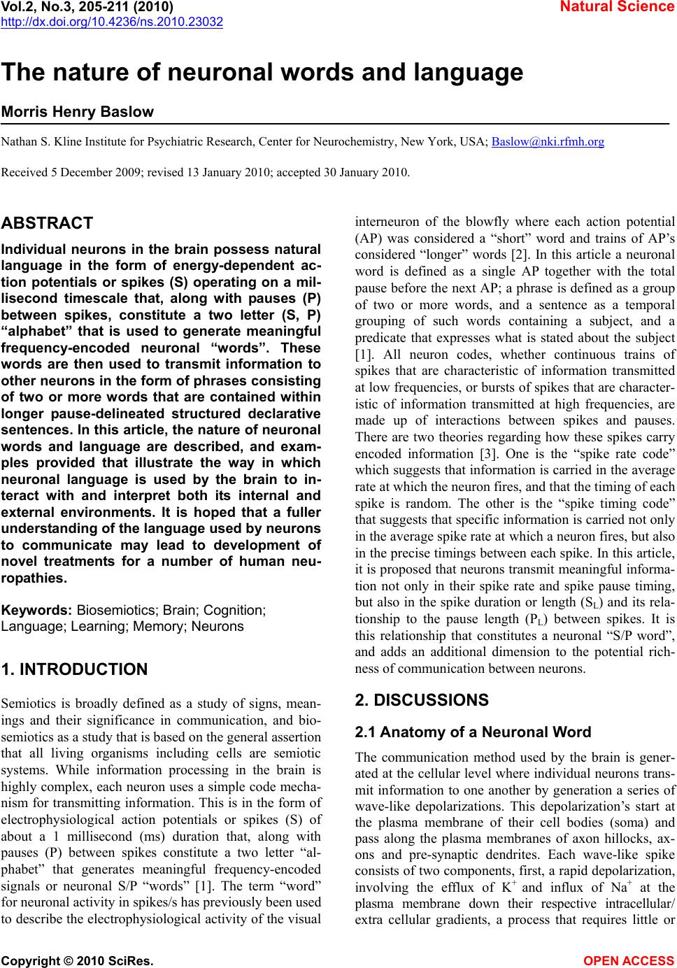
only two spikes, as presented in Figure 1, it is clear that
the time between individual spikes treated as point
sources, and the pause time between spikes will always
be the same. On the other hand, the neuronal word code
hypothesis as detailed above takes into consideration
that individual spikes have a significant time dimension
and shows that even at the level of only two spikes, there
can be an enormous number of specific S/P words that
can be generated and used to transmit different kinds of
information. This is because each neuronal word is
formed by individual components of the AP process as-
sociated with spike generation, and differences in each
component can alter the ratio between the total spike
length and the total pause length. For the three examples
provided above, S0.5/P 9.5, S1.0/P9.0 and S1.5/P8.5, their S/P
ratios are 0.052, 0.111 and 0.176 respectively, and all are
capable of being transmitted at the same frequency (Hz)
where both the inter-spike time and pause time are the
same. While all three hypotheses reflect frequency-based
neuronal codes, the neuronal word code with its spike
time dimension is the most physiologically realistic
since it is the only one that identifies a mechanism
whereby the AP can interact with the rate of depolariza-
tion-induced release of neurotransmitters from pre-
synaptic vesicles. Thus, only the neuronal word code
appears to have the potential for an enhanced richness in
information transfer, especially at the low frequencies
and short periods dictated by ongoing bioenergetic and
temporal physiological constraints. Moreover, measure-
ment of the ratios that comprise S/P words may provide
a new investigational tool, not available using the other
“codes”, with which to assess differences in neuronal
function between normal and pathological conditions.
2.4. Nature of Neuronal Language
There are three components to neuronal language. First,
neurons are “wired” in the sense that each neuron in the
corpus occupies a specific place on a brain “map” of a
given organism. Second, by virtue of its phenotype, each
neuron sends specific kinds of messages for interpreta-
tion within the CNS network. For environmental sensing
neurons, these may identify the qualitative nature of the
messages being communicated such as where in the
brain or corpus it originates, and whether the message is
a general call for stored information, or whether it is
from a specific region associated with some specific
factor such as sound, light or availability of “glucose”
(Glc). Lastly, the messages sent by neurons must also
concern the quantitative nature of sensed information
such as the importance of message itself, or the specific
levels of light, sound, pain, pressure, stretch, or sub-
stance concentrations sensed. It is this last component,
the quantitative nature of information within the brain,
where specific frequency-encoded neuronal words ap-
pear to play an important role.
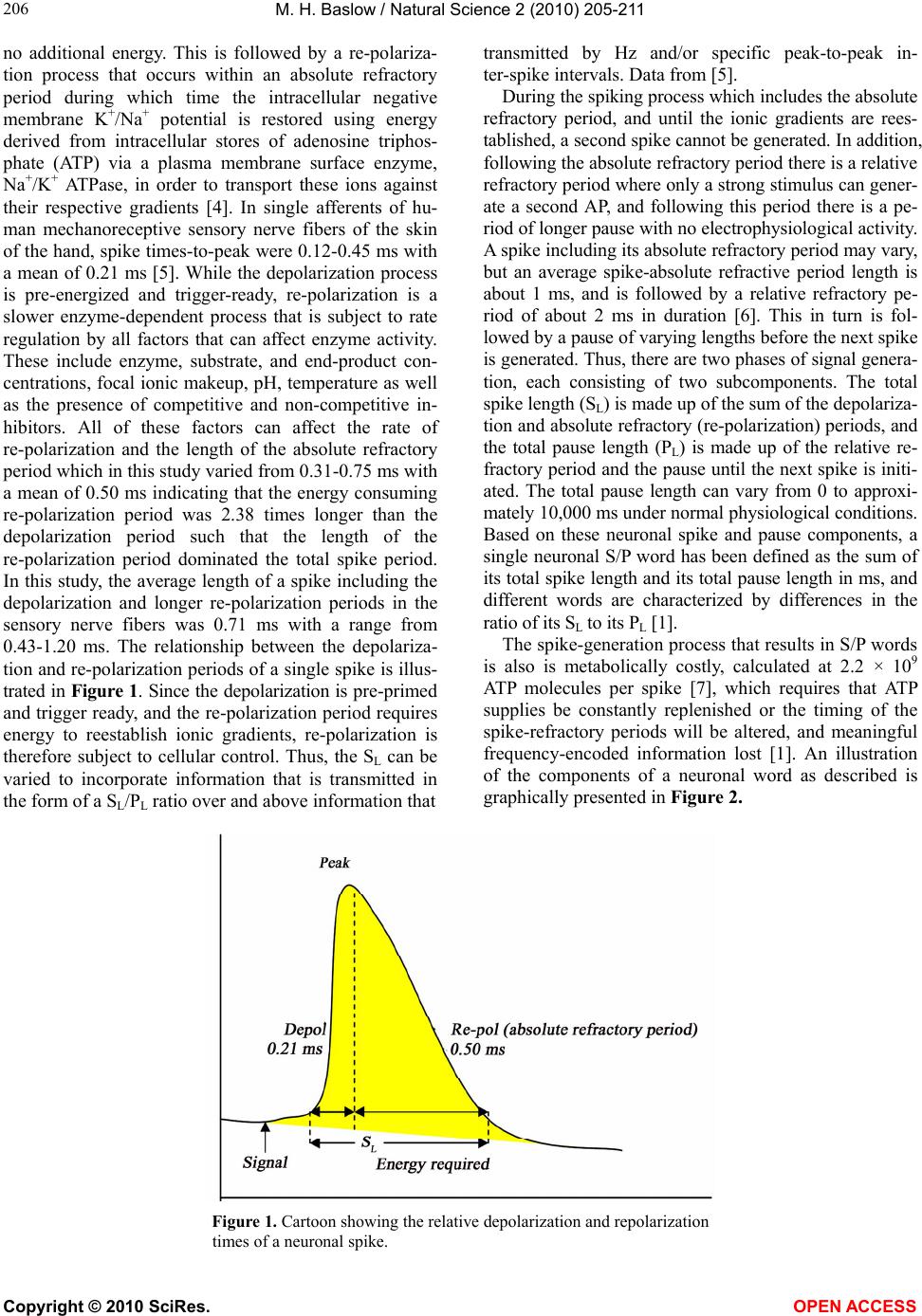
As an example, based on a recorded neuronal fre-
quency of 100 Hz, a neuronal spike with a total spike
length of 1 ms and a total pause length of 9 ms would
constitute the neuronal S/P word ( S1P9) of 10 ms dura-
tion. When this word is repeated, it becomes a phrase. In
the blowfly, this phrase corresponds to immersion of its
isolated salt receptor in 2.0 M NaCl [8]. Based on using
a value for total SL of 1 ms, the specific words in “fly-
speak” for immersion in additional NaCl concentrations
of 0.5 and 1.0 M are S1P18 (52 Hz) and S1P12 (80 Hz)
respectively. In addition, if the SL is known along with
the total S + P time in ms derived from Hz, the total
“pause” time in ms can be derived using the following
general relationship: P = [1000 ms (Hz × SL)]/Hz.
It has also been proposed, that the language of neu-
rons can be written in any of a number of fre-
quency-encoded formats including Hz, S/P and musical
notations and can be translated into any of the oral, writ-
ten or symbolic human languages as well [1]. Impor-
tantly, using artificial electronically generated frequen-
cies that mimic the frequencies of recorded natural neu-
ronal words, it has been demonstrated that it is possible
not only to record these neuronal words, but also to
communicate directly with the central nervous system
(CNS) of rats. Where this has been done, the manufac-
tured words have been shown to elicit the same behav-
ioral responses in rats that would have been generated by
exposure to a specific environmental stimulus [9].
2.5. The Dictionary of Neuronal Words and
their Representational Nature
The dictionary of neuronal S/P words in an ms time-
frame is relatively short, being limited at its upper level
by the absolute and relative neuronal refractory periods
and at its lower level by physiological requirements such
as maintenance of the human heart rate at 68 beats/min
(1.13 Hz). With the average spike including an absolute
refractory period of 1 ms duration followed by a relative
refractory period of about 2 ms, the highest possible
frequency would be S1P2 or 333 Hz. If the system were
driven by a strong enough input eliminating the relative
refractory period altogether, the highest possible fre-
quency might be S1P0 or 1,000 Hz.
Since the brain’s primary form of communication,
both internally and with the outside world, is in the form
of these electrophysiological messages, it follows that
neuronal words must also be highly representational in
nature. For example, audible sound in many animals
may range up to 20,000 Hz and visible light is in the
M. H. Baslow / Natural Science 2 (2010) 205-211
Copyright © 2010 SciRes. OPEN ACCESS
209
Figure 3. A “dark room” paradigm of brain function.
Table 1. List of several neuronal S/P words and their meanings.
Organism Sensor Word Meaning Reference
Blowfly
sensillum salt S
1P499 NaCl 0.10 M [8]
S
1P45 0.20
S
1P27 0.25
S
1P18 0.50
S
1P12 1.00
S
1P9 2.00
S
1P7 3.00
Ground beetle
Sensillum salt S
1P1000 NaCl 0.001 M [11]
S
1P71 0.01
S
1P29 0.10
S
1P23 1.00
Hamster
taste cells acid S
1P286 pH 2.5 [12]
S
1P312 3.0
S
1P1999 4.3
S
1P4000 5.0
Cat brain
hypothalamus glucose
glucosensing steady-state S
1P10000 normal Glc level [13]
Glc i.v. 30 min S
1P2000 elevated Glc level
Rat
taste cells quinine S
1P21 quinine 0.01 mM [9]
Rat brain
hippocampal sound S1P499 frequency 2 kHz [10]
neuron S
1P142 10 kHz
Guinea pig brain
GABAergic H
2O2 S
1P53 H
2O2 0.0 mM [14]
neurons S
1P40 1.5
range of 1014 Hz. For the brain to comprehend its exter-
nal environment, these frequencies must first be con-
verted into representations of those wavelengths at less
than 1000 Hz, with the same meanings as the original
signals, which can then be transmitted by neurons within
the CNS network. These representations can also be
“memorized” in the form of structural and biochemical
changes in the dendrite-synapse-dendrite (DSD) fields
M. H. Baslow / Natural Science 2 (2010) 205-211
Copyright © 2010 SciRes. OPEN ACCESS
210
between neurons, and later recalled to “imagine” the
nature of the original wavelengths even without external
input. For example, one can imagine “in the minds eye”
the color “red” with the eyes closed, and with eyes open
can also compare a newly observed representation of red
with that which was previously seen and stored in mem-
ory. This phenomenon is illustrated in Figure 3 where a
specific sound tone of 2 kHz impinging on the ear is
converted into a representational neuronal word at 2 Hz
for CNS network processing.
In this illustration, the translation by a sense organ of
the specific frequency-encoded environmental informa-
tion for the approximate musical tone B6 (1975.53 Hz) is
illustrated. The sound sense organ first converts the tone
into a brain frequency-encoded S/P word representation
for B6 at 2 Hz (S1P499) that is then compared within the
CNS network to auditory area DSD memory constructs.
During this comparison related historical documents in
other brain areas may also be probed for relevant infor-
mation. Upon an executive command, appropriate mus-
cles can also be activated to reproduce the 1975.53 Hz
frequency, exporting it to another organism and also
sending it back to the sense organ where it can be com-
pared to the original tone. Importantly, the S/P word for
the tone B6 or any other musical tone can also be synthe-
sized de novo by the brain from memory engrams with-
out external input and can be “heard” within the brain as
recalled sounds or melodies. All of these functions occur
in a ms/sub-ms timeframe and apply to any other sensed
parameter, whether derived internally or external to the
brain. The specific auditory data presented in Figure 3 is
adapted from rat experiments [10].
2.6. Examples of Neuronal Words and their
Meanings
In Table 1 descriptions of some S/P words and their
meanings are presented. In each of these cases, the S/P
words have been derived from frequency data by using an
average SL including an absolute refractory period of 1.0
ms, and rounded to the nearest integer. However, it is also
apparent that neurons can operate in a sub-millisecond
timeframe [10], which is in keeping with the microsecond
timeframe dictated by physiological demands and com-
munication networking requirements of the brain.
3. CONCLUSIONS
In this paper it is proposed that neurons possess natural
language in the form of specific neuronal words, phrases
and sentences, and evidence is provided that when spe-
cific neuronal words are known, it is possible to mimic
them and generate electronic words to communicate
intelligently with the CNS. While most investigative
recordings of neuronal activity are in the form of Hz, the
differences between Hz and word formats have been
described, and a case has been made that the word for-
mat is the only format that can be physiologically asso-
ciated with the release of specific quanta of neurotrans-
mitters at synapses and is therefore more representative
of the potential richness of information that can be
transmitted by neurons. In addition, based on physio-
logical limitations associated with the bioenergetic na-
ture of neuronal AP’s, it has been shown that much of
the information about the external environment that is
transmitted to the brain must first be reduced to a fre-
quency-encoded representation of that information that
can be communicated at < 1000 Hz. These representa-
tions can be acted upon immediately, memorized when
appropriate, and the memorized information recalled as
needed in the form of such representations within the
brain. Of note, in humans and other animals these inter-
nal representations can also be converted back into their
original electromagnetic spectrum frequencies by exter-
nally re-creating the original environmental source such
as by reproducing a particular memorized sound fre-
quency. Finally, it is hoped that this paper will encourage
the continued analysis of the nature of neuronal words
and language, and that such studies will lead to addi-
tional development of novel electronic methods for
communicating directly and intelligently with the CNS
as has already been attempted in treatment of several
human neuropathies [15]. With such specific fre-
quency-encoded words as tools, it may be possible to
alter or induce new behaviors, and thus lead to success-
ful treatment stratagems for a variety of human psycho-
logical and neurological conditions.
REFERENCES
[1] Baslow, M.H. (2009) The languages of neurons; An
analysis of coding mechanisms by which neurons com-
municate, learn and store information. Entropy, 11,
782-797.
[2] Borst, A. (2003) Noise, not stimulus entropy, determines
neuronal information rate. Journal of Computational
Neuroscence, 14, 23-31.
[3] Clifford, C.W. and Ibbotson, M.R. (2009) Response vari-
ability and information transfer in directional neurons of
the mammalian horizontal optokinetic system. Visual
Neuroscience, 17, 207-215.
[4] Baslow, M.H. and Guilfoyle, D.N. (2007) Using proton
magnetic resonance imaging and spectroscopy to under-
stand brain activation. Brain and Language, 102,
153-164.
[5] Brink, E.E. and Mackel, R.G. (1993) Time course of
action potentials recorded from single human afferents.
Brain, 116, 415-432.
[6] Berry II, M.J. and Meister M. (1998) Refractoriness and
neural precision. Journal of Neuroscience, 18, 2200-
2211.
[7] Lennie, P. (2003) The cost of cortical computation. Cur-
rent Biology, 13, 493-497.
M. H. Baslow / Natural Science 2 (2010) 205-211
Copyright © 2010 SciRes. OPEN ACCESS
211
[8] Gillary, H.L. (1966) Stimulation of the salt receptor of
the blowfly. II. Temperature. Journal of General Physi-
ology, 50 351-357.
[9] Di Lorenzo, P.M., Leshchinskiy, S., Moroney, D.N. and
Ozdoba, J.M. (2009) Making time count: Functional evi-
dence for temporal coding of taste sensation. Behavioral
Neuroscience, 123, 14-25.
[10] Takahashi, S. and Sakurai, Y. (2009) Sub-millisecond
firing synchrony of closely neighboring pyramidal neu-
rons in hippocampal CA1 of rats during delayed non-
matching to sample task. Frontiers in Neural Circuits,
3(9)
[11] Merivee, E., Renou, M., Mand, M., Luik, A., Heidemaa,
M. and Ploomi, A. (2004) Electrophysiological responses
to salts from antennal chaetoid taste sensilla of the
ground beetle Pterostichus aethiops. Journal of Insect
Physiology, 50, 1001-1013.
[12] Gilbertson, T.A., Avenet, P., Kinnamon, S.C. and Roper,
S.D. (1992) Proton currents through amiloride-sensitive
Na channels in hamster taste cells. Journal of General
Physiology, 100, 803-824.
[13] Anand, B.K., Chhina, G.S., Sharma, K.N., Dua, S. and
Singh, B. (1964) Activity of single neurons in the hypo-
thamic feeding centers: effect of glucose. American
Journal of Physiology, 207, 1146-1154.
[14] Lee, C.R. and Rice, M.E. (2008) Hydrogen peroxide
increases the excitability of substantia nigra pars reticu-
lata GABAergic neurons. Society for Neuroscience
Meeting, November 16, Program 179.2, Poster # QQ37.
[15] George, M.S. and Aston-Jones, G. Noninvasive (2010)
techniques for probing neurocircuitry and treating illness:
Vagus nerve stimulation (VNS), transcranial magnetic
stimulation (TMS) and transcranial direct current stimulation
(tDCS). Neuropsychopharmacology, 35, 301-316.