J. Biomedical Science and Engineering, 2010, 3, 300-303
doi:10.4236/jbise.2010.33040 Published Online March 2010 (http://www.SciRP.org/journal/jbise/
JBiSE
).
Published Online March 2010 in SciRes. http://www.scirp.org/journal/jbise
Effect of static magnetic field on erythrocytes characterizations
Mohamed A. Elblbesy
Medical Biophysics Department, Medical Research Institute, Alexandria University, Alexandria, Egypt.
Email: mimizizo@yahoo.com
Received 29 November 2009; revised 15 December 2009; accepted 28 December 2009.
ABSTRACT
The interaction of static magnetic field (SMF) with
living organisms is a rapidly growing field of investi-
gation. Recently, exposure to moderate intensity
SMFs (1 mT – 1 T) has attracted much attention for
its various medical applications. This study was de-
signed to show the microscopic effect of SMF on
erythrocytes in vitro. For this purpose SMF system
was constructed in my lab on basis of the idea of cell
tracking velocimetry system. The changes in eryth-
rocytes surface area, sphericity, and adhesion num-
ber for erythrocytes were calculated to quantify the
effect of SMF on erythrocytes characterizations. The
results showed that SMF increased erythrocytes sur-
face area and reduced their sphericity. The adhesion
number of erythrocytes under the influence of SMF
showed the tendency of erythrocytes to adhere with
each other. These findings indicate that more study
on microscopic scales must be carried out in order to
investigate the effect of SMF on erythrocytes.
Keywords: Static Magnetic Field; Erythrocytes;
Surface Area; Shericity, Adhesion
1. INTRODUCTION
During the last few years there has been an increasing
interest in the influence of magnetic field on biological
cell [1]. Many attempts have been made to measure the
magnetic effects. Historically, there are several signifi-
cant studies in the field of erythrocyte rheology [2].
Reports from in vitro research indicate that low level
high frequency fields may alter membrane structural and
functional properties that trigger cellular responses. It
was hypothesized that the cell membrane may be sus-
ceptible to low level high frequency fields, especially
when these fields are amplitude modulated at extremely
low frequencies [3]. There are abundant data on the ef-
fects of weak physical signals on cell functional activity.
However cell targets for these signals are not established
yet. Since water is the main component of biological
systems and all metabolic processes take place in aque-
ous medium it was supposed that even minor changes in
the physico-chemical properties of water could signifi-
cant modify cell functional activity. The data about the
effect of EMF on water specific electrical conductivity
and wheat sprouting serve as an additional evidence for
the hypothesis according to which the EMF-induced
water structure changing is an important pathway
through which the biological effect of EMF is realized
[4].
Most of the theories addressing the mechanism of in-
teraction between biological systems and MFs suggest
that the plasma membrane, by virtue of its bioelectrical
properties, is the site, where MFs exert their primary
effects [5]. To investigate the effect of static magnetic on
biological system, it is necessary to clarify interaction
mechanisms of magnetic field with biological systems.
Simple components of biological system, as cells, bio-
molecules, artificial membrane, are used in vitro studies
that allow reducing of biological variables and more
precisely defining and controlling the exposure parame-
ters, compared with in vivo exposure. Erythrocytes are
often used as model in exploring the structure and func-
tion of the biological cells as well as studying the effect
of different physical and chemical factors on the cell.
The present study was established to elucidate the
changes in biological cells interaction and morphological
characterization through in vitro exposure of human
erythrocytes to SMF.
2. MATERIALS AND METHODS
2.1. Samples Collection and Preparation
Twenty blood samples of 5 ml were collected from
healthy volunteers of the same age and gender. The
blood samples were centrifuged and erythrocytes were
separated. Erythrocytes were washed three times in
0.155 M NaCl (2,000 rpm for 10 min). After the final
centrifugation suspension were diluted to final erythro-
cytes concentration 2% in phosphate buffer slain (PBS).
2.2. SMF System
The SMF system was constructed in my lab. SMF sys-
tem is based on the idea of cell tracking velocimetry
system. In this system the movement of cells in a well
M. A. Elblbesy / J. Biomedical Science and Engineering 3 (2010) 300-303
Copyright © 2010 SciRes
301
JBiSE
defined magnetic field is videotaped [6]. Erythrocytes
suspension were placed in a glass sample holder and
viewed using an inverted optical microscope in conjunc-
tion with a computer linked via digital camera and image
capture software. Two rod of iron to produce a magnetic
field with large gradient, were placed on both sides of
sample holder. The iron rods were alternately magnet-
ized by solenoids controlled by a DC power supply.
When magnetized, the magnetic field produced by each
rod exerted a force on erythrocytes directed towards the
edge of the rod.
2.3. Erythrocytes Surface Area and Sphericity
Erythrocytes were suspended in phosphate buffer saline
(PBS) at concentration of 5%. The erythrocytes suspen-
sion was placed on the sample holder of SMF system.
Image of unexposed erythrocytes (control) was taken
then the magnetic field was turned on and the image of
exposed erythrocytes was captured. For each value of B
images of exposed erythrocytes were compared with
control by imaging processing software in order to cal-
culate the changes in erythrocytes surface area Ach and
sphericity of each erythrocyte in the image.
The change in erythrocytes surface area was calcu-
lated as the following:
Ex
ch
0
A
AA
(1)
where Ach is the change in erythrocytes surface area, AEx
is the surface area of the erythrocytes after exposed to
SMF, and A0 is the surface area of unexposed erythro-
cytes.
Ach was calculated as the average of 100 erythrocytes
for each image. It was taken that if Ach greater than one
there was an increase in erythrocytes surface area, and if
Ach smaller than 1 there was a decrease in erythrocytes
surface area.
The sphericity of an erythrocyte is a dimensionless
parameter first described by Canham and Burton [7] and
is a measure of how spherical a cell is, with values be-
tween 0 and 1, for a sphere and laminar disk, respec-
tively. It was calculated as the following:
23
Ex
V
Sphericity4.84 A
(2)
where V is the volume of erythrocytes and is taken to be
88.5 µm3.
2.4. Erythrocytes Adhesion Number
Five volume fractions (0.00001, 0.00003, 0.00005,
0.00007, 0.00009) of erythrocytes suspensions in PBS
were prepared by further dilution of the main erythro-
cytes suspension. The erythrocytes suspension was
placed in sample holder of SMF system. For each sam-
ple counting of doublets erythrocytes (N2) and singlet
erythrocytes (N1) was performed. The plot of doublet to
singlet ratio versus particle volume fraction should yield
a straight line passing through the origin. The gradient of
the line is a measure of the adhesion number. The same
previous procedure was performed with SMF turned on
to calculate the adhesion number of exposed erythro-
cytes.
2.5. Statistical Analysis
The data was present as the mean ± SD. For each plot,
correlation (R2) was calculated using Microsoft Excel.
3. RESULTS
The erythrocytes surface area increased as the value of
magnetic field intensity increased (Figure 1). A strong
correlation was found between Ach and B (mT) (R2 =
0.95). At low magnetic field the increased in erythro-
cytes surface area was slightly small. At higher values of
magnetic field the increased in erythrocytes surface area
raised.
The sphericity of the erythrocytes is inversely propor-
tional to the applied SMF (Figure 2). Under the influ-
ence of SMF, the sphercity of erythrocytes was reduced
by about 35 % in comparison with unexposed erythro-
cytes. The decrease in erythrocytes sphericity was re-
duced rapidly when SMF was applied. A small decrease
in sphericity was recorded at higher values of B.
As indicated in Figure 3, The adhesion number in-
creased under the influence of SMF. Acute increase in
adhesion number was indicated at 15 mT. A strong rela-
tion between adhesion number and B was pointed out (R2 =
0.96).
4. DISCUSSION
It is generally known that morphological and structural
changes to the plasma membrane interfere with many
functional and structural features of the cells, leading,
Figure 1. Erythrocytes surface area increased as the values of
SMF intensity increased. Strong correlation between Ach and B
was observed (R2 = 0.95).
302 M. A. Elblbesy / J. Biomedical Science and Engineering 3 (2010) 300-303
Copyright © 2010 SciRes JBiSE
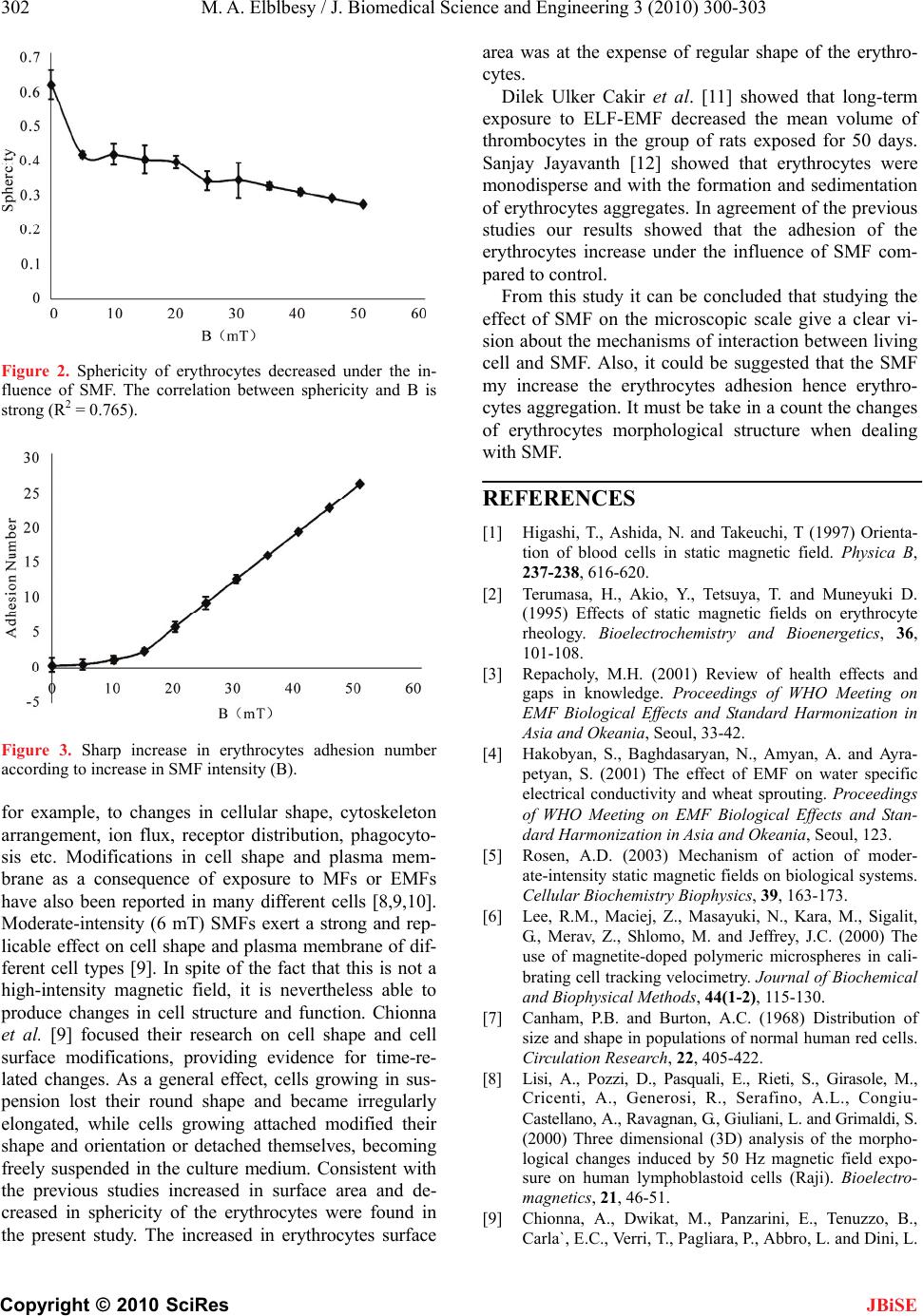
Figure 2. Sphericity of erythrocytes decreased under the in-
fluence of SMF. The correlation between sphericity and B is
strong (R2 = 0.765).
Figure 3. Sharp increase in erythrocytes adhesion number
according to increase in SMF intensity (B).
for example, to changes in cellular shape, cytoskeleton
arrangement, ion flux, receptor distribution, phagocyto-
sis etc. Modifications in cell shape and plasma mem-
brane as a consequence of exposure to MFs or EMFs
have also been reported in many different cells [8,9,10].
Moderate-intensity (6 mT) SMFs exert a strong and rep-
licable effect on cell shape and plasma membrane of dif-
ferent cell types [9]. In spite of the fact that this is not a
high-intensity magnetic field, it is nevertheless able to
produce changes in cell structure and function. Chionna
et al. [9] focused their research on cell shape and cell
surface modifications, providing evidence for time-re-
lated changes. As a general effect, cells growing in sus-
pension lost their round shape and became irregularly
elongated, while cells growing attached modified their
shape and orientation or detached themselves, becoming
freely suspended in the culture medium. Consistent with
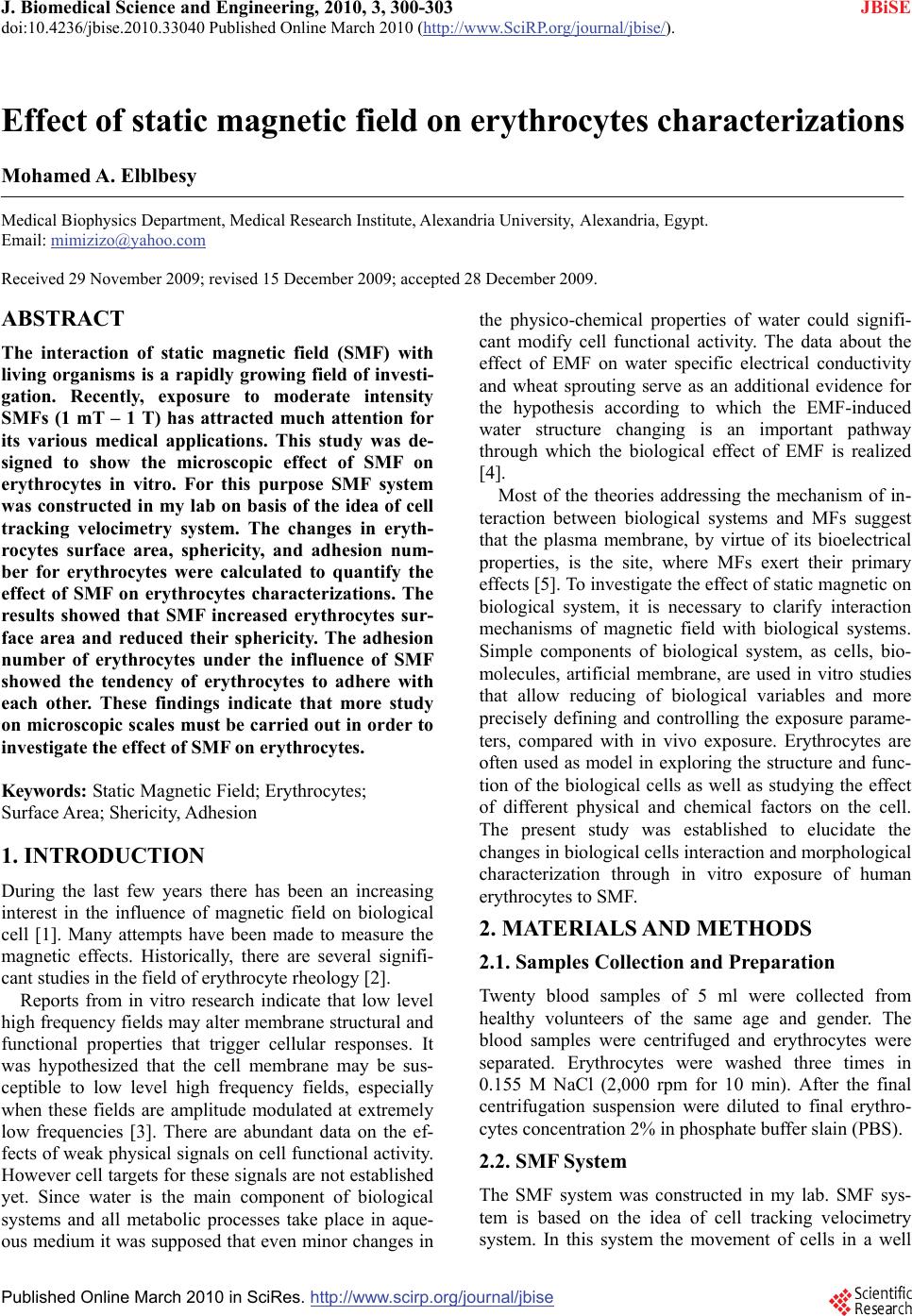
the previous studies increased in surface area and de-
creased in sphericity of the erythrocytes were found in
the present study. The increased in erythrocytes surface
area was at the expense of regular shape of the erythro-
cytes.
Dilek Ulker Cakir et al. [11] showed that long-term
exposure to ELF-EMF decreased the mean volume of
thrombocytes in the group of rats exposed for 50 days.
Sanjay Jayavanth [12] showed that erythrocytes were
monodisperse and with the formation and sedimentation
of erythrocytes aggregates. In agreement of the previous
studies our results showed that the adhesion of the
erythrocytes increase under the influence of SMF com-
pared to control.
From this study it can be concluded that studying the
effect of SMF on the microscopic scale give a clear vi-
sion about the mechanisms of interaction between living
cell and SMF. Also, it could be suggested that the SMF
my increase the erythrocytes adhesion hence erythro-
cytes aggregation. It must be take in a count the changes
of erythrocytes morphological structure when dealing
with SMF.
REFERENCES
[1] Higashi, T., Ashida, N. and Takeuchi, T (1997) Orienta-
tion of blood cells in static magnetic field. Physica B,
237-238, 616-620.
[2] Terumasa, H., Akio, Y., Tetsuya, T. and Muneyuki D.
(1995) Effects of static magnetic fields on erythrocyte
rheology. Bioelectrochemistry and Bioenergetics, 36,
101-108.
[3] Repacholy, M.H. (2001) Review of health effects and
gaps in knowledge. Proceedings of WHO Meeting on
EMF Biological Effects and Standard Harmonization in
Asia and Okeania, Seoul, 33-42.
[4] Hakobyan, S., Baghdasaryan, N., Amyan, A. and Ayra-
petyan, S. (2001) The effect of EMF on water specific
electrical conductivity and wheat sprouting. Proceedings
of WHO Meeting on EMF Biological Effects and Stan-
dard Harmonization in Asia and Okeania, Seoul, 123.
[5] Rosen, A.D. (2003) Mechanism of action of moder-
ate-intensity static magnetic fields on biological systems.
Cellular Biochemistry Biophysics, 39, 163-173.
[6] Lee, R.M., Maciej, Z., Masayuki, N., Kara, M., Sigalit,
G., Merav, Z., Shlomo, M. and Jeffrey, J.C. (2000) The
use of magnetite-doped polymeric microspheres in cali-
brating cell tracking velocimetry. Journal of Biochemical
and Biophysical Methods, 44(1-2), 115-130.
[7] Canham, P.B. and Burton, A.C. (1968) Distribution of
size and shape in populations of normal human red cells.
Circulation Research, 22, 405-422.
[8] Lisi, A., Pozzi, D., Pasquali, E., Rieti, S., Girasole, M.,
Cricenti, A., Generosi, R., Serafino, A.L., Congiu-
Castellano, A., Ravagnan, G., Giuliani, L. and Grimaldi, S.
(2000) Three dimensional (3D) analysis of the morpho-
logical changes induced by 50 Hz magnetic field expo-
sure on human lymphoblastoid cells (Raji). Bioelect ro-
magnetics, 21, 46-51.
[9] Chionna, A., Dwikat, M., Panzarini, E., Tenuzzo, B.,
Carla`, E.C., Verri, T., Pagliara, P., Abbro, L. and Dini, L.
M. A. Elblbesy / J. Biomedical Science and Engineering 3 (2010) 300-303
Copyright © 2010 SciRes
303
JBiSE
(2003) Cell shape and plasma membrane alterations after
static magnetic fields exposure. European Journal of
Histochemistry, 47, 299-308.
[10] Rieti, S., Manni, V., Lisi, A., Giuliani, L., Sacco, D.,
D’Emilia, E., Cricenti, A., Generosi, R., Luce, M. and
Grimaldi, S. (2004) SNOM and AFM microscopy tech-
niques to study the effect of non-ionizing radiation on the
morphological and biochemical properties of human
keratinocytes cell line (HaCaT). Journal of Microscopy,
213, 20-28.
[11] Dilek, U.C., Beran, Y., Mehmet, Z.A., Cemil, S. and
Nuriye, M. (2009) Alterations of hematological varia-
tions in rats exposed to extremely low frequency mag-
netic fields (50 Hz). Archives of Medical Research, 40,
352-356.
[12] Sanjay, J. and Megha, S. (2002) Influence of an inhomo-
geneous magnetic field on erythrocyte aggregation
mechanism an analysis by He–Ne laser aggregometer.
Journal of Magnetism and Magnetic Materials, 252,
412-414.