Pharmacology & Pharmacy
Vol.4 No.2(2013), Article ID:29889,4 pages DOI:10.4236/pp.2013.42033
Diurnal Variation of Nuclear Receptors in Mice with or without Fasting
![]()
1Department of Pharmacy, School of Pharmacy, Kinki University, Osaka, Japan; 2Department of Physiology, Hyogo College of Medicine, Nishinomiya, Japan.
Email: *iwaki@phar.kindai.ac.jp
Copyright © 2013 Atsushi Kawase et al. This is an open access article distributed under the Creative Commons Attribution License, which permits unrestricted use, distribution, and reproduction in any medium, provided the original work is properly cited.
Received January 19th, 2013; revised March 1st, 2013; accepted April 7th, 2013
Keywords: CAR; Circadian Rhythm; Diurnal Variation; Fasting; Nuclear Receptor; PXR
ABSTRACT
Nuclear receptors such as pregnane X receptor (PXR) and constitutive androstane receptor (CAR) regulate the transcription of transporter and cytochrome P450 (CYP). The diurnal variation was observed in some transporters regulated by nuclear receptors. We investigated whether diurnal variation in PXR and CAR exists in mice. We also examined the effect of food intake on the diurnal rhythm of hepatic PXR and CAR using fed and fasted mice. In liver and small intestine of fed mice, the mRNA levels of PXR and CAR were unchanged between 7:00 AM and 7:00 PM. In contrast to fed mice, fasting mice partly exhibited the diurnal variation in PXR, not in CAR. The mRNA levels of PXR at 7:00 AM were significantly higher than that those at 7:00 PM in liver of fasting mice. These results indicated the different effects of fasting in mice on diurnal variation of PXR in each tissue.
1. Introduction
The nuclear receptors pregnane X receptor (PXR, NR1I2: GenBank NM_010936) and constitutive androstane receptor (CAR, NR1I3: GenBank NM_009803) are involved in the primary response to xenobiotics and endogenous toxins. These receptors respond to ligands by activating the expressions of genes encoding enzymes involved in phase I (functionalization reactions) and phase II (conjugation reactions) metabolism and transporters [1-3]. Recent studies have shown that the gene expression of several transporters such as H+/peptide cotransporter PEPT1 [4] and P-glycoprotein [5] exhibited the diurnal variation. It is also reported that fasting changes both the quantity of cytochrome P450 (CYP) and the metabolic activity of rat hepatic microsomes, inducing CYP2B1, 2E1, and 3A2, and reducing CYP1A2 and 2C11 [6-9]. However, little is available on the correlation between the diurnal variation of transporter or CYP and that of nuclear receptor. The diurnal variation of PXR or CAR is an important factor to design the experiments for transporters or CYP if the diurnal variation were shown. It is known that the expression of transporters and CYP were affected on pathological condition. We also previously demonstrated that the mRNA or protein levels of transporters and CYP regulated by nuclear receptors were changed on arthritis condition [10,11].
In the present study, we investigated the diurnal variation of PXR and CAR in mice liver and small intestine. We also determined whether the fasting influences the diurnal variation of PXR and CAR. This information is important for the determination of alteration in transporters regulated by PXR and CAR.
2. Materials and Methods
2.1. Animals
Five to 6 weeks old male ddY mice were purchased from Japan SLC Co. (Shizuoka, Japan). Mice were housed in an air-conditioned room at 22˚C ± 0.5˚C with a 12-h lighting schedule (7:00 AM-7:00 PM) and were fed on the light/dark schedule for 1 week before they were divided into fed and fasted groups. Four different groups of mice were used in this study: 1) control group fed normal chow (MF, Oriental Yeast Co., Tokyo, Japan) sacrificed at 7:00 AM; 2) control group fed normal chow sacrificed at 7:00 PM; 3) fasted for 3 days sacrificed at 7:00 AM and 4) fasted for 3 days sacrificed at 7:00 PM. Water was available to all groups throughout the experiments. The experiments were approved by the Committee for the Care and Use of Laboratory Animals at Kinki University School of Pharmaceutical Science.
2.2. Tissue Preparation
The liver and small intestine were removed from mice with/without fasting after anesthesia by inhalation of ether and euthanasia by cervical dislocation. The small intestine was flushed with 50 mL of ice-cold saline, and was excised. The small intestine was a length of about 5 cm removed from about 5 cm below the ligament of Treitz. Each sample was preserved at −80˚C until use after flash freezing with liquid nitrogen.
2.3. Real-Time Reverse Transcriptase Polymerase Chain Reaction
Total RNA was extracted from approximately 100 mg of each rat liver and small intestines using TRIzol Reagent (Invitrogen, Carlsbad, CA, USA) according to the manufacturer’s instructions. Following RNase-free DNase I treatment (TaKaRa, Shiga, Japan), approximately 500 ng total RNA, as evaluated by UV absorption at 260 nm, was reverse-transcribed to complementary DNA (cDNA) using a PrimeScript-RT reagent Kit (TaKaRa) according to the manufacturer’s instructions. The reactions were incubated for 15 min at 37˚C and 5 s at 85˚C. The reverse-transcribed cDNA was used as template for realtime polymerase chain reaction (PCR). Amplification was performed in 50-μL reaction mixtures containing 2 × SYBR Premix Ex Taq (TaKaRa), 0.2 μM primer set of target gene or ribosome 28S ribosomal RNA (28S rRNA) as endogenous reference. Amplification and detection were performed with an ABI PRISM 7000 (Applied Biosystems, Foster City, CA, USA). The PCR reactions were incubated at 95˚C for 10 s, and amplified by a 40 three-step cycles at 95˚C for 5 s, 55˚C for 20 s, and 72˚C for 31 s. The amount of 28S rRNA in each sample was also measured for normalization. For all PCR amplifications, we used the following oligonucleotide sequences designed by Primer Express 2.0 (Applied Biosystems): PXR: 5’-CCCATCAACGTAGAGGAGGA-3’ and 5’-GGGGGTTGGTAGTTCCAGAT-3’; CAR: 5’-GGAGGACCAGATCTCCCTTC-3’ and 5’-ATTTCATTGCCACTCCCAAG-3’; and 28S rRNA: 5’-CGGCTCTTCCTATCATTGTG-3’ and 5’-CCTGTCTCACGACGGTCTAA-3’. Data were analyzed using the ABI Prism 7000 SDS Software (Applied Biosystems) particularly for the multiplex comparative method. The relative quantitation of the amount of target mRNA in the tested tissue samples was accomplished by measuring Cycle thresholds (Ct). To determine the quantity of the target gene-specific transcripts present in the liver and small intestines, their respective Ct values were first normalized by subtracting the Ct value obtained from the ribosome 28S rRNA control (ΔCt = Ct, target – Ct, control). The concentration of gene-specific mRNA in the liver and small intestines of PM relative to each tissue of AM mouse was calculated by subtracting the normalized Ct values obtained for each tissue of AM mouse from those obtained from each tissue of PM mouse (ΔΔCt = ΔCt, PM – ΔCt, AM) and the relative concentration was determined (2−∆∆Ct).
2.4. Statistical Analysis
Significant differences between mean values of the gene expression levels were estimated using Student’s unpaired t-test.
3. Results and Discussion
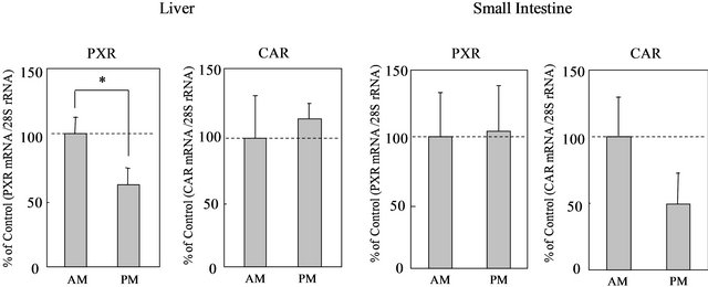
Figure 1 showed the diurnal variation of PXR and CAR in liver and small intestine derived from fed mice at 7:00 AM and 7:00 PM. PXR and CAR exhibited similar mRNA levels in liver and small intestine, suggesting that little diurnal variation of PXR (but not fasting) and CAR exists. To clarify the effect of fasting in mice on diurnal variation of nuclear receptors, the mRNA levels of PXR and CAR were determined in liver and small intestine of fasting mice. Figure 2 showed the diurnal variation of PXR and CAR in liver and small intestine derived from fasting mice at 7:00 AM and 7:00 PM. Interestingly, the mRNA levels of PXR exhibited diurnal variation by fasting for 3 days in liver. The PXR mRNA levels at 7:00 AM were significantly higher than those at 7:00 PM in liver. In contrast to PXR, the diurnal variation could not be observed in the CAR mRNA levels. Little change on diurnal variation of PXR and CAR was observed in small intestine. These results indicated that PXR and CAR exhibited little diurnal variation except for PXR in liver derived from fasting mice. The diurnal variation of transporters regulated by PXR and CAR could not be altered by the change in the mRNA levels of PXR or CAR under normal conditions. Therefore, diurnal variation in some transporters such as PEPT1 [4] or P-glycoprotein [5] could occur by change of other factors. These results also indicated that the consideration for diurnal variation in PXR and CAR is unimportant for experimental design under normal conditions. We assumed that fasting influenced the diurnal variation of nuclear recaptors because Pan et al. demonstrated that the expression of PEPT1 was affected by fasting [4]. In fasting condition, little diurnal variation of PXR or CAR was shown expect for PXR in liver. It is possible that the alterations in some factors such as lipid or glucose metabolisms under fasting condition affect the liver compared to the small intestine. However, it is unclear the precise mechanisms for diurnal variation induced in liver by fasting for

Figure 1. Diurnal variation of PXR and CAR in liver and small intestine at 7:00 AM and 7:00 PM. The mRNA levels of PXR and CAR were determined in the livers using an RT-PCR method. The results are expressed as the mean ± S.D. of each group following normalization by the corresponding level of 28S rRNA (n = 4).

Figure 2. Effect of fasting for 3 days on the diurnal variation of PXR and CAR in liver and small intestine. The mRNA levels of PXR and CAR were determined in the livers using an RT-PCR method. The results are expressed as the mean ± S.D. of each group following normalization by the corresponding level of 28S rRNA (n = 4). *p < 0.05, significant difference between the values for 7:00 AM and 7:00 PM.
3 days. Further studies are needed to clarify the details of PXR diurnal variation need to be on fasting condition. CAR and PXR have been reported to have distinct target genes, although they regulate the overlapping of some target genes involved in all phases of xenobiotic metabolism [12]. Thus, it is difficult to connect these results and the alterations of transporter and CYP in liver and small intestine.
This study provides important information regarding the diurnal variation of PXR and CAR with/without fasting. Little diurnal variation was observed on normal condition, whereas, it is possible that the expression of transporter or CYP regulated by PXR is affected by alteration in diurnal variation of PXR on fasting condition.
REFERENCES
- S. A. Kliewer, J. T. Moore, L. Wade, J. L. Staudinger, M. A. Watson, S. A. Jones, D. D. McKee, B. B. Oliver, T. M. Willson, R. H. Zetterstrom, T. Perlmann and J. M. Lehmann, “An Orphan Nuclear Receptor Activated by Pregnanes Defines a Novel Steroid Signaling Pathway,” Cell, Vol. 92, No. 1, 1998, pp. 73-82. doi:10.1016/S0092-8674(00)80900-9
- P. Honkakoski, I. Zelko, T. Sueyoshi and M. Negishi, “The Nuclear Orphan Receptor CAR-Retinoid X Receptor Heterodimer Activates the Phenobarbital-Responsive Enhancer Module of the CYP2B Gene,” Molecular and Cellular Biology, Vol. 18, 1998, pp. 5652-5658.
- D. J. Waxman, “P450 Gene Induction by Structurally Diverse Xenochemicals: Central Role of Nuclear Receptors CAR, PXR, and PPAR,” Archives of Biochemistry and Biophysics, Vol. 369, No. 1, 1999, pp. 11-23. doi:10.1006/abbi.1999.1351
- X. Pan, T. Terada, M. Okuda and K. Inui, “Altered Diurnal Rhythm of Intestinal Peptide Transporter by Fasting and Its Effects on the Pharmacokinetics of Ceftibuten,” Journal of Pharmacology and Experimental Therapeutics, Vol. 307, No. 2, 2003, pp. 626-632. doi:10.1124/jpet.103.055939
- H. Ando, H. Yanagihara, K. Sugimoto, Y. Hayashi, S. Tsuruoka, T. Takamura, S. Kaneko and A. Fujimura, “Daily Rhythms of P-Glycoprotein Expression in Mice,” Chronobiology International, Vol. 22, No. 4, 2005, pp. 655-665. doi:10.1080/07420520500180231
- C. S. Chaurasia, M. A. Alterman, P. Lu and R. P. Hanzlik, “Biochemical Characterization of Lauric Acid OmegaHydroxylation by a CYP4A1/NADPH-Cytochrome P450 Reductase Fusion Protein,” Archives of Biochemistry and Biophysics, Vol. 317, No. 1, 1995, pp. 161-169. doi:10.1006/abbi.1995.1149
- J. Y. Hong, J. M. Pan, F. J. Gonzalez, H. V. Gelboin and C. S. Yang, “The Induction of a Specific Form of Cytochrome P-450 (P-450j) by Fasting,” Biochemical and Biophysical Research Communications, Vol. 142, No. 3, 1987, pp. 1077-1083. doi:10.1016/0006-291X(87)91525-7
- S. Imaoka, Y. Terano and Y. Funae, “Changes in the Amount of Cytochrome P450s in Rat Hepatic Microsomes with Starvation,” Archives of Biochemistry and Biophysics, Vol. 278, No. 1, 1990, pp. 168-178. doi:10.1016/0003-9861(90)90245-T
- Q. Ma, G. A. Dannan, F. P. Guengerich and C. S. Yang, “Similarities and Differences in the Regulation of Hepatic Cytochrome P-450 Enzymes by Diabetes and Fasting in Male Rats,” Biochemical Pharmacology, Vol. 38, No. 19, 1989, pp. 3179-3184. doi:10.1016/0006-2952(89)90611-4
- A. Kawase, Y. Tsunokuni and M. Iwaki, “Effects of Alterations in CAR on Bilirubin Detoxification in Mouse Collagen-Induced Arthritis,” Drug Metabolism and Disposition, Vol. 35, No. 2, 2007, pp. 256-261. doi:10.1124/dmd.106.011536
- A. Kawase, I. Yoshida, Y. Tsunokuni and M. Iwaki, “Decreased PXR and CAR Inhibit Transporter and CYP mRNA Levels in the Liver and Intestine of Mice with Collagen-Induced Arthritis,” Xenobiotica, Vol. 37, No. 4, 2007, pp. 366-374. doi:10.1080/00498250701230534
- J. M. Maglich, C. M. Stoltz, B. Goodwin, D. HawkinsBrown, J. T. Moore and S. A. Kliewer, “Nuclear Pregnane X Receptor and Constitutive Androstane Receptor Regulate Overlapping but Distinct Sets of Genes Involved in Xenobiotic Detoxification,” Molecular Pharmacology, Vol. 62, No. 3, 2002, pp. 638-646. doi:10.1124/mol.62.3.638
NOTES
*Corresponding author.

