Open Journal of Obstetrics and Gynecology
Vol.2 No.2(2012), Article ID:20342,3 pages DOI:10.4236/ojog.2012.22032
Successful laparoscopic management of hemoperitoneum due to spontaneous venous rupture overlying a uterine fibroid*
![]()
Department of Obstetrics and Gynecology, Kobe Medical Center, Kobe, Japan
Email: #kyousuket@dolphin.ocn.ne.jp
Received 29 March 2012; revised 27 April 2012; accepted 9 May 2012
Keywords: laparoscopy; uterine fibroid; venous rupture; hemoperitoneum; intraoperative autologous blood transfusion
ABSTRACT
Spontaneous venous rupture overlying a uterine fibroid is a rare cause of hemoperitoneum. A 38-yearold woman presented to the emergency department with acute onset of abdominal pain. The ultrasound revealed multiple fibroids and free fluid in the peritoneum. There was a significant drop of the hemoglobin and hematocrit. The patient underwent emergent exploratory laparoscopy. A subserosal uterine leiomyoma was found, with a bleeding vein on its basis and massive hemoperitoneum. Laparoscopic myomectomy was successfully performed with local injection of vasopressin and intraoperative autologous blood transfusion. This case suggests that spontaneous intraperitoneal haemorrhage associated with uterine fibroids, although rare, should be considered in women with hypovolemic shock and a pelvic mass.
1. INTRODUCTION
Spontaneous venous rupture overlying a uterine fibroid is an unusual cause of hemoperitoneum, which may be catastrophic if it is not promptly diagnosed and treated [1-3]. We herein report the unusual case of hemoperitoneum caused by venous rupture of a uterine fibroid, which was successfully treated by laparoscopy.
2. CASE
A 38-year-old woman, gravida 2, para 2, presented to the emergency department with acute onset of epigastric pain. The past medical and surgical history was positive for a uterine fibroid without symptoms. Vital signs revealed that she was afebrile, with a pulse of 107 beats per minute, respiratory rate of 20/min and blood pressure of 90/58 mmHg. Pregnancy test was negative. Her hemoglobin concentration was 7.8 g/dl. The patient was nauseated but denied vomiting or having constapitation, abdominal trauma or exertion. She developed diffuse abdominal tenderness, guarding and rebound. Because of the location of the pain, an upper abdominal ultrasound was performed and showed moderate amount of free fluid in Morrison’s pouch, and multiple uterine fibroids. The amount of fluid was apparently dominant in the upper abdomen. Magnetic resonance (MR) imaging revealed multiple fibroids (mainly two fibroids of 6 cm in diameter) occupying the pelvic cavity and hemorrhagic ascites, which was prominent around the basis of the well-circumscribed pedunculated fundal fibroid (Figure 1). Both ovaries were intact. Although the cause of hemoperitoneum was still unknown, bleeding from a uterine fibroid was tentatively suspected.
To investigate the origin of the hemoperitoneum, laparoscopy was performed, and revealed marked hemoperitoneum. A bleeding vein was identified on the basis of a 6-cm pedunculated fibroid with tortuous blood vessels across its surface (Figure 2). Intramural fibroid of 5 cm in diameter was also noted in anterior uterine wall. Total amount of 20 ml diluted vasopressin 0.2 IU/ml was used to infiltrate the fibroid around the bleeding vessel, which was simultaneously electrocoagulated. The bleeding was remarkably decreased with these procedures. A thorough search of the pelvic and abdominal cavities revealed no other source of bleeding or pathology. Because marked hemoperitoneum was noted and no symptoms due to uterine fibroid existed, laparoscopic myomectomy for the bleeding fibroid was performed. During surgery, 2800 ml of blood were suctioned from the abdominal cavity and the processed blood was reinfused for autologous transfusion during surgery with Cell Saver 5 (Haemonetics
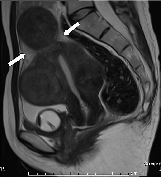
Figure 1. Multiple fibroids and hemorrhagic ascites, which was prominent around the basis of the well-circumscribed pedunculated fundal fibroid (arrows).

Figure 2. A bleeding vein on the basis of a 6-cm pedunculated fibroid with tortuous blood vessels (arrow).
Corporation, Braintree, Massachusetts). The postoperative course was uncomplicated and the patient was discharged on postoperative day 3.
3. DISCUSSION
Hemoperitoneum is usually caused by ectopic pregnancy, ruptured ovarian mass or adnexal torsion. Acute bleeding from a blood vessel overlying a uterine fibroid occurs rarely. In most cases there is a history of violent coitus, hard work, defecation and examination of anesthesia [1]. Direct pelvic trauma can induce avulsion of a pedunculated fibroid [2,3]. Posterior location of the bleeding vein favors the theory of direct contact injury from the promontory of the sacrum.
Spontaneous rupture of a superficial vein is extremely uncommon. Several theories are considered responsible for spontaneous venous rupture of a uterine fibroid. Increased congestion of a superficial vein of the fibroid owing to menstruation or pregnancy has been suggested [4]. Spontaneous bleeding without any history of trauma, increased abdominal pressure, recent pregnancy or menstruation as our case is extremely rare. Dahan et al. [5] reported that risk of uterine fibroid rupture was independent of patient age and parity, and the size of the fibroid.
Treatment consists of ligation of bleeding vessels, myomectomy and/or hysterectomy. In most reported cases, laparotomy was chosen for the presence of active bleeding and hemodynamic instability. Estrade-Huchon et al. [3] reported a case of avulsion of a pedunculated uterine bibroid, where laparoscopy performed for severe hemoperitoneum revealed bleeding originated from the base of a subserosal fibroid, and subtotal hysterectomy via laparotomy was subsequently carried. In the present case, laparoscopic myomectomy was successfully performed because the patient had only one bleeding vein which was immediately identified and controlled by injection of vasopressin.
Although uncommon, spontaneous bleeding from uterine fibroids should be in the differential diagnosis of hemoperitoneum in a premenopausal woman with subserosal and/or pedunculated uterine fibroids. Laparoscopy is useful in investigating the origin of hemoperitoneum and consequent conservative surgery (i.e., myomectomy) can be conducted with injection of vasopressin if prompt preoperative diagnosis was made before deterioration of hemodynamic parameters.
REFERENCES
- Danikas, D., Theodorou, S.J., Kotrotsios, J., Sills, C. and Cordero, P.E. (1999) Hemoperitoneum from spontaneous bleeding of a uterine leiomyoma: A case report. The American Journal of Surgery, 65, 1180-1182.
- Drutman, J. and Fruechte, D.M. (1992) Hemoperitoneum due to traumatic avulsion of a pedunculated uterine leiomyoma. American Journal of Roentgenology, 158, 1410.
- Estrade-Huchon, S., Bouhanna, P., Limot, O., Fauconnier, A. and Bader, G. (2010) Severe life-threatening hemoperitoneum from posttraumatic avulsion of a pedunculated uterine leiomyoma. Journal of the Minimally Invasive Gynecology, 17, 651-652. doi:10.1016/j.jmig.2010.04.006
- Mattison, D.R. and Yeh, S.Y. (1980) Hemoperitoneum from rupture of a uterine vein overlying a leiomyoma. American Journal of Obstetrics and Gynecology, 136, 415-416.
- Dahan, M.H. and Ahmadi, R. (2002) Spontaneous subserosal venous rupture overlying a uterine leiomyoma. A case report. The Journal of Reproductive Medicine, 47, 419-420.
NOTES
*Conflict of interest: the authors have no conflicts of interest in connection with submitted material.
#Corresponding author.