Vol.2, No.3, 246-252 (2010)
doi:10.4236/health.2010.23035
Copyright © 2010 SciRes Openly accessible at http://www.scirp.org/journal/HEALTH/
Health
The increase of adenylate kinase activity in the blood
can control aggregation of platelets in coronary or
peripheral arterial ischemia
Bożena Studzińska, Anna Seroka, Marta Łępic ka, Katarzyna Roszek, Michał Ko moszyński
Biochemistry Department, Institute of General and Molecular Biology, Nicolaus Copernicus University, Toruń, Poland;
michkom@chem.uni.torun.pl
Received 15 November 2009; revised 21 December 2009; accepted 24 December 2009.
ABSTRACT
Activation and aggregation of blood platelets is
crucial for hemostasis and thrombosis. In the
vascular system adenine nucleotides are im-
port ant signaling molecules playing a key role in
hemostasis. ADP was the first low molecular
weight agent recognized to cause blood plate-
lets activation and aggregation. NTPDases and
adenylate kinase (AK) are the main enzymes
involved in metabolism of extracellular adenine
nucleotides. The majority of studies concen-
trated on the role of NTPDase1 (apyrase) in the
inhibition of platelets aggregation. Up to now,
there are still insufficient data concerning the
role of AK in this process. We found that ade-
nylate kinase activity in the serum of patients
with myocardial infarction is significantly in-
creased when compared to the healthy volun-
teers. The elevated activity of AK is connected
to appearance of another isoform of that en-
zyme, expressed in patients with myocardial
infarction. The influence of AK on the pig blood
platelets aggregation induced by 20 μM ADP or
7.5 μg/ml rat collagen was examined. 1U of
adenylate kinase added to platelet-rich plasma
(PRP) before ADP or collagen, inhibited the
platelets aggregation. One minute after induc-
tion of platelets activation by ADP as much as
5U of adenylate kinase was necessary to stop
the platelet aggregation. In the case of collagen
activated aggregation, only 2U of AK added 1 or
5 minutes after initiation of the aggregation
process were sufficient for disaggregation of
platelets. The increase of ATP: ADP ratio is
probably responsible for the initiation of dis-
aggregation process. We conclude that ade-
nylate kinase is involved in regulation of plate-
lets aggregation. Anticoagulative role of AK in-
dicates the possibility of using this enzyme in
the treatment of cardiovascular diseases.
Keywords: Hemostasis; Platelets Aggregation;
Adenylate Kinase
1. INTRODUCTION
It is well known that platelets aggregation is the key
process in thrombosis. Platelets hyperaggregability is
associated with the risk factors for cerebral, coronary or
peripheral arterial ischemia causing stroke, cardiovascu-
lar disease and venous thrombosis [1,2]. The WHO es-
timated that 12.6 p ercent of deaths worldwide are caused
by the ischemic heart disease, that is the leading cause of
death in the develop ed countries [3]. The novel th erapies
against platelet-dependent thrombosis are targeted at
purinergic receptor (P2Y12) activating the platelets ag-
gregation. In the last few years the thienopyridyne de-
rivatives (ticlopidine, clopidogrel, prasugrel) are mainly
used.
Adenine nucleotides play an important role in blood
platelets function. Platelets possess three purinergic re-
ceptors for ecto-nucleotides: P2Y1 and P2Y12, which
interact with ADP, and P2X1, which interacts with ATP
[4-6]. The interaction of adenine nucleotides with their
receptors activates platelets and leads t o their aggregation.
Among the nucleotides, ADP is a key mo lecule involved
in hemostasis and development of arterial thrombosis
[7-9]. Furthermore, ATP stimulation of P2X1 triggers off
the platelets shape change and helps to amplify platelet
responses mediated by agonists such as collagen. Acti-
vation of each of these nucleotide receptors results in
unique si g nal t rans d uct i on pat h way s that are im port a nt i n
the regulation of hemostasis and thrombosis [10].
The most important enzymes metabolizing ecto-
ncleotides in the vascular system are: NTPDase1 (EC
3.6.1.5), NTPDase2 (EC 3.6.1.5), 5’-nucleotidase (EC
2.7.4. 6), ade nos ine deam inase ( EC 3.5. 4.4 ) and a deny late
kinase (EC 2.7.4.3) [8-10]. These enzymes maintain the
B. Studzińska et al. / HEALTH 2 (2010) 246-252
Copyright © 2010 SciRes Openly accessible at http://www.scirp.org/journal/HEALTH/
247
proper level of purine nucleotides.
NTPDase1 (apyrase, CD39) metabolizes ATP and ADP
to AMP. Its role in blocking the platelet aggregation
triggered by ADP is well recognized [14,15]. The de-
creased activity of NTPDase1 in the atherosclerotic ar-
teries proves its important regulatory function in throm-
bosis [13].
Adenylate kinase (AK) also participates in controlling
the adenine nucleotides concentration due to transferring
the phosphate group between ATP, ADP and AMP [17-
19]. Some authors suggest, that serum adenylate kinase is
responsible for the clearance of circulating ADP and
therefore blocks activation of platelets [11,20]. Based on
these information, adenylate kinase may be useful in the
regulation of platelets activity. The research results pub-
lished in the 1970s show the increased AK activity in
serum and urine of patients in the early phase of myo-
cardial infarction [21,22]. However, there are still insuf-
ficient data concerning the adenylate kinase influence on
the platelets aggregation [23].
The aim of our research was to determine the role of
adenylate kinase in the regulation of platelets aggrega-
tion.
2. MATERIALS AND METHODS
Reagents. The following reagents were used: ethanola-
mine, n-heptane, KCl, HClO4 and EDTA (POCh Gliwice,
Poland, p.a. grade), KH2PO4, K2HPO4, tetrabutylammo-
nium hydrogen sulphate (TBA) and isocratic methanol
(Baker Phillipsburg, USA, HPLC grade). Adenylate
kinase (AK) from Bacillus stearothermophilus, potato
NTP-Dase1 (apyrase), ADP, rat collagen and other re-
agents were purchased from Sigma Chemical Co.
(USA).
Materials. Human serum was obtained from blood
samples taken from healthy volunteers and patients with
myocardial infarction collected not later than 3 hours
after the incident in the WSZ Hospital, Torun (Poland).
Platelet-rich plasma (PRP) was prepared from fresh
pig blood samples. The whole citrated blood was centri-
fuged at 200 × g for 20 min in temperature 10ºC and the
resulted supernatant was used in the experiments.
Platelet aggregation. All the experiments were con-
ducted in 37ºC. To 800 l sample of PRP (containing
246 × 103 to 458 × 103 platelets in μl counted by Sysmex
K-1000 hematology analyzer) the appropriate am- ounts
of ADP, rat collagen and adenylate kinase was added.
The platelet aggregation was monitored by spectropho-
tometric measurement at λ = 600nm.
The appropriate amount of the adenylate kinase was
added to the PRP together with ADP or collagen, 1 min-
ute or 5 minutes after initiation of the aggregation, re-
spectively.
Assay of nucleotides. The qualitative and quantitative
analysis of purines in the reaction mixtures was made by
the HPLC method [21]. The samples were separated on
the Nova-Pack C18 column, 3.9 mm × 150 mm (Waters
Co., Milford, MA, USA). The presence of purines was
detected at λ=260 nm.
Adenylate kinase activity determination. 50 μl diluted
human serum was added to 50 μl reaction mixture (2mM
ADP, 1.5 mM MgCl2 and 0.1 mM suramin in 50 mM
Hepes, pH 7.6). The enzymatic reaction was terminated
with 100 μl 1M HClO4. Then the samples were neutral-
ized with KOH and delipidated with n-heptane. 20 μl
aliquots were analysed by the HPLC method [21].
Partial purification of adenylate kinase. Human se-
rum from healthy volunteers and patients with myocar-
dial infarction was precipitated with ammonium sulfate
(35% saturation and the obtained supernatant up to 85%
saturation). The partially purified enzyme with the activ-
ity of 2.27 U (healthy volunteers serum) or 17.3 U (se-
rum after myocardial infarction) was used in further ex-
periments.
Affinity chromatography on Blue-Sepharose. The Blue
-Sepharose column (Ø 1.5 cm × 13 cm) was equilibrated
with 10 mM Tris-HCl buffer pH 7.4 containing 1 mM
dithiotreitol and 0.1 mM EDTA. Approximately 15 mg
of partially purified protein from the previous step was
applied to the column. The proteins were eluted with the
linear gradient of 0÷2M NaCl. One-milliliter fractions
were collected and used to the determination of AK ac-
tivity and protein concentration.
Statistical analysis. All data presented in this paper
derive from 3 to 6 independent experiments. The results
are expressed as mean SD.
3. RESULTS
Our experiments on the purine nucleotides metabolism
in the human serum confirmed that adenylate kinase
activity in patients with myocardial infarction is signi-
ficantly increased when compared to the healthy volun-
teers (Figure 1).
The elution profile after Blue-Sepharose chroma-
tography of partially purified human serum obtained
from healthy volunteers and patients with myocardial
infarction is also different (Figure 2). There were no
significant changes in the NTPDase activity (data not
shown). These data suggest that the elevated activity of
AK is connected to appearance of another isoform of
that enzyme, expressed only in patients with myocardial
infarction.
To precisely explain the role of adenylate kinase in
blood, the AK influence on the platelets aggregation
process was determined. Simultaneously with the plate-
lets aggregation we analyzed: a/ the concentration of
purines and b/ the adenylate kinase influence of this
process. These experiments provoked that a minimal vo-
B. Studzińska et al. / HEALTH 2 (2010) 246-252
Copyright © 2010 SciRes Openly accessible at http://www.scirp.org/journal/HEALTH/
248
lume of PRP analyzed was 800 μl. Therefore, the meas-
urement of platelet aggregation required using the spec-
trophotometer. The degree of platelet aggregation in pig
blood is dependent on the ADP and rat collagen concen-
trations (Figure 3). Based on the experiments, we found
that 20 μM ADP and 7.5 μg/ml rat collagen were used in
further experiments as sufficient to the efficient aggrega-
tion of pig blood platel e ts.
Figure 1 . Comparison of th e aden ylate kinase activity in human
serum of patients with myocardial infarction () and healthy
volunteers ().
(a)
(b)
Figure 2. Elution profile after Blue-Sepharose chromatography
of human serum preparation from healthy volunteers (a) and
patients with myocardial infarction (b). Adenylate kinase activ-
ity of eluted fractions (—o—) was estimated wit h 2mM ADP as
substrate. The protein concentration (——) was measured
spectrophotometrically at λ = 280 nm. NaCl concentration
(——) ranged from 0 to 2M.
(a)
(b)
Figure 3. The influence of different concentrations of ADP (a)
and rat co l la g en (b ) on the platelets aggr egation in PRP. Control
PRP (—o—), PRP + 10 M ADP (——), PRP + 20 M ADP
(——), PRP + 30 M ADP (——),PRP + 5 g/ml collagen
(——), PRP + 7.5 g/ml collagen (——), PRP + 10 g/ml
collagen (——).
The purines concentration was controlled after 10
minutes of incubation in 37ºC in control PRP and in PRP
activated by 20μM ADP or 7.5μg/ml rat collagen (Table
1). Concentration of nucleotides and nucleosides differs
between PRP activated by 20μM ADP and by 7.5μg/ml
rat collagen.
Table 1. Concentration of purines after 10 min incubation of
control PRP, PRP activated by 20 μM ADP and PRP activated
by 7. 5 μg/ml rat collagen.
Purines concentration [μM]
Ado AMP ADP ATP
Control PRP292.9±27.8 0 0 0
PRP+20μM
ADP 1042.0±100.4 46.0±6.7 18.5±3.3 14.3±2.4
PRP+7,5μg/m
l rat col lagen 2084.9±140. 666.7 ± 6.3 10.9 ± 4. 7 26.3±1 1.3
B. Studzińska et al. / HEALTH 2 (2010) 246-252
Copyright © 2010 SciRes Openly accessible at http://www.scirp.org/journal/HEALTH/
249
Then the adenylate kinase influence on the aggrega-
tion process induced with the addition of 20 M ADP
was analysed. The appropriate amount of the enzyme
was added to the PRP together with ADP, 1 minute or 5
minutes after initiation of the aggregation, respectively–
Figures 4-7.
The presented results show that addition of 1U ade-
nylate kinase before initiation of the aggregation proc-
esss by ADP effectively blocks aggregation (Figure 4).
Addition of 5U ad enylate kinase in 1 minute after plate-
lets activation (Figure 6) or 6U in 5 minutes after acti-
vation (Figure 7), respectively, is necessary to stop the
aggregation process. The purines concentration in the
control PRP, in PRP after activating platelets with 20μM
ADP and in the presence of adenylate kinase are sum-
marized in Table 2.
Time [s]
0100 200 300 400 500 600
% Aggregation
0
20
40
60
80
100
120
Figure 4. Influence of 1U adenylate kinase (AK) on the plate-
lets aggregation in PRP induced by 20μM ADP. Control PRP +
20 M ADP (—o—), PRP + 1U AK + 20 M ADP (——).
Time [s]
0100 200 300 400 500 60
0
% Aggregati on
0
20
40
60
80
100
120
140
160
Figure 5. Influence of 1U and 2U adenylate kinase (AK) added
1 min after initiation of the platelets aggregation in PRP by 20
μM ADP. Control PRP + 20 M ADP (—o—), PRP + 20 M
ADP + 1U AK after 1 min. (——), PRP + 20 M ADP + 2U
AK after 1 min. (——).
Time [s]
0100 200300 400500 600
% Aggregati on
0
20
40
60
80
100
120
Figure 6. The influence of 5U adenylate kinase (AK) added 1
min after initiation of the pla t elets aggregation in PRP by 20μM
ADP. Control PRP + 20 M AD P (—o—), PRP + 20 M ADP +
5U AK after 1 min. (——).
Time [s]
0100 200 300 400 500 600
% Aggregati on
0
20
40
60
80
100
120
Figure 7. The influence of 6U adenylate kinase (AK) added 5
min after initiation of the pla t elets aggregation in PRP by 20μM
ADP. Control PRP + 20 M AD P (—o—), PRP + 20 M ADP +
6U AK after 5 min. (——).
Table 2. Influence of AK on the concentration of purine nu-
cleotides in PRP activated by 20 μM ADP.
Nucleotides concentration [μM]
Amount of
AK
Time of
enzyme
addition AMP ADP ATP
No enzyme- 46.0 ± 6.7 18.5 ± 3.3 14.3 ± 2.4
1U Before activa-
tion by ADP 92.9 ± 11.3 21.5 ± 4.6 55.2 ± 3.3
1U 1 min after
activation by
ADP 40.4 ± 3.3 5 2.3 ± 5.8 45.0 ± 1.5
2U 1 min after
activation by
ADP 49.3 ± 0.7 6 3.2 ± 2.6 67.9 ± 1.9
5U 1 min after
activation by
ADP 100.8 ± 2. 1 23.6 ± 4.5 50.2 ± 2.1
6U 5 min after
activation by
ADP 81.2 ± 13.9 22.9 ± 1.3 56.7 ± 2.0
B. Studzińska et al. / HEALTH 2 (2010) 246-252
Copyright © 2010 SciRes Openly accessible at http://www.scirp.org/journal/HEALTH/
250
Inhibition of adenylate kinase activity by 20μM dia-
denosine pentaphosphate (AP5A) in the presence of
ADP causes the platelets aggregation as in the control
with ADP only ( Figure 8). It proves that the aggregation
process is inhibited in fact due to the adenylate kinase
activity.
The adenylate kinase influence on the aggregation
process induced by the ad ditio n of 7.5 μg/ml rat collagen
was also analyzed. The appropriate amount of the en-
zyme was added to the PRP simultaneously with colla-
gen, 1 minute or 5 minutes after initiation of the aggre-
gation respectively (Figures 9-11).
The presented results show that only 2U of adenylate
kinase added to PRP within 1 or 5 minute after initiation
of the aggregation process cause an almost complete
disaggregation of platelets. The purines concentration in
control PRP, in PRP after activating the platelets with 7.5
μg/ml rat collagen and in the presence of adenylate
kinase are summarized in Table 3.
Time [s]
0100 200 300400 500 600
% Aggregation
0
20
40
60
80
100
120
Figure 8. The influence of 20μM AP5A on the platelets ag-
gregation in PRP in the presence of 1U adenylate kinase (AK).
Control PRP + 20 M ADP (—o—), PRP + 1U AK + 20 M
ADP (——), PRP + 20μM AP5A + 1U AK + 20 M ADP
(——).
Time [s]
0100 200 300 400 500 600
% A ggregation
0
20
40
60
80
100
120
Figure 9. The influence of 1U adenylate kinase (AK) on the
platelets aggregation in PRP induced by 7.5μg/ml rat collagen.
Control PRP + 7.5 g/ml collagen (—o—), PRP + 1U AK + 7 .5
g/ml collagen (——).
Time [s]
0100 200 300400 500600
% Aggregation
0
20
40
60
80
100
120
Figure 10. The influence of 1U and 2U adenylate kinase (AK)
added 1 min after initiation of the platelets aggregation in PRP
by 7.5μg/ml rat collagen. Control PRP + 7.5 g/ml collagen
(—o—), PRP + 7.5 g/ml collagen + 1U AK after 1 min.
(——), PRP + 7.5 g/ml collagen + 2U AK after 1 min.
(——).
Time [s]
0100 200 300 400 500600
% Aggregation
0
20
40
60
80
100
120
Figure 11. The influence of 2U adeny late kinase (AK) added 5
min after initiation of the platelets aggregation in PRP by
7.5μg/ml rat collagen. Control PRP + 7.5 g/ml collagen
(—o—), PRP + 7.5 g/ml collagen + 2U AK after 5 min.
(——).
Table 3. Influence of AK on the concentration of purine nu-
cleotides in PRP activated by 7.5 μg/ml rat collagen.
Nucleotides concentration [μM]
Amount
of AK Time of enzyme
addition AMP ADP ATP
No
enzyme - 66.7 ± 6.3 10.9 ± 4.7 26.3 ± 11.3
1U Before activation
by rat collagen 68.0 ± 1.3 12.9 ± 0.3 47.7 ± 4.3
1U 1 min after
activation
by rat collagen 35.9 ± 6.2 36.0 ± 1.1 41.4 ± 2.3
2U 1 min after
activati o n by rat
collagen 78.0 ± 4.3 14.4 ± 0.3 49.4 ± 0.8
2U 5 min after
activati o n by rat
collagen 100.3 ±18.2 8.8 ± 8.9 51.1 ± 0.1
B. Studzińska et al. / HEALTH 2 (2010) 246-252
Copyright © 2010 SciRes Openly accessible at http://www.scirp.org/journal/HEALTH/
251
4. DISCUSSION
The nucleotide hydrolases and kinases are both engaged
in the precise regulation of purines concentration in the
vascular system therefore controlling the platelets agg-
regation process. Previous studies on platelets aggrega-
tion and thrombosis revealed that NTPDase1 (apyrase)
inhibits activation of blood platelets [14,15]. The potato
apyrase was used most often. However the latest ex-
periments concerned human NTPDase1 produced by
using a yeast expression system, purified, and reconsti-
tuted within lipid vesicles [25] or recombined human
NTPDase1 expressed in bacteria [26].
The results of our studies indicated that in addition to
antithrombotic effects of NTPDase1, also adenylate
kinase is involved in regulation of aggregation. The in-
creased activity of AK in patients after myocardial in-
farction is connected with ap pearance of another isoform
of the enzyme. The occurence of another active isoform
of AK can constitute an emergency mechanism main-
taining the proper blood flow due to the clearance of
circulating ADP. Likewise in Duchenne dystrophic pa-
tients, an additional isoform of AK was identified and
separated on Blue-Sepharose column chromatography
[27]. The increase of AK activity observed for patients
after myocardial infarction confirmed its correlation with
platelets aggregation.
The minimal efficient ADP concentration required for
human platelet aggregation is 1.5 μM [28]. We found
that in pig blood the ADP and collagen concentrations
required for the platelets aggregation in vitro are higher
than in human blood. However, in the literature there is
very little data concerning the pig platelets aggregation
process. It cannot be excluded that there are differences
between species in minimal ADP and collagen concen-
trations required to the platelets aggregation.
The decrease in ADP con centration may lead to termi-
nating the platelets aggregation. Therefore NTPDase1
(apyrase), which hydrolyses ATP and ADP to AMP, has
an antithrombotic effect due to inhibition of aggregation
but not to disaggregation of platelets [28]. Our experi-
ments proved that aggregation process initiated by ADP
as well as by rat collagen was blocked but not reversed
in the presence of potato NTPDase1 (results not shown).
Unexpectedly, AK causes almost complete disaggre-
gation of activated platelets up to five minutes after ini-
tiation of the aggregation process by collagen. These
results are in agreement with the paper of Rysanek et al.
[23], but their experiments concerned myokinase prepa-
ration containing ammonium sulphate. It is not clear that
myokinase itself caused the disaggregation of platelets.
In our experiments myokinase did not affected in neither
inhibition of aggreg ation nor disaggregation of activated
platelets (results not shown). The main difference be-
tween myokinase and adenylate kinase is their substrate
specificity. The preferred substrate for myokinase is ATP
while for adenylate kinase from B. stearothermophilus
the KM for ATP is the same as for ADP. The value is
KM(ADP) = 0,037 mM, KM(ATP) = 0,036 mM respectively
[29]. Therefore, during activation and aggregation of
platelets, when high concentrations of ADP are present
in the blood, adenylate kinase from B. stearothermophi-
lus preferably uses two ADP molecules to synthesis of
ATP and AMP. The decrease in ADP concentration si-
multaneous with the increase in ATP concentration may
be the main requirement for disaggregation of platelets.
In the aggregation process induced by 20 μM ADP,
concentration of the nucleotide is high enough for the
activated platelets to partially undergo irreversible ag-
gregation [30,31]. In this case, NTPDase1 as well as
adenylate kinase stop the aggregation, but do not trigger
the disaggregation of platelets. ADP concentration in the
samples activated by rat collagen is lower. The ATP:
ADP ratio after the AK add ition to th e platelets activated
by collagen is higher than that observed for platelets
activated by ADP. The increase of ATP:ADP ratio is
probably responsible for the initiation of disaggregation
process. That explains why the aggregation may be re-
versed only in the case of platelets activation initiated by
rat collagen.
In conclusion, the disaggregation process is possible
in the appropriately low ADP concentration and only in
the presence of adenylate kinase. However, the effects of
adenylate kinase activity are diverse and multidirectional.
The enzyme not only decreases the ADP level but also
produces ATP and AMP. Dilation of blood vessels in the
response to ATP decreases the blood pressure. AMP is
hydrolysed by 5’-nucleotidase to adenosine that inhibits
blood platelets aggregation [32,33].
The platelet activation and aggregation occurs in the
early stage of the thrombosis. The occlusive thrombus
formation is a main cause of acute coronary syndromes
and stroke in humans. Anticoagulative role of adenylate
kinase indicates the possibility of using this enzyme as
antithrombotic agent in the treatment of cardiovascular
diseases, including myocardial infarction and ischemic
stroke.
REFERENCES
[1] Jurk, K. and Kehrel, B.E. (2 005) Platelets: Physiolog y and
Biochemistry. Seminars in thr ombosis and hemostasis, 31,
381-392.
[2] Willoughby, S., Holmes, A. and Loscalzo, J. (200 2) Plate-
lets and cardiovascular disease. European Journal of
Cardiovascular Nursing, 1, 273-288.
[3] www.americanheart.org/presenter.jhtml?identifier=4478.
[4] Heptinstall, S., Johnson, A., Glenn, J.R. and White, A.E.
(2005) Adenine nucleotide metabolism in human blood–
important roles for leukocytes and erythrocytes. Journal
of Thrombosis a nd Haemostasis, 3, 2331-2339.
B. Studzińska et al. / HEALTH 2 (2010) 246-252
Copyright © 2010 SciRes http://www.scirp.org/journal/HEALTH/Openly accessible at
252
[5] Cattaneo, M. (2007) Platelet P2 receptors: old and new
targets for antithrombotic drugs. Expert Review of Car-
diovascular Therapy, 5, 45-55.
[6] Erlinge, D. and Burnstock, G. (2008) P2 receptors in
cardiovascular regulation and disease. Purinergic Signal,
4, 1-20.
[7] Woulfe, D., Yang, J. and Brass, L. (2001) ADP and
platelets: The end of the beginning. Journal of Clinical
Investigation, 107, 1591-1598.
[8] Gachet, C. (2001) ADP receptors of platelets and their
inhibition. Journal of Thrombosis and Haemostasis, 86,
222-232.
[9] Daniel, J.L., Dangelmaier, C., Jin, J., Ashby, B., Smith,
J.B. and Kanapuli, S.P. (1998) Molecular Basis for ADP-
induced platelet activation. Journal of Biological Chem-
istry, 273, 2024-2029.
[10] Kahner , B.N., Shankar, H., Murugappan, S., Prasad, G.L.
and Kunapuli, S.P. (2006) Nucleotide receptor sign aling in
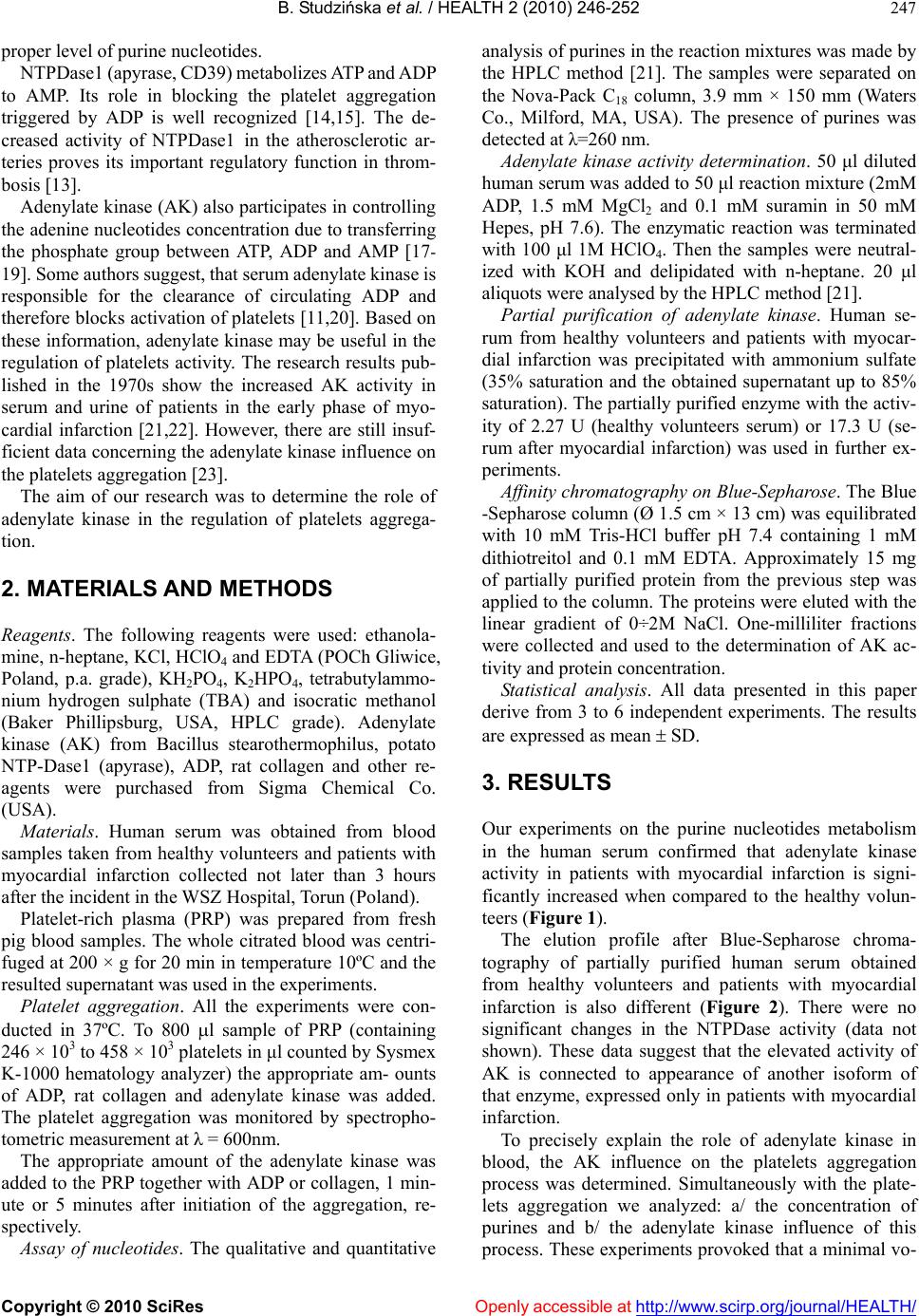
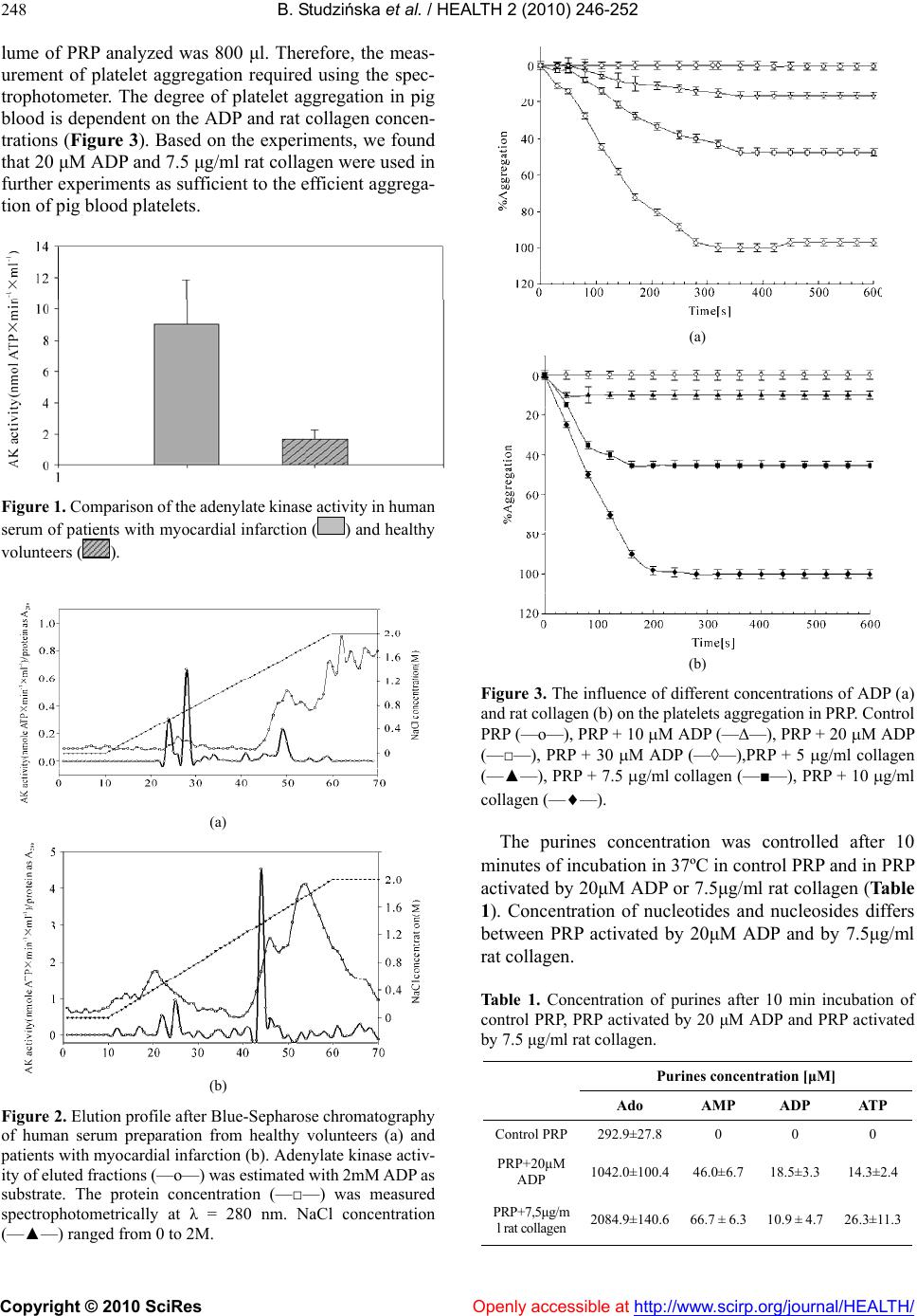
platelets. Journal of Thrombosis and Haemostasis, 4,
2317-2326.
[11] Yegutkin, G.G., Samburski, S.S. and Jalkanen S. (2003)
Soluble purine-converting enzymes circulate in human
blood and regulate extracellular ATP level via counter-
acting pyrophosphatase and phosphotransfer reactions.
Journal of the Federation of American Societies for Ex-
perimental Biology, 17, 1328-1330.
[12] Yegutkin, G.G. (2008) Nucleotide and nucleoside
-converting ectoenzymes: Important modulators of puri-
nergic signalling cascade. Biochimica et Biophysica Acta,
1783, 673- 694.
[13] Quillen, E.E., Haslam, G.C., Samra, H.S., Amani-Taleshi,
D., Knight, J.A., Wyatt, D.E., Bishop, S.C., Colvert, K.K,
Richter, M.L. and Kitos, P.A. (2006) Ecto-adenylate
kinase and plasma membrane ATP synthase activities of
human vascular endothelial cells. Journal of Biological
Chemistry, 281, 20728-20737.
[14] Marcus, A.J., Broekman, M.J., Drosopoulos, J.H., Olson,
K.E., Islam, N., Pinsky, D.J. and Levi, R. (2005) Role of
CD39 (NTPDase-1) in thromboregulation, cerebropro-
tection, and cardioprotection. Seminars in Thrombosis
and Hemostasis, 31, 234-246.
[15] Atkinson, B., Dwyer, K., Enjyoji, K. and Robson, S.C.
(2006) Ecto-nucleotidases of the CD39/NTPDase family
modulate platelet activation and thrombus formation:
Potential as therapeutic targets. Blood Cells Molecules
and Diseases, 36, 217-222.
[16] Łęcka, J., Molski, S. and Komoszyński, M. (1999) Al-
teration of ectopurine metabolism in vascular disease. In
Vanduffel L., Lemmens R. (ed.), Ecto-ATPases and re-
lated ectonucleotidases. Maastricht, Shaker Publishing
BV.
[17] Gellerich, F.N. (1992) The role of adenylate kinase in
dynamic compartmentation of adenine nucleotides in the
mitochondrial intermembrane space. FEBS Letters, 297,
55-58.
[18] Picher, M. and Boucher, R.C. (2003) Human airway
ecto-adenylate kinase. A mechanism to propagate ATP
signaling on airway surfaces. Journal of Biological
Chemistry, 278, 11256 -11264.
[19] Noma, T. (2005) Dynamics of nucleotide metabolism as a
supporter of life phenomena. Journal of Medical Inves-
tigation, 52, 127-136.
[20] Haslam, R.J. and Mills, D.C.B. (1967) The adenylate
kinase of human plasma, erythrocytes and platelets in re-
lation to the degradation of adenosine diphosphate in
plasma. Biochemical Journal, 103 , 773-784.
[21] Kędrowa, S. and Worsztynowicz-Jałowiec, E. (1971)
Myokinase activity in myocardial infarction. Polish
Medical Journal, 10, 805-811.
[22] Frithz, G., Ericsson, P. and Ronquist, G. (1976) Serum
adenylate kinase activity in the early phase of acute
myocardial infarction. Upsala Journal of Medical Sci-
ences, 81, 155-158.
[23] Rysánek, K., Svehla, C., Spánková, H. and Mlejnková M.
(1969) The effect of myokinase on the aggregation and
disaggregation of thrombocytes. Experientia, 25, 31-32.
[24] Czarnecka, J., Cieślak, M. and Komoszyński, M. (2005)
Application of solid phase extraction and high-perform-
ance liqu id chroma tography to q ualitative and quantit ative
analysis of nucleotides and nucleosides in human cere-
brospinal fluid. Journal of Chromatography B, 822, 85- 90.
[25] Haller, C.A., Cui, W., Wen, J., Robson, S.C. and Chaikof,
E.L. (2006) Reconstitution of CD39 in liposomes ampli-
fies nucleoside triphosphate diphosphohydrolase activity
and restores thromboregulatory properties. Journal of
Vascular Surgery, 43, 816-823.
[26] Yang, M. and Kirley, T.L. (2008) Engineered human sol-
uble calcium-activated nucleotidase inhibits coagulation
in vitro and thrombosis in vivo. T hr ombosis Resear ch, 122,
541-548.
[27] Hamada, M., Sumida, M., Kurokawa Y., Sunayashiki
-Kusuzaki, K., Okuda, H., Watanabe, T. and Kuby, S.A.
(1984) Studies on the adenylate kinase isozymes from the
serum and erythrocyte of normal and Duchenne dys-
trophic patients. Journal of Biological Chemistry, 260,
11595-11602.
[28] Łęcka, J. and Komoszyński, M. (2003) The role of
ecto-aden osine a nd ect o-a denin e nucleo ti des i n r egul ation
of blood pressure, haemostasis and etiology of athero-
sclerosis. Progress in Medical Research, 1, 48-74.
[29] Glaser, P., Presecan, E., Delepierre, M, Surewicz, W.K.,
Mantsch, H.H., Barzu, O. and Gilles, A.M. (1992) Zinc, a
novel structural element found in the family of bacterial
adenylate kinases. Biochemistry, 31, 3038-3043.
[30] Furie, B. and Furie, B.C. (2005) Thrombus formation in
vivo. Journal of Clinical Investigation, 115 , 3355-3362.
[31] Kuwahara, M., Sugimoto, M., Tsuji, S., Matsui, H., Mi-
zuno, T., Miyata, S. and Yoshioka, A. (2002) Platelet
shape changes and adhesion under high shear flow. Arte-
riosclerosis, Thrombosis, and Vascular Biology, 22, 329-
334.
[32] Wan, J., Ristenpart, W.D. and Stone, H.A. (2008) Dy-
namics of shear-induced ATP release from red blood cell.
Proceedings of National Academy of Sciences, USA, 105,
16432-16437.
[33] Kawashima, Y., Nagasawa, T. and Ninomiya, H. (2000)
Contribution of ecto-5’-nucleotidase to the inhibition of
platelet aggregat ion by human endothelial cells. Blood, 96,
2157-2162.